| disease | Platybasia and Basilar Invagination |
Basilar invagination refers to the upward indentation of the skull base bone around the foramen magnum of the occipital bone into the cranial cavity, causing the underlying atlas and axis, particularly the odontoid process, to elevate or even enter the skull base. This deformity rarely occurs in isolation and is often associated with other malformations in the foramen magnum region, such as occipitalization of the atlas, cervicalization of the occipital bone, stenosis of the foramen magnum, and developmental abnormalities of the odontoid process.
bubble_chart Pathological Changes
Basilar invagination refers to the inward depression of the bony structures around the foramen magnum into the cranial cavity, with the upward displacement of the atlas and odontoid process, which protrude into the foramen magnum, compressing neural structures such as the brainstem (Figure 1).
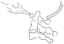
Figure 1 Basilar invagination
Basilar invagination is generally classified into two types: primary and secondary. The former refers to congenital malformations, which are more common. It is often associated with anomalies of the atlas and axis, atlanto-occipital fusion, hypoplasia of the anterior or posterior arches or lateral masses of the atlas, odontoid process abnormalities, and the so-called Klippel-Feil syndrome, among other common associated malformations. Occasionally, severe rickets, osteomalacia, osteoporosis, renal osteodystrophy, and other factors can also lead to basilar invagination. Due to the softening of the bone, the skull sinks under the force of gravity, causing basilar invagination, which is termed secondary. This type is extremely rare and its clinical significance is far less important than the congenital form.
Platybasia refers to the higher developmental position of the posterior cranial fossa, where the basal angle—formed by the lines from the center of the sella turcica to the anterior edge of the foramen magnum and from the nasion to the sella turcica—increases, resulting in a flattened cranial base. In normal adults, this angle ranges from 132° to 140°. A decreased basal angle has no clinical significance, while an increased angle indicates a developmental malformation of the cranial base.bubble_chart Clinical Manifestations
Congenital basilar invagination often gradually manifests neurological symptoms in middle age, typically after 20 to 30 years of age. It is frequently triggered by minor trauma or falls, leading to damage to the brainstem or spinal cord. At this stage, even young children may develop symptoms. However, most patients experience gradual symptom progression due to aging, degeneration of intervertebral joints, and ligament laxity.
Congenital basilar invagination tends to affect the cerebellum, brainstem, and vestibular function. It not only presents with motor and sensory disturbances in the limbs and ataxia but may also include vertigo, nystagmus, and symptoms or signs of damage to the fifth, ninth, tenth, and twelfth cranial nerves. Sexual dysfunction, sphincter abnormalities, and clinical symptoms of vertebrobasilar artery insufficiency may also occur.
Weakening of respiratory muscle function often causes patients to experience shortness of breath and weak speech. In severe cases, varying degrees of central respiratory depression or sleep-related breathing difficulties may arise.
This condition is often accompanied by atlantoaxial deformity or Arnold-Chiari malformation, in which case the manifestations of nerve damage are more complex.

A lateral craniocervical X-ray centered on the atlas can be used for the following measurements.
Chamberlain's line: A line from the posterior edge of the foramen magnum of the occipital bone to the upper edge of the posterior end of the hard palate. If the tip of the odontoid process exceeds this line by more than 3mm, it is considered abnormal. Sometimes, the posterior edge of the foramen magnum is not clearly visible on plain X-rays, or it may be affected by inward invasion due to basilar invagination, compromising the measurement accuracy.
McGregor's line: A line from the lowest point of the posterior edge of the foramen magnum of the occipital bone to the posterior end of the hard palate. Normally, the tip of the odontoid process lies above this line but less than 4.5cm. Exceeding this value indicates basilar invagination. This line avoids the shortcomings of Chamberlain's line.
McRae's line: A line from the posterior edge of the foramen magnum to the lowest point of the clivus. This line is not useful for diagnosis but indicates the degree of odontoid process protrusion into the foramen magnum. According to McRae, symptoms rarely occur when the odontoid process lies below this line, whereas symptoms are more common when it is above.
Sometimes, due to facial deformities, changes in the position of the hard palate, or odontoid dysplasia, the accuracy of the above measurements is affected. Performing the following measurements on coronal tomograms can aid in diagnosis.

Klaus height index: A line is drawn from the tuberculum sellae to the internal occipital protuberance, and the length of the perpendicular line from the apex of the odontoid process to this line is the height index. A normal value is 40mm–41mm, 36mm–40mm indicates platybasia, and >30mm indicates basilar invagination.
Tomography and CT scans are helpful in understanding the morphology and relationships of the bony structures in this region and identifying developmental defects. CTM (CT myelography) and MRI are necessary to determine the location and severity of nerve compression. MRI can also reveal internal pathological changes in neural structures and may sometimes replace CTM and myelography.
bubble_chart Treatment Measures
Asymptomatic basilar invagination does not require treatment but should be monitored regularly. Patients with symptoms of nerve compression require surgical intervention. Compression at the posterior edge of the foramen magnum necessitates posterior decompression via foramen magnum enlargement. If the posterior arch of the atlas is removed, occipitocervical fusion should also be performed. However, ventral compression of the brainstem or spinal cord is more common and often accompanied by congenital atlanto-occipital fusion or odontoid process abnormalities. In such cases, anterior decompression is preferable. The transoral approach allows direct visualization for resection of the anterior arch of the atlas and the odontoid process, with the possibility of including the axis vertebral body and the lower clivus if necessary. However, this surgical approach offers limited visibility and requires specialized instruments such as self-retaining retractors, light sources, and pneumatic drills. Since anterior decompression compromises stability, posterior occipitocervical fusion is typically performed first.


