| disease | Placenta Previa |
| alias | Placenta Previa |
The normal placenta attaches to the posterior, anterior, or lateral walls of the uterine body. If the placenta attaches to the lower segment of the uterus, or even if the lower edge of the placenta reaches or covers the internal cervical os, and its position is lower than the fetal presenting part, it is called placenta previa. Placenta previa is one of the main causes of bleeding in the advanced stages of pregnancy and is a serious complication during pregnancy. Improper management can endanger the lives of both the mother and the fetus. The reported incidence in China is 0.24% to 1.57%, while internationally it is 1.0%. Among patients with placenta previa, 85-90% are multiparous women, especially grand multiparas, with an incidence rate as high as 5%.
bubble_chart Etiology
The exact cause is unclear, but it may be related to the following factors:
1. Uterine corpus endometrial lesions, such as puerperal infections, multiparity, multiple curettages, or cesarean sections, which can lead to endometritis or endometrial damage. This results in incomplete vascular growth of the uterine decidua. When the fertilized egg implants, insufficient blood supply forces the placenta to expand its area to obtain adequate nutrients, extending to the lower uterine segment.
2. Excessive placental area. For example, the placental area in twin pregnancies is larger than in singleton pregnancies, reaching the lower uterine segment. The incidence of placenta previa in twin pregnancies is twice as high as in singleton pregnancies.
3. Placental abnormalities, such as a succenturiate placenta. While the main placenta is located in the uterine corpus, the accessory placenta may extend to the lower uterine segment near the internal cervical os.
4. Delayed development of the fertilized egg's trophoblast. When the fertilized egg reaches the uterine cavity, it may not yet have developed sufficiently for implantation and continues to descend, implanting in the lower uterine segment, where it grows and develops into placenta previa.bubble_chart Clinical Manifestations
1. Symptoms: The main symptom of placenta previa is painless, recurrent vaginal bleeding without any inducement during the advanced stage of pregnancy or labor. Occasionally, it may occur around 20 weeks of pregnancy. The bleeding occurs because, as the lower segment of the uterus gradually stretches during the advanced stage of pregnancy or labor, the cervical canal disappears or the cervix dilates, but the placenta attached to the lower segment of the uterus or the internal cervical os cannot stretch accordingly. This causes the previa portion of the placenta to detach from its attachment site, leading to the rupture of blood sinuses and subsequent bleeding. The initial bleeding is usually not excessive, and the bleeding may temporarily stop once the blood at the detachment site clots. However, there are occasional cases where the first episode of bleeding is heavy. As the lower segment of the uterus continues to stretch, bleeding tends to recur, and the amount of bleeding increases. The timing of vaginal bleeding, the frequency of recurrence, and the amount of bleeding are closely related to the type of placenta previa. Complete placenta previa often presents with early initial bleeding, around 28 weeks of pregnancy, with frequent recurrent episodes and heavier bleeding. Sometimes, a single massive bleeding episode can plunge the patient into shock. Marginal placenta previa usually presents with later initial bleeding, mostly around 37–40 weeks of pregnancy or during labor, with less bleeding. Partial placenta previa falls between the two in terms of the timing and amount of initial bleeding. For patients with partial or marginal placenta previa, rupture of membranes can facilitate the compression of the placenta by the presenting part of the fetus. If the presenting part descends rapidly after membrane rupture and directly compresses the placenta, the bleeding may stop.
Due to recurrent or massive vaginal bleeding, the patient may develop anemia, the severity of which is proportional to the amount of bleeding. Severe bleeding can lead to shock, and the fetus may experience hypoxia, distress, or even death.2. Signs: The patient's general condition depends on the amount of bleeding. In cases of massive bleeding, symptoms such as pale complexion, weak pulse, and decreased blood pressure may occur, indicating shock. Abdominal examination: The size of the uterus corresponds to the gestational age. Because the lower segment of the uterus is occupied by the placenta, the descent of the presenting part into the pelvis is hindered, resulting in a high-floating presenting part. About 15% of cases are complicated by abnormal fetal positions, especially breech presentation. During labor examination: Uterine contractions are intermittent, and the uterus can fully relax during the intervals. Sometimes, a placental murmur may be heard above the pubic symphysis.
1. History: If sudden, painless, and recurrent vaginal bleeding occurs without any inducement during advanced pregnancy or labor, placenta previa should be considered. If the bleeding occurs early and is heavy, the likelihood of complete placenta previa is high.
2. Signs: Symptoms vary depending on the amount of blood loss. Repeated bleeding may lead to anemia, while acute massive bleeding can result in shock. Except for the occasional high floating of the fetal presenting part, abdominal examination is the same as in a normal pregnancy. Excessive blood loss may cause fetal hypoxia, and in severe cases, intrauterine fetal death. A placental souffle may sometimes be heard above the pubic symphysis, but it is not audible if the placenta is attached to the posterior wall of the lower uterine segment.
3. Vaginal Examination: Generally, only vaginal speculum examination and palpation of the fornix should be performed. Digital examination of the cervical canal should be avoided to prevent massive bleeding caused by placental detachment. In cases of complete placenta previa, this could even be life-threatening. Vaginal examination is suitable for confirming the diagnosis and determining the mode of delivery before terminating the pregnancy. It must only be performed under conditions where intravenous fluids, blood transfusion, and surgical intervention are available. If the diagnosis is already clear or bleeding is excessive, vaginal examination should not be repeated. In recent years, the widespread use of B-mode ultrasound has significantly reduced the need for vaginal examination.Examination Method: After strict disinfection of the vulva, a vaginal speculum is used to inspect for vaginal wall varices, cervical polyps, cervical carcinoma, or other lesions causing bleeding. Following speculum examination, gentle palpation of the vaginal fornix around the cervix is performed with the index and middle fingers. If the fetal presenting part is clearly palpable, placenta previa can be ruled out. If thick soft tissue (placenta) is detected between the fingers and the fetal presenting part, placenta previa should be considered. If the cervical os is partially dilated and there is no active bleeding, the index finger may be gently inserted into the cervix to check for spongy tissue (placenta); blood clots, if present, are easily crushed. The relationship between the placental edge and the cervical os should be noted to determine the type of placenta previa. If the fetal membranes are felt and rupture is decided, the membranes may be punctured. The procedure must be performed gently to avoid further separation of the placental tissue from its attachment site, which could lead to massive bleeding. If severe bleeding occurs during the examination, it should be stopped immediately, and a cesarean section should be performed to terminate the delivery.
4. Ultrasound Examination: B-mode ultrasound tomography can clearly visualize the uterine wall, fetal presenting part, placenta, and cervical position. The relationship between the placental edge and the internal cervical os further clarifies the type of placenta previa (Figure 1). The accuracy of placental localization is as high as 95%, and the examination can be repeated. In recent years, this method has been widely adopted both domestically and internationally, largely replacing other techniques such as radioisotope scanning and indirect placentography.
| | |
| | |
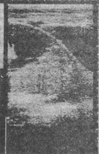


Figure 1: Ultrasound Image of Placenta Previa.
When diagnosing placenta previa with B-ultrasound, attention must be paid to the gestational weeks. During the mid-pregnancy [second trimester], the placenta occupies half of the uterine cavity. Therefore, the chances of the placenta being near or covering the internal cervical os are higher. By the late-pregnancy stage, the placental area reduces to one-third or one-fourth of the uterine cavity. Meanwhile, the formation and stretching of the lower uterine segment increase the distance between the internal cervical os and the placental edge. Thus, a placenta that initially appears to be in the lower uterine segment may shift upward with the expansion of the uterine body and revert to a normal position. For this reason, if a low-lying placenta is detected during mid-pregnancy [second trimester] via B-ultrasound, a diagnosis of placenta previa should not be made prematurely. Regular follow-ups are recommended. Unless vaginal bleeding occurs, a diagnosis of placenta previa is generally not made before 34 weeks of gestation.
5. Postpartum examination of the placenta and membranes For patients with antepartum hemorrhage, the delivered placenta should be carefully examined after delivery to verify the diagnosis. The placenta at the previa site has dark purple old blood clots attached. If the distance from the rupture of the membranes to the placental margin is <7cm, it is a partial placenta previa.
bubble_chart Treatment Measures
The treatment principle is to stop bleeding and tonify blood. Decisions should be made based on the amount of vaginal bleeding, the presence or absence of shock, the gestational week of pregnancy, parity, fetal position, whether the fetus is alive, and whether labor has occurred.
1. Expectant Management The goal of expectant management is to preserve the pregnancy while ensuring the safety of the mother. Preserving the pregnancy aims to prolong the gestational age, allowing the fetus to reach or approach full term, thereby improving perinatal survival rates. This approach is suitable for pregnancies before 37 weeks or when the estimated fetal weight is <2300g, with minimal vaginal bleeding, good maternal condition, and a viable fetus. The patient should be hospitalized for observation, with strict bed rest, emphasizing the left lateral position to minimize disturbances and reduce the risk of bleeding. Intermittent oxygen therapy should be administered regularly, three times a day for one hour each, to enhance fetal oxygen supply. The goal is to sustain the pregnancy until 36 weeks. During this period, close monitoring for bleeding is essential, with blood cross-matching on standby. Sedatives and blood tonics may be administered, and tocolytics such as salbutamol sulfate or magnesium sulfate may be used if necessary. Auxiliary tests should be conducted during expectant management to confirm the diagnosis. If a partial or complete placenta previa is diagnosed, continued hospitalization is mandatory. During observation, fetal maturity should be assessed based on the estimated due date and B-ultrasound measurements of the biparietal diameter. If massive vaginal bleeding or recurrent bleeding occurs during observation, termination of pregnancy is necessary.
2. Termination of Pregnancy
(1) Cesarean Section: Cesarean section can promptly conclude childbirth, delivering the fetus in a short time, ensuring safety for both mother and child. It is currently the primary method for managing placenta previa.
Complete placenta previa must be resolved by cesarean section. In recent years, there has been a tendency to perform cesarean sections for partial or marginal placenta previa in primiparous women. Timely and decisive cesarean section can immediately end childbirth, achieving rapid hemostasis. It reduces fetal trauma, decreases perinatal morbidity, and allows direct visualization to manage postpartum metrorrhagia. It is the safest and most effective method for managing placenta previa and a critical emergency measure for severe bleeding.
Preoperatively, shock should be actively corrected with fluid infusion and blood transfusion to replenish blood volume. These measures not only rescue the patient but also improve fetal hypoxia in utero.
A lower uterine segment incision is typically chosen for cesarean section, ideally avoiding the placenta. The surgical approach should be determined based on placental attachment location, confirmed by preoperative B-ultrasound. If the placenta is attached to the posterior wall, a transverse lower segment incision is made; if attached to the anterior wall, a longitudinal lower segment incision may be used. If the placenta is attached to the uterine incision site, the placenta should be gently displaced to rupture the membranes.
Due to poor contractility of the lower uterine segment, if the placenta does not deliver immediately after the fetus, manual removal is necessary. Concurrently, 0.2–0.4mg of ergometrine should be injected into the uterine wall to enhance lower segment contraction, supplemented by uterine massage to reduce postpartum metrorrhagia.
(2) Vaginal Delivery: This is only suitable for marginal placenta previa, occiput presentation, minimal bleeding, and cases where childbirth can be concluded quickly. After deciding on vaginal delivery, artificial rupture of membranes is performed. Rupture of membranes allows the fetal head to descend and compress the placenta for hemostasis while promoting uterine contractions to accelerate childbirth. If the presenting part does not descend adequately after membrane rupture, bleeding persists, or labor progress is unsatisfactory, an immediate switch to cesarean section is required.
(3) Management During Emergency Transfer: If a patient experiences massive vaginal bleeding and local treatment is unavailable, intravenous fluids or blood transfusion should be administered. Vaginal packing under sterile conditions can provide temporary hemostasis, and the patient should be rapidly transferred to a hospital for definitive treatment.
Whether after cesarean section or vaginal delivery, attention should be paid to correcting anemia and preventing infection.
Promote family planning, advocate contraception, prevent excessive childbirth, and avoid multiple curettages or intrauterine infections to prevent uterine membrane injury or endometritis. Strengthen prenatal check-ups and education, and seek medical attention promptly for any bleeding during pregnancy, regardless of the amount, to ensure early diagnosis and proper treatment.
In the advanced stage of pregnancy, bleeding should primarily be differentiated from placental abruption. Other causes of antepartum hemorrhage, such as rupture of vasa previa in velamentous placenta, rupture of marginal placental sinus, and cervical conditions like polyps, erosion, or cervical carcinoma, can be diagnosed through a combination of medical history, vaginal examination, B-ultrasound, and placental examination after childbirth.


