| disease | Non-Hodgkin Lymphoma |
| alias | Non-Hodgkin Lymphoma, Lymph Fleshy Tumor, Lymphoreticular Fleshy Tumor |
Non-Hodgkin's lymphoma (lymph fleshy tumor and reticular cell fleshy tumor) is a group of lymphomas with varying histological changes, sites of onset, and clinical manifestations. This group of lymphomas differs from Hodgkin's disease in terms of clinical symptoms, pathology, modes of spread, and response to treatment. They also differ from adult non-Hodgkin's lymphoma in terms of cellular differentiation, often consisting of poorly differentiated tumor cells that are already widely disseminated at the time of diagnosis. Controlling the progression of the primary disease is challenging, and treatment outcomes are generally poor. In recent years, advances in radiotherapy and combination chemotherapy have led to some improvement in prognosis. Non-Hodgkin's lymphoma is more common in children than Hodgkin's disease, with an average age higher than that of acute leukemia. The incidence is higher in males than in females, with a ratio of approximately 3:1.
bubble_chart Pathological Changes
The affected lymph nodes are enlarged, and the capsule is initially intact but later disrupted due to tumor infiltration, leading to the loss of normal structure. Tumor cells breach the lymph node capsule and infiltrate the surrounding adipose and connective tissues. Fibrous hyperplasia occurs within the lymph nodes, and hemorrhage and necrosis may sometimes be observed.
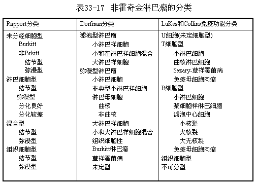
In the past, lymphomas were classified into two major categories based on the predominant cells in the affected lymph nodes: lymphosarcoma and reticulum cell sarcoma. In recent years, three classifications have been commonly used internationally based on microscopic and immunological observations (Table 1).
The classification discussed at the 1982 National Lymphoma Research Symposium in China is as follows:
B-cell lineage lymphomas
1. B small lymphocyte-type lymphoma
2. Plasmacytoid lymphocyte-type lymphoma
3. (Large and small) cleaved cell-type lymphoma
4. Mixed cell-type lymphoma
5. Large non-cleaved cell-type lymphoma
6. B immunoblastic lymphoma
7. Plasma cell-type lymphoma
8. Burkitt lymphoma
T-cell lineage lymphomas
1. Lymphoblastic lymphoma
2. Immunoblastic lymphadenopathy-like T-cell lymphoma
3. T immunoblastic sarcoma
4. Clear cell-type lymphoma
5. Polymorphic cell-type lymphoma
6. Mycosis fungoides-Sezary syndrome-cutaneous T-cell lymphoma
7. T small lymphocyte-type lymphoma
8. Monocytic T-cell lymphoma
Histiocytic sarcoma
Unclassifiable lymphoma
The Rappaport classification has limited prognostic significance for childhood non-Hodgkin lymphoma, as the majority of childhood non-Hodgkin lymphomas are poorly differentiated diffuse lymphocytic types, while the mixed nodular type, which has the best prognosis, is extremely rare in children. According to modern immunological concepts, lymphocytes can be divided into T, B, and U (undifferentiated) cells. Non-Hodgkin lymphomas originating in the mediastinum often test positive in the E-rosette assay and are likely derived from T cells, whereas those originating in the abdominal cavity may derive from B cells. The Rappaport classification is based on the morphological typing of Dorfman cells. Convoluted lymphocytic cells are poorly differentiated lymphoblasts, most of which originate from T cells. Histiocytic or large lymphocytic cells mostly belong to the B-cell lineage.

Non-Hodgkin lymphoma spreads early via the bloodstream or lymphatic channels, with about 50% of patients showing bone marrow infiltration. Those originating in mediastinal lymph nodes almost always (100%) spread to the bone marrow and progress to lymphocytic leukemia. In patients with lymphocytic leukemia, over one-third develop meningeal infiltration, whereas those without leukemia do not exhibit meningeal lesions. Of those with meningeal infiltration, 80–90% are derived from T lymphocytes. Additionally, T-cell lymphomas frequently infiltrate the testes.
bubble_chart Clinical Manifestations
Symptoms vary depending on the location and extent of tumor spread. It commonly occurs in the neck, mediastinum, armpits, and mesentery. Painless and non-tender enlargement of cervical or supraclavicular lymph nodes is typical, with a firm texture and no adhesion between nodes. The condition progresses rapidly, with lymph nodes enlarging within 1–2 weeks, often forming a massive conglomerate in advanced stages. Local lymph node enlargement may coincide with widespread dissemination, infiltrating lymphoid tissues of the tonsils, adenoids, sinuses, and invading surrounding tissues or sites like the orbit, breast, skin, and bones.
Enlarged lymph nodes may cause compressive symptoms, such as mediastinal lymphadenopathy compressing the trachea and bronchi, leading to respiratory obstruction, dyspnea, or even superior vena cava syndrome. Pleural invasion may cause effusion, with tumor cells detectable in aspirated fluid. Primary abdominal lymph node involvement, often in the ileocecal region, may cause abdominal pain, intestinal obstruction, or ascites. Abdominal wall lymphoma may induce chronic, recurrent intussusception. Bone invasion can lead to bone pain.

Leukemic transformation is more frequent in poorly differentiated lymphocytic types (historically termed lymphosarcoma), often T-cell lymphomas originating in mediastinal lymph nodes, whereas histiocytic and Burkitt types rarely develop leukemia.
Although Burkitt lymphoma is prevalent in Africa, sporadic cases occur worldwide, including in China. Its hallmark is jaw involvement, often presenting with bloody nasal discharge and facial swelling, prompting visits to ENT specialists, alongside abdominal distension, hemiplegia, or other extranodal tumors. It rarely involves the spleen, mediastinal, or peripheral lymph nodes. Over 10% of pediatric non-Hodgkin lymphomas are undifferentiated cell types, morphologically and histochemically resembling Burkitt lymphoma but differing in cell size, classified as undifferentiated non-Burkitt type.
Clinical staging follows Hodgkin lymphoma, though its utility is limited due to rapid systemic spread. Bone marrow or CNS involvement automatically classifies as stage IV.
The diagnosis of such tumors is primarily based on the location of tumor growth, a history of rapid short-term enlargement, and confirmation through pathological biopsy. For mediastinal lymphoma, X-ray examination reveals a central tumor shadow that extends bilaterally. When the tumor compresses the trachea, causing respiratory obstruction, immediate radiotherapy can be administered. If the tumor shrinks, it also aids in diagnosis. The detection of tumor cells in pleural effusion or ascites further supports the diagnosis.
In cases without concurrent leukemia or bone marrow involvement, blood counts are generally normal. For patients with extensive infiltration, plasma uric acid and lactate dehydrogenase levels are elevated. Whenever possible, T- and B-cell typing should be performed.
bubble_chart Treatment Measures
Non-Hodgkin's lymphoma is primarily treated with chemotherapy, and the treatment regimen for acute lymphocytic leukemia can be adopted, such as the COAP regimen: vincristine 1–2 mg/m2 intravenously on days 1, 8, 15, and 22; prednisone 40–60 mg/m2/day orally; cyclophosphamide 600 mg/m2 on day 1; and doxorubicin 30–40 mg/m2 intravenously on day 1 or repeated on day 21. Alternatively, doxorubicin can be replaced with daunorubicin or idarubicin (10 mg/m2 per dose, administered for 2–3 consecutive doses). L-asparaginase 10,000 IU (m2·d) is administered intravenously or intramuscularly for 10 consecutive days, which is particularly effective for stage III–IV patients, especially those with bone marrow infiltration. Additionally, bleomycin and 6-mercaptopurine can be used alternately.
In recent years, high-dose methotrexate (HD-MTX) at 3–7.5 g/m2 per dose has been recommended internationally for the treatment of non-Hodgkin's lymphoma. When using this method, leucovorin rescue is required, along with urine alkalinization and adequate fluid intake. This approach plays a crucial role in reducing complications in the central nervous system, testes, and ovaries.
New drug applications: ① Epirubicin and pirarubicin (THP-ADM): Their antitumor spectrum and cytotoxicity are similar to doxorubicin, but they have less cardiotoxicity, milder alopecia areata and gastrointestinal reactions, with the main side effect being myelosuppression. They are more expensive; ② VM26: A podophyllotoxin-derived antitumor drug, administered intravenously at 100–150 mg/m2 per dose. Its counterpart is etoposide (VP16), and both can be used alone or in combination with cytarabine or cyclophosphamide, with myelosuppression being the main side effect; ③ Other drugs include mitoxantrone and aclarubicin.
Radiotherapy has a limited scope of application, restricted to cases where the lesion is confined to a specific area or when severe symptoms are caused by tumor compression. It can be used for palliative relief, while for stage III–IV cases with widespread lesions, radiotherapy is generally performed after chemotherapy.
Surgery for non-Hodgkin's lymphoma is primarily diagnostic. For enlarged lymph nodes primarily located in the abdominal cavity and/or accompanied by visceral infiltration or compression symptoms, chemotherapy is usually administered first to shrink the tumor, followed by surgical removal, with continued chemotherapy postoperatively.
Chemotherapy generally lasts 1.5–2 years, and for cases combined with leukemia, it should be extended to 3 years or longer, depending mainly on the disease stage and pathological classification.
Autologous bone marrow transplantation is an excellent method for treating advanced-stage lymphoma (excluding cases combined with leukemia), significantly increasing the dose of chemotherapy and radiotherapy and providing a new approach for refractory cervical malignancy with cachexia. Allogeneic bone marrow transplantation, although effective, faces challenges such as donor matching difficulties, rejection reactions, and high costs, making it difficult to implement in pediatric cases.
Immunotherapy: With advancements in cell biology, molecular biology, and bioengineering technology, immunotherapy has gained momentum. The proposal of the biological response modifier (BRM) theory has established tumor biotherapy as the fourth-generation treatment modality beyond surgery, radiotherapy, and chemotherapy. Interferon (INF) has been successively applied clinically, either alone or in combination, opening new prospects for tumor treatment.
Since pediatric non-Hodgkin's lymphoma is mostly of the poorly differentiated lymphocyte diffuse type, the prognosis is poor. About 50% of cases achieve long-term remission with combination chemotherapy. With proper treatment, relapses mostly occur within 6 months, and recurrence is rare if remission lasts over a year. Cases with primary mediastinal or abdominal involvement and large tumor masses generally have a poor prognosis.
It should be differentiated from fleshy tumors of striated muscle, Ewing's osteosarcoma, neuroblastoma, Hodgkin's disease, and other benign or malignant tumors of the lymph nodes.


