| disease | Wolff-Parkinson-White Syndrome |
| alias | Pre-excitation Syndrome, WPW, Wolf-Parkinson-White Syndrome |
Pre-excitation is an abnormal phenomenon of atrioventricular conduction where impulses travel through an accessory pathway, prematurely exciting part or all of the ventricles, causing some ventricular muscles to be activated early. The condition with pre-excitation is called pre-excitation syndrome or WPW (Wolf-Parkinson-White) syndrome, often accompanied by episodes of paroxysmal supraventricular tachycardia. Pre-excitation is a relatively rare arrhythmia, and diagnosis mainly relies on electrocardiography.
bubble_chart Etiology
It is now widely accepted that the disease cause of pre-excitation is the presence of congenital atrioventricular accessory pathways (referred to as bypass tracts) outside the normal atrioventricular conduction system. Most patients have no organic heart disease. It is also seen in certain congenital and acquired heart diseases, such as Ebstein's anomaly and obstructive cardiomyopathy.
Electrophysiological studies have demonstrated that bypass tracts conduct impulses rapidly. Atrial impulses partially travel quickly down the bypass tract, arriving prematurely at the ventricular end of the tract, exciting adjacent myocardium, thereby causing premature ventricular activation and altering the normal excitation sequence of the ventricular muscle. The result is a deformed QRS complex on the electrocardiogram, with an initial delta wave (δ wave). The remaining portion of the atrial impulse may travel down the normal pathway, merging with the ventricular activation caused by the bypass tract to form a ventricular fusion beat. The morphology of the ventricular fusion beat is determined by the relative refractory periods of the normal pathway and the bypass tract. If the normal pathway has a long refractory period or most of the impulse travels via the bypass tract, the QRS complex appears significantly deformed; if the bypass tract has a long refractory period, the ventricular fusion beat appears closer to normal.
Patients with pre-excitation syndrome have two conduction pathways between the atria and ventricles, making them prone to reentry and reentrant tachycardia. During tachycardia episodes, most impulses travel retrograde via the bypass tract and antegrade down the normal pathway, resulting in a normal QRS complex morphology during tachycardia. Occasionally, impulses travel antegrade via the bypass tract and retrograde via the normal pathway, causing the QRS complex to appear pre-excited during tachycardia. Pre-excitation patients may also experience atrial fibrillation or atrial flutter, which are often caused by retrograde impulses arriving at the atria during the vulnerable period. During atrial flutter or fibrillation, concealed conduction within the junctional tissue promotes impulse transmission mostly or entirely via the bypass tract to the ventricles. Atrial flutter or fibrillation with an extremely rapid ventricular rate and deformed QRS complexes may sometimes progress to ventricular fibrillation.Unidirectional block (mostly anterograde block) in the bypass tract may result in no pre-excitation on the electrocardiogram but recurrent supraventricular tachycardia; electrophysiological studies can confirm the involvement of the bypass tract in the tachycardia reentry circuit. Second-degree conduction block in the bypass tract may lead to intermittent pre-excitation on the electrocardiogram.
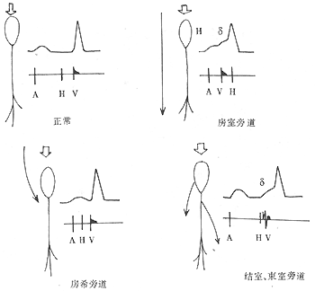
The following types of bypass tracts are known, and a single patient may have multiple tracts: ① Atrioventricular bypass tract (Kent bundle). Most are located along the left or right atrioventricular groove or septum, connecting atrial and ventricular myocardium; ② Atrio-nodal bypass tract (James pathway). This is a connection between the atria and the lower part of the atrioventricular node or the bundle of His, possibly formed by fibers of the posterior internodal tract; ③ Nodo-ventricular or fasciculoventricular connections (Mahaim fibers). These are pathways connecting the distal atrioventricular node, bundle of His, or proximal bundle branches to the ventricular septum. Among these, the atrioventricular bypass tract is the most common. (Figure 1).

Figure 1 Anatomical, electrocardiographic, and His bundle electrogram manifestations of different bypass tracts.
bubble_chart Clinical Manifestations
Preexcitation alone is asymptomatic. The associated supraventricular tachycardia is similar to typical supraventricular tachycardia. In cases complicated by atrial flutter or atrial fibrillation, the ventricular rate is often around 200 beats per minute, and in addition to discomfort such as palpitations, shock, heart failure, or even sudden death may occur. When the ventricular rate is extremely fast, such as 300 beats per minute, the auscultated heart rate may be only half of the ventricular rate seen on the electrocardiogram, indicating that half of the ventricular activations fail to produce effective mechanical contractions.
bubble_chart Auxiliary Examination
The electrocardiographic manifestations {|###|} of various accessory pathways causing preexcitation are as follows. {|###|} (1) Atrioventricular bypass tract: ① The PR interval (essentially the P-δ interval) is shortened to less than 0.12 seconds, mostly 0.10 seconds; ② The QRS duration is prolonged to more than 0.11 seconds; ③ The initial part of the QRS complex is slurred, forming a notch with the remaining part, known as preexcitation; ④ Secondary ST-T wave changes (Figure 2). {|###|}{|###|}{|###|} Figure 2 Preexcitation syndrome {|###|} The above ECG changes can also be classified into types A and B. In type A, the preexcitation wave and QRS complex in lead V{|###|}1{|###|} are both upward (Figure 3), while in type B, the preexcitation wave and the main wave of the QRS complex in lead V{|###|}1{|###|} are both downward. The former suggests preexcitation in the left ventricle or the posterior basal part of the right ventricle, while the latter suggests preexcitation in the anterior lateral wall of the right ventricle. Although this classification is limited by the variable QRS complexes caused by different locations of the accessory pathways, it helps distinguish whether the ventricular end of the pathway is on the left or right, anterior or posterior, and thus remains in use today. {|###|}{|###|}{|###|} Figure 3 Type A preexcitation syndrome, showing the preexcitation wave and the main wave of the QRS complex in lead V{|###|}1{|###|} are upward {|###|} (2) Atrio-nodal and atrio-His bypass tracts: The PR interval is less than 0.12 seconds, mostly 0.10 seconds; the QRS complex is normal without a preexcitation wave. This ECG manifestation is also called short PR, normal QRS syndrome or Lown-Ganong-Levine (LGL) syndrome (Figure 4). {|###|}{|###|}{|###|} Figure 4 LGL syndrome {|###|} (3) Nodo-ventricular and fasciculo-ventricular connections: The PR interval is normal, the QRS complex is widened, and there is a preexcitation wave. {|###|} During episodes of supraventricular tachycardia in preexcitation syndrome, the preexcitation manifestations often disappear, and the ECG shows supraventricular tachycardia with normal QRS morphology (Figure 5). When atrial flutter or atrial fibrillation is present, it is not uncommon for the QRS to retain preexcitation characteristics (Figure 6). The ECG shows atrial flutter or fibrillation with abnormally wide QRS complexes; the ventricular rate often exceeds 200 beats/min and may even reach 300 beats/min. In atrial flutter, 1:1 atrioventricular conduction may occur, and flutter waves may be identifiable. In atrial fibrillation, the ventricular rhythm is irregular, and after long pauses, occasional QRS complexes with normal morphology may be seen (possibly due to prolonged refractory period of the accessory pathway, with impulses conducted entirely or mostly through the AV node after disappearance of concealed conduction), and fibrillation waves may be identifiable. When the ventricular rate is extremely rapid, frequency-dependent intraventricular conduction changes may also occur. {|###|}{|###|}{|###|} Figure 5 Preexcitation syndrome with supraventricular tachycardia. Left: Preexcitation manifestations during sinus rhythm. Right: Episode of supraventricular tachycardia with normal QRS morphology. {|###|}{|###|}{|###|} Figure 6 Preexcitation syndrome with atrial fibrillation. Left: Atrial fibrillation episode with ventricular rate 167–300 beats/min, average 210 beats/min, markedly irregular. Right: Sinus rhythm with typical preexcitation manifestations.
bubble_chart DiagnosisIn addition to the aforementioned ECG characteristics, vectorcardiograms can serve as diagnostic criteria. Their hallmark is a slow, linear initial segment of the QRS loop across all planes, lasting up to 0.08 seconds, followed by an abrupt turn and continuation at normal speed. The QRS loop duration may exceed 0.12 seconds. His bundle electrograms and body surface or epicardial mapping aid in differentiating various types of preexcitation and locating accessory pathways, playing a crucial role in determining whether the accessory pathway participates in the tachycardia reentry circuit.
On ECG, preexcitation patterns should be distinguished from bundle branch block, ventricular hypertrophy, or myocardial infarction. Shortened PR intervals and the presence of delta waves confirm preexcitation. Accelerated idioventricular rhythm with interference atrioventricular dissociation from sinus rhythm (especially when ventricular and sinus rates are similar) may exhibit brief episodes of shortened PR intervals and widened, bizarre QRS complexes, mimicking intermittent preexcitation. However, prolonged recordings often reveal variable PR intervals and atrioventricular dissociation, making differentiation from preexcitation straightforward.
When preexcitation is complicated by supraventricular tachycardia, QRS complexes typically remain narrow. However, except in cases of concealed preexcitation, characteristic ECG changes appear after termination of the episode. During preexcitation complicated by atrial fibrillation or flutter, QRS complexes often widen and must be differentiated from ventricular tachycardia.
bubble_chart Treatment Measures
Preexcitation itself does not require special treatment. When complicated by supraventricular tachycardia, the treatment is the same as for general supraventricular tachycardia. In cases complicated by atrial fibrillation or atrial flutter with a rapid ventricular rate and circulatory impairment, synchronized direct current cardioversion should be performed as soon as possible. Lidocaine, procainamide, propafenone, and amiodarone can slow conduction through the accessory pathway, thereby reducing the ventricular rate or converting atrial fibrillation and atrial flutter back to sinus rhythm. Digitalis accelerates accessory pathway conduction, while verapamil and propranolol slow conduction within the atrioventricular node, both of which may significantly increase the ventricular rate or even lead to ventricular fibrillation, and thus should not be used. If episodes of supraventricular tachycardia, atrial fibrillation, or atrial flutter are frequent, long-term oral administration of the aforementioned antiarrhythmic drugs is recommended to prevent recurrence. For cases where medication fails to control the condition, electrophysiological studies confirm a short refractory period of the accessory pathway or shortening of the refractory period during rapid atrial pacing, or when the ventricular rate during atrial fibrillation reaches around 200 beats per minute, ablation (via electrical, radiofrequency, laser, or cryotherapy) or surgical interruption of the accessory pathway is indicated to prevent recurrence after localization.


