| disease | Priapism |
| alias | Priapism |
Priapism refers to a persistent erection of the penis unrelated to sexual desire. An erection lasting more than 6 hours is considered abnormal. Traditionally, priapism is classified as primary (idiopathic) or secondary. Based on hemodynamics, it is divided into low-flow (ischemic) and high-flow (non-ischemic) types. The former is caused by venous obstruction (veno-occlusive), while the latter results from abnormal arterial blood inflow (arterial). Priapism can also be categorized as acute, intermittent (recurrent or episodic, such as in sickle cell anemia), or chronic (usually high-flow type). In the initial stage of priapism, it is always a physiological erection, which may later develop into the high-flow type.
bubble_chart Etiology
Statistics show that 30-40% of cases of priapism are primary, with most disease causes unknown. Secondary causes of sexually transmitted diseases include: thromboembolic disorders (sickle cell anemia, fat embolism, etc.), neurological diseases (spinal cord injuries and lesions, spinal stenosis, etc.), tumors (metastatic cancers such as prostate cancer and kidney cancer, leukemia, melanoma, etc.), trauma (perineal or genital injuries, etc.), infections or poisoning (dysentery, rabies, etc.), medications (antidepressants, α-adrenergic blockers, anticoagulants, etc.), total parenteral nutrition, and intracavernosal injection of vasoactive agents.
Low-flow priapism is caused by various factors that impair the detumescence mechanism of the penis, including excessive secretion of neurotransmitters, obstruction of venous return, and prolonged relaxation of intracavernosal smooth muscle. As a result, the intracavernosal pressure remains persistently elevated at 10.06–15.60 kPa (80–120 mmHg) and gradually worsens, progressing to an ischemic state. Pain typically occurs after 6–8 hours of ischemia. The severity of ischemia and the number of affected draining veins are related to the duration of venous occlusion. Experimental studies have shown that under hypoxic conditions, the autonomous contractility and tension of cavernosal smooth muscle decrease, and normal contractile responses to α-adrenergic agonists are impaired. After several days of priapism, thrombosis becomes less likely to form in the penile blood flow (even in low-flow priapism) because the activity of fibrinolytic enzymes in the cavernosal tissue is three times higher than in peripheral blood.
The frequency of recurrent priapism episodes ranges from several times a day to once every few months. After the initial episode of ischemic priapism, functional changes occur in the adrenergic or endothelial-mediated mechanisms that control penile detumescence.All cases of priapism initially present as non-ischemic high-flow priapism. However, in most cases, venous thrombosis, acidosis, and hypoxia develop within 6 hours, eventually progressing to the typical low-flow type. In some cases, high blood flow persists, with unimpeded cavernosal venous return and adequate blood oxygenation. Due to the open venous pathways, the erect penis remains compressible, with erection hardness ranging from mild to grade II, and sexual stimulation can increase penile rigidity.
bubble_chart Clinical Manifestations
Priapism commonly occurs between the ages of 5-10 and 20-50. It usually involves only the corpora cavernosa of the penis, with most cases occurring during nocturnal penile tumescence.
Low-flow priapism causes pain due to tissue ischemia if it persists for several hours, with the penis becoming rigidly erect. In high-flow priapism, the penis is rarely painful and cannot achieve full erection hardness. There is usually a history of perineal or penile trauma. In most cases of this type, the penis can still regain full erectile function after stirred pulse embolization or surgical vascular ligation, but this typically takes several weeks to months.
bubble_chart Auxiliary Examination
Analysis of cavernous blood qi aspect can distinguish between high-flow and low-flow types. The former has qi aspect values similar to stirred pulse blood, while the latter resembles venous blood. Notably, early-stage priapism is always of the high-flow type.
Cavernosography can also differentiate the two types. In venous occlusion, blood flow stagnates; in the stirred pulse type, cavernous blood reflux is rapid. Color Doppler ultrasound shows minimal stirred pulse flow and cavernous expansion in the low-flow type, whereas the high-flow type displays stirred pulse rupture and abnormal blood pools at vascular injury sites.bubble_chart Treatment Measures
Examination and Treatment Procedures for Priapism:
1. Non-surgical Treatment
(1) Low-flow priapism: The goal of treatment is to increase venous blood return, reduce penile swelling, prevent persistent ischemia-induced injury to the corpora cavernosa, and alleviate pain. Medication should be attempted before surgical intervention. It should be noted that prolonged drug therapy extends treatment duration and may increase the risk of cavernosal fibrosis and erectile dysfunction. Reports indicate that the incidence of erectile dysfunction in low-flow priapism is as high as 50%. However, if treated with medication within 12–24 hours, erectile function can almost always be restored. Kulmala and Tamella (1995) observed that in most cases treated with aspiration and α-adrenergic agonists within 36 hours, cavernosal fibrosis could be avoided. Beyond 36 hours, α-adrenergic drugs become ineffective, and varying degrees of fibrosis may develop within the corpora cavernosa.
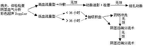
Reports suggest injecting diluted α-adrenergic agonists into the corpora cavernosa—for example, 1 mg of epinephrine in 1000 ml of saline. First, aspirate stagnant blood from the corpora cavernosa using a 21-gauge needle, then inject 20 ml of the diluted solution. After 2 minutes, aspirate the blood again, repeating the injection and aspiration several times until the swelling subsides. Alternatively, phenylephrine (10 mg in 500 ml of saline) can be used, with 10–15 ml injected each time. Satisfactory results can be achieved if treatment is initiated within 12 hours of onset.
Recurrent priapism often occurs in patients with sickle cell anemia or a history of priapism. Young patients may be treated with diluted phenylephrine solution. For patients without sexual function, anti-androgens or gonadotropin-releasing hormone agonists can be used to suppress nocturnal erections and prevent recurrence.Complications of drug therapy include acute hypertension, headache, palpitations, and arrhythmias induced by α-adrenergic agents, as well as infections, bleeding, and urethral injury caused by aspiration.
(2) High-flow priapism: Early application of an ice pack can induce vasoconstriction, potentially leading to spontaneous thrombosis in damaged vessels. Most cases of ruptured cavernosal arteries do not heal on their own and often require internal pudendal arteriography and embolization. Reports indicate successful treatment with methylene blue injection or autologous blood clot embolization into the arterial lumen.
High-flow priapism has a better prognosis, with an erectile dysfunction rate of 20%.
2. Surgical Treatment Currently, fewer cases meet the criteria for surgical intervention.
Non-ischemic priapism is typically managed non-surgically. Early ischemic cases may convert to non-ischemic after thorough penile irrigation.
For ischemic priapism, if aspiration and irrigation fail, direct incisions can be made from the glans into the corpora cavernosa, or a biopsy needle can be used to create shunt channels between the glans and corpora cavernosa. Sacher (1972) introduced a perineal proximal urethral-cavernosal anastomosis, emphasizing staggered anastomotic sites to prevent urethral strictures.
In some high-flow priapism cases, if arterial embolization alone is ineffective, ligation of the torn artery’s supplying vessels may be necessary.


