| disease | Juvenile Osteochondrosis of the Vertebral Body |
| alias | Scheuermann's Disease |
Juvenile vertebral osteochondrosis, also known as Scheuermann's disease or juvenile kyphosis. The onset age is between 13 and 17 years, with males slightly outnumbering females. 75% of cases occur in the thoracic spine, particularly the lower thoracic region; 25% may involve the thoracic spine and upper lumbar spine, especially the lower thoracic region. The incidence rate is unclear, but statistics show it accounts for 6% in military personnel and 8% in industrial workers.
bubble_chart Etiology
The vertebral body has three ossification centers: the primary ossification center in the middle of the vertebral body and the secondary ossification centers at the upper and lower ends of the vertebral body. The latter, known as the ring apophysis, appears after the age of 4 and is located at the edge of the cartilaginous plate, separating the vertebral body from the intervertebral disc. Scheuermann believed that this disease was due to aseptic necrosis of the ring apophysis, but this view has been refuted. Currently, Schmorl's explanation is more widely accepted. The main pathology lies in the intervertebral cartilage. It is caused by congenital or developmental defects at the junction of the spongy bone and cartilage. When excessive load is applied, the nucleus pulposus of the intervertebral disc herniates into the vertebral body, damaging the cartilaginous plate of the vertebral body and causing growth imbalance. At the same time, the intervertebral disc loses its cushioning (protective) function, subjecting the anterior edge of the vertebral body to excessive pressure, resulting in delayed growth, wedge-shaped deformation, and fragmentation of the vertebral body. The posterior edge of the vertebral body maintains its original height due to the protection of the posterior articular processes, leading to the development of kyphotic deformity in the spine.
bubble_chart Clinical ManifestationsKyphosis is the main symptom, accompanied by spinal stiffness. The neck is often flexed, the shoulders droop, the thorax is narrow and flat, and the scapulae protrude. The pain is not severe and is often a dull ache. The kyphotic deformity progresses until after the age of 20.
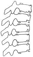
**X-ray findings** ① Irregular indentations on the anterior upper and lower edges of the vertebral bodies, with uneven morphology and size of the ring-shaped epiphyses in corresponding areas, which are separated from the vertebral bodies; ② Multiple vertebral bodies show anterior wedging, accompanied by Schmorl’s nodes; ③ Grade I narrowing of the intervertebral spaces; ④ Kyphotic deformity of the thoracic or thoracolumbar spine exceeding the normal range of 25°–40°; ⑤ Early appearance of osteoarthritic osteophytes on the anterior edges of vertebral bodies in adulthood (Figure 1).

**Figure 1** Juvenile vertebral osteochondrosis (Scheuermann’s disease)
The diagnostic criteria are: ① adolescent; ② kyphotic deformity must exceed 35°; ③ wedge-shaped deformation of at least one vertebral anterior edge must be greater than 5°; ④ must affect 3 to 5 consecutive vertebrae.
bubble_chart Treatment Measures
This disease is a self-limiting condition with an active phase lasting about two years. If kyphosis deformity has already developed, complete correction is impossible, and early secondary osteoarthritis may occur in adulthood. The goal of treatment is to prevent deformity and protect the spine from compressive damage until the epiphyseal plates mature. The previous practice of prolonged bed rest in a plaster cast is now rarely used. If the disease is severe at onset, immobilization in a plaster cast or plaster jacket for 2–3 months may be considered, followed by bracing and back muscle exercises. If the child is pain-free, treatment can be determined based on the degree of deformity: for kyphosis less than 45–50°, corrective exercises alone are sufficient; for deformities between 50–80°, bracing combined with back muscle training is required. In very few cases, severe kyphosis may necessitate spinal correction and fusion surgery for cosmetic reasons. Rarely, spinal cord compression symptoms may require decompression.
It should be differentiated from the following diseases.
1. Spinal subcutaneous node: This is a progressive bone destructive disease, where the vertebral edges are blurred rather than whitened as in osteochondrosis. Most patients develop paravertebral abscesses.
2. Postural kyphosis: This type of kyphosis is not fixed and can be easily corrected either passively or actively. X-rays show no wedge-shaped deformation of the vertebrae.


