| disease | Felon |
Suppurative finger inflammation is a subcutaneous suppurative infection of the terminal segment of the finger, often caused by puncture wounds. The causative bacteria are mostly Staphylococcus aureus.
bubble_chart Pathological Changes
There are many longitudinal fibrous cords between the skin on the palmar surface of the distal phalanx and the nail bone membrane, dividing the soft tissue into numerous enclosed small compartments. These compartments contain adipose tissue and a rich network of nerve endings. During infection, pus does not easily spread to surrounding areas, so swelling is not significant. However, the formation of a high-pressure pus-filled cavity can not only cause extremely severe pain but also compress the nutrient vessels of the distal phalanx, leading to bone ischemia and necrosis (Figure 1). Additionally, direct invasion of pus into the bone can also cause osteomyelitis.
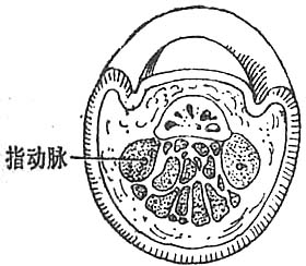
(1) The distal longitudinal septum of the distal phalanx, showing the subcutaneous tissue as enclosed small compartments
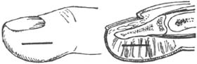
(2) Schematic diagram of the surgical incision for felon
Figure 1 Felon
bubble_chart Clinical Manifestations
Initially, there is a needle-like pain at the fingertip. Later, as the tissue swells and the pressure within the small cavity increases, rapidly intensifying pain develops. When the finger's pulse is pressed, the pain becomes throbbing and worsens when the affected limb is lowered. Severe pain often causes the patient to become restless and unable to sleep through the night. The redness and swelling of the finger are not very noticeable, and sometimes the skin may appear yellowish-white, but the tension is significantly increased, with even a light touch of the fingertip causing severe pain. At this stage, systemic symptoms such as fever, general malaise, and increased white blood cell count are often present. In the advanced stage, most of the tissue becomes ischemic and necrotic, and the nerve endings become numb due to compression and nutritional impairment, leading to a reduction in pain. However, this does not indicate an improvement in the condition. If felon is not treated promptly, it can often lead to ischemic necrosis of the finger bone, resulting in chronic osteomyelitis and prolonged non-healing of the wound.
bubble_chart Diagnosis1. The distal phalanx often shows swelling on the surface, presenting a snake-head-like appearance, accompanied by severe throbbing pain that worsens when the hand is lowered.
2. The palmar skin is under high tension, slightly red with significant tenderness, and local fluctuation is usually not obvious.
3. If left untreated, it may rupture spontaneously, resulting in a wound that is difficult to heal. X-ray may reveal necrosis of the distal phalanx.
4. Systemic symptoms such as fever and headache may also occur.
bubble_chart Treatment Measures
When there is pain in the fingertip and swelling is not obvious upon examination, it can be soaked multiple times in hot saline water, for about 20 minutes each time; alternatively, topical medication can be applied (refer to the treatment of paronychia). Sulfonamides or antibiotics may be used as appropriate. After the above treatments, the inflammation often subsides. If throbbing pain occurs and the tension in the fingertip significantly increases, incision for decompression and drainage should be performed immediately, without waiting for fluctuation to appear before surgery. Although there may be little or no pus after the incision, it can reduce the pressure in the enclosed cavity of the fingertip, alleviating pain and complications.
During the surgery, a longitudinal incision is made on the side of the affected finger, as long as possible but not exceeding the junction of the distal and middle phalanges, to avoid injuring the tendon sheath (Figure 11-4). When making the incision, forcefully sever the fibrous septa in the subcutaneous tissue and trim any protruding fat tissue to avoid obstructing drainage. If the abscess cavity is large, counter-drainage can be performed, but a bubo-shaped incision should not be made to prevent postoperative scarring from affecting the infected finger. A latex sheet is placed in the incision for drainage. During incision and drainage, if there are necrotic bone fragments, they should be removed. Postoperative systemic treatment follows the general management of suppurative infections. Another method involves making a straight incision in the center of the palmar side of the distal phalanx, draining all the pus without placing a drain, and applying a thick layer of zinc oxide ointment before bandaging. The dressing is changed every 2–3 days until healing. This type of incision is superior to a lateral incision, as it provides direct and unobstructed drainage; causes less injury to the fibrous septa and fat pad, preserving postoperative grasping function; avoids the complication of injuring the digital nerve and subsequent loss of sensation in the ipsilateral fingertip, which is common with lateral incisions; and results in painless scarring.


