| disease | Primary Hyperparathyroidism (Surgical) |
There are usually four parathyroid glands, closely attached to the inner backside of the left and right lobes of the thyroid. These glands are oval-shaped, flat, measuring 5–6 mm in length, 3–4 mm in width, and about 2 mm in thickness; they weigh 30–45 mg, are yellowish-brown in color, and have a soft texture. The parathyroid glands secrete parathyroid hormone, whose physiological function is to regulate calcium metabolism in the body and maintain the balance of calcium and phosphorus. Primary hyperparathyroidism is mostly caused by a single parathyroid adenoma (90%), less commonly by multiple adenomas or hyperplasia of the parathyroid glands, and rarely by adenocarcinoma.
bubble_chart Pathogenesis
Normally, a person has four parathyroid glands, with upper and lower glands on each side, left and right. The upper parathyroid glands are usually located on the medial side of the posterior surface of the thyroid lobes, at the level of the cricoid cartilage, near where the recurrent laryngeal nerve enters the larynx. The lower parathyroid glands are situated on the lateral side of the posterior surface of the thyroid lobes, near the level where the inferior thyroid artery intersects with the recurrent laryngeal nerve. A minority of people have only three parathyroid glands (13%, with two glands on one side fused into one) or as many as five parathyroid glands (6%, with the extra gland often located in the mediastinum).
The upper parathyroid glands and the thyroid share a common origin from the fourth pharyngeal pouch. The lower parathyroid glands and the thymus originate from the third pharyngeal pouch. During embryonic development, the upper parathyroid glands descend to the neck along with the thyroid primordium, while the lower parathyroid glands descend with the thymic primordium to the level of the thyroid root before separating. The thymic primordium continues to descend into the thoracic mediastinum. If the parathyroid primordium stops midway or continues descending with the thymic primordium, it can lead to ectopic parathyroid glands. Even when ectopic, the upper parathyroid glands usually remain near the thyroid, whereas the lower parathyroid glands can vary significantly in location, ranging from the angle of the mandible to the thymus. Common ectopic locations for the upper parathyroid glands include the superior pole of the thyroid, near the thyroid vessels, within the tracheoesophageal groove, or behind the pharynx and esophagus. The lower parathyroid glands may be found at the mandibular angle, near the carotid artery bifurcation, within the mid-carotid sheath, in the adipose tissue below the thyroid root, within the thymic tongue, near the great vessels of the mediastinum, or adjacent to the pericardium. If embedded within the thyroid or thymus, the parathyroid glands can be difficult to locate during surgery. If located in the mediastinum, the upper parathyroid glands are typically in the posterior mediastinum, while the lower ones are in the anterior mediastinum.
The upper parathyroid glands are supplied by the superior or inferior thyroid artery, and the lower parathyroid glands are supplied by the inferior thyroid artery. If a lower parathyroid adenoma descends into the mediastinum due to its weight and cannot be found behind the thyroid, tracing the branches of the inferior thyroid artery often leads to its discovery. However, lower parathyroid glands that descend into the mediastinum during embryonic development may be supplied by branches of the internal mammary artery or the aorta.Normal parathyroid glands vary in shape, appearing oval, rod-like, spherical, disc-shaped, or leaf-like. Their average size is 5×3×1 mm, with a minimum of 2×2×1 mm and a maximum of 12×2×1 mm. Elongated glands tend to be narrow and thin, while shorter ones are broader and thicker. Their average weight is 35–40 mg. Their color ranges from reddish-brown to yellowish-brown, depending on blood supply and fat content. The texture is soft, and the cut surface resembles that of chicken liver, which helps distinguish them from lymph nodes. Normal parathyroid glands are mobile and easily separated from surrounding adipose tissue; if fixed or adherent to adjacent tissues, malignancy should be suspected.
The parathyroid glands consist mainly of chief cells, a small number of oxyphil cells, and stroma. Chief cells secrete parathyroid hormone (PTH). Oxyphil cells are likely aged chief cells and normally have no secretory function. Chief cells contain fat granules, and the stroma contains adipocytes. In hyperfunctioning parathyroid glands, chief cells lose fat, making the gland denser than normal. This difference in density can be used to distinguish normal from hyperfunctioning glands (hyperplasia or adenoma).
The parathyroid glands secrete parathyroid hormone (PTH), which has the following effects:1. Promotes calcium reabsorption in the proximal renal tubules, reducing urinary calcium and increasing blood calcium.
2. Inhibits phosphate reabsorption in the proximal renal tubules, increasing urinary phosphate and decreasing blood phosphate.
3. Stimulates osteoclastic decalcification, releasing calcium phosphate (Ca3PO4) from the bone matrix, thereby increasing blood calcium and phosphate levels.
4. Promotes the hydroxylation of vitamin D to generate the active 1,25-dihydroxy D3, which enhances intestinal absorption of dietary calcium.
The synthesis and release of parathyroid hormone are regulated by serum calcium ion concentration, exhibiting a negative feedback relationship between the two. Hypocalcemia stimulates the synthesis and release of parathyroid hormone, thereby increasing blood calcium levels, whereas hypercalcemia inhibits the synthesis and release of parathyroid hormone, promoting calcium transfer to bones and reducing blood calcium levels. These mechanisms maintain blood calcium within the normal range in healthy individuals.
In healthy individuals, there is an antagonistic relationship between blood calcium and phosphorus levels: high blood calcium corresponds to low blood phosphorus, and the product of blood calcium and phosphorus remains constant, maintained between 35 and 40.
Hyperparathyroidism can be classified as primary or secondary.
Primary hyperparathyroidism results from the autonomous overproduction of PTH due to parathyroid hyperplasia, adenoma, or adenocarcinoma, which is unresponsive to the feedback regulation of blood calcium, leading to persistently elevated calcium levels.
Secondary hyperparathyroidism typically arises from compensatory hypertrophy and hyperfunction of the parathyroid glands caused by hypocalcemia, which may result from severe renal insufficiency, vitamin D deficiency, bone disorders, or gastrointestinal malabsorption.Both conditions involve bone decalcification, but primary hyperparathyroidism is characterized by elevated blood calcium, whereas secondary hyperparathyroidism is associated with decreased blood calcium.
This article primarily focuses on primary hyperparathyroidism. Primary hyperparathyroidism can be caused by hyperplasia (12%), adenoma (80%), or adenocarcinoma.
(1) Hyperplasia: The chief cells are primarily involved in hyperplasia. Typically, all four glands are affected, but the degree of hyperplasia varies among them. Some glands may only be slightly enlarged, making it difficult to assess normality based on size alone. Occasionally, one gland may exhibit marked hyperplasia, often misdiagnosed as an adenoma.
(2) Adenoma: An adenoma may occupy part or all of a gland. Usually, only one gland is affected, and the simultaneous occurrence of adenomas in two glands is extremely rare. Both hyperplasia and adenoma involve densely packed cell clusters, making pathological distinction challenging. However, glands exceeding 2 cm in size are more likely to be adenomas.
(3) Adenocarcinoma: It is difficult to distinguish between adenoma and adenocarcinoma based solely on cellular morphology. The following conditions suggest adenocarcinoma: ① Adhesion of the gland to surrounding tissues. ② Presence of metastasis. ③ Recurrence after resection.
bubble_chart Clinical Manifestations
(1) Skeletal System In the early stages, there is only pain, localized tenderness, and aggravated pain with movement or weight-bearing, leading to difficulty walking or even being bedridden. Over time, vertebral compression occurs, limbs become bent, height decreases, and pathological fractures are prone to occur. The earliest X-ray changes include subperiosteal cortical resorption (commonly seen in the phalanges, outer third of the clavicle, and distal ulna) and generalized decalcification with osteopenia (such as mottled radiolucency in the skull, resorption of the mandibular ramus and alveolar bone). In the advanced stage, osteoclastomas form bone cysts (commonly seen in the central medullary cavity of long bones, metacarpals, or ribs).
(2) Urinary System Due to excessive PTH secretion, bone decalcification occurs, leading to increased blood calcium levels and elevated glomerular filtration of calcium. Even though PTH promotes renal tubular reabsorption of calcium, urinary calcium excretion remains high. Simultaneously, PTH inhibits renal tubular reabsorption of phosphorus, increasing urinary phosphorus excretion. The high levels of calcium and phosphorus in the urine predispose to the formation of urinary tract stones or nephrocalcinosis. Stones can cause obstruction or secondary infections. During infections, the urine becomes alkaline, further promoting the deposition of calcium phosphate and calcium oxalate, forming stones. The characteristic feature of urinary tract stones in this condition is their bilateral and multiple nature, presenting as renal colicky pain, hematuria, or secondary urinary tract infections. Recurrent episodes can lead to renal impairment and eventually renal failure.
(3) Hypercalcemia Syndrome Hypercalcemia can cause fatigue, lethargy, anorexia, weakened gastrointestinal motility, nausea, vomiting, abdominal distension and fullness, constipation, generalized weakness, sluggish reflexes, and difficulty swallowing. Hypercalcemia stimulates the gastric mucosa, increasing gastrin secretion and gastric acid production, often leading to duodenal ulcers in patients. Due to hypercalcemia, calcium salt deposition in pancreatic ducts can cause obstruction, resulting in pancreatitis. In addition to nephrocalcinosis, patients may exhibit ectopic calcification in the cornea, cartilage, pleura, tendons, muscles, nerves, subcutaneous tissues, and around joints. Increased urinary calcium and phosphorus can lead to polyuria, polydipsia, and excessive drinking.
bubble_chart Auxiliary Examination
Patients with hyperparathyroidism have hypercalcemia (>11 mg/dl) and hypophosphatemia (<3 mg/dl). In early cases, serum calcium levels fluctuate and may sometimes be normal, requiring repeated measurements. In advanced-stage cases with renal insufficiency, phosphorus excretion is impaired, and serum phosphorus levels may rise. In 90% of patients, serum PTH is higher than normal (255±46 pg/ml by radioimmunoassay). Serum alkaline phosphatase may be normal in early cases with only urinary tract stones but is elevated to varying degrees (>12 King-Armstrong units) in cases with bone disease manifestations. In units where serum PTH measurement is unavailable, the renal tubular phosphorus reabsorption rate 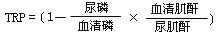 can be used to assess parathyroid function. The normal range is 85–95%, averaging 90%, while in hyperparathyroidism, it ranges from 45–83%, averaging 75%.
Serum calcium often exceeds 12mg%, serum phosphorus usually drops to 2-3mg%, serum alkaline phosphatase increases; urinary calcium excretion significantly rises, exceeding 20mg per 24 hours. This confirms the diagnosis.
bubble_chart Treatment Measures
Once the diagnosis of hyperparathyroidism is confirmed, preoperative localization of the affected gland should be performed whenever possible. Tumors located in the neck are generally not palpable. Ultrasound can detect neck tumors, and CT can identify tumors in the neck and mediastinum, but the positive detection rates are not high. Inserting a catheter from the femoral vein into the superior vena cava, innominate vein, and the superior, middle, and inferior thyroid veins draining the parathyroid glands, followed by blood sampling from these veins to measure PTH levels, can differentiate between hyperplasia and tumors and determine the tumor's location. However, this method requires complex equipment, is technically challenging, and carries certain risks, so it is not routinely performed. It is only used when no tumor is found in the neck during the first surgery and is conducted before the second operation.
Primary hyperparathyroidism requires surgical treatment. If it is a tumor, the affected gland should be excised. If it is hyperplasia, a subtotal parathyroidectomy should be performed, removing all three glands and part of the fourth, leaving behind a portion of normal-sized gland tissue (approximately 30–50 mg). The blood supply to the remaining gland must be preserved and not injured. If it is a carcinoma, the affected gland and the surrounding adherent tissues (such as the thyroid lobe and recurrent laryngeal nerve) should be resected en bloc. Biopsy should not be performed on carcinomas, as it can lead to local spread of cancer cells and recurrence.
Since 98% of parathyroid glands are located in the neck, the initial surgical exploration focuses on the neck. Tumors are more likely to occur on the right side, so if preoperative localization is unsuccessful, the right side should be explored first. Typically, the superior and inferior parathyroid glands on one side are identified behind the thyroid lobe. A significantly enlarged gland is definitely pathological, but a non-enlarged gland may not necessarily be normal. Normal glands and pathological glands (adenomas or hyperplasia) differ in density (specific gravity), so a density difference test can be used for differentiation. The method involves taking 1–2 mm thick slices from the superior and inferior glands and placing them in a test tube containing 20% mannitol solution. Initially, both specimens float at the top of the solution. Water is gradually added and mixed to dilute the solution until one specimen sinks to the bottom. If only one specimen sinks, it indicates that only one gland is pathological (adenoma), and exploration of the contralateral parathyroid glands is unnecessary. If both specimens sink simultaneously, it suggests similar densities—either both glands are normal or both are hyperplastic—requiring further exploration of the contralateral superior parathyroid glands.
If the superior and inferior parathyroid glands cannot be found behind the thyroid lobe, the tracheoesophageal groove and the posterior pharynx and esophagus should be exposed to locate the superior parathyroid gland. In 10–20% of cases, parathyroid adenomas are located in the mediastinum, but almost all can be removed via a cervical incision by pulling the thymus from behind the sternum into the neck and excising it to locate the inferior parathyroid gland. If the missing parathyroid gland is still not found in these locations, some advocate blindly resecting the ipsilateral thyroid lobe, though the success rate is very low. If the inferior parathyroid gland is not found during cervical exploration, initial sternotomy to explore the mediastinum is generally not recommended. Instead, postoperative selective venous catheterization to measure PTH levels in parathyroid venous blood for localization is advised, followed by a second surgery to re-explore the neck or perform sternotomy for mediastinal exploration.
In patients with parathyroid tumor diseases, the function of the unaffected parathyroid glands is suppressed. Hypocalcemic symptoms may appear 2–3 days after adenoma removal, but this condition is temporary, and blood calcium levels will normalize even without calcium supplementation, with symptom relief. Postoperative hypocalcemia is generally less pronounced in hyperplasia patients. If the adenoma is not removed or insufficient hyperplastic tissue is excised, postoperative blood calcium levels will not drop significantly. If a patient diagnosed with an adenoma does not exhibit hypocalcemic symptoms postoperatively, it suggests misdiagnosis, and the condition may actually be hyperplasia.
The main differentiation is from hypercalcemia caused by other reasons (such as kidney cancer, bronchogenic carcinoma, multiple myeloma, sarcoidosis, vitamin D intoxication) and secondary hyperparathyroidism. Cancers like kidney cancer and bronchogenic carcinoma can secrete parathyroid hormone-like polypeptides, which can cause hypercalcemia even without bone metastasis. Secondary hyperparathyroidism due to renal insufficiency or vitamin D deficiency each has specific manifestations of the original disease. In cases of hypercalcemia caused by cancer, myeloma, or sarcoidosis, PTH levels are not elevated. Hypercalcemia due to multiple myeloma, sarcoidosis, milk-alkali syndrome, or vitamin D intoxication can all be suppressed by oral corticosteroids. The alkaline phosphatase level in myeloma is normal.


