| disease | Tubal Pregnancy |
| alias | Tubal Pregnancy |
The egg is fertilized in the ampulla of the fallopian tube. If the fertilized egg is blocked in the fallopian tube for certain reasons and implants and develops in a part of the tube, a tubal pregnancy occurs. The most common site is the ampulla, accounting for 50–70% of cases, followed by the isthmus at 30–40%. The fimbriae and interstitial portions are the least common, accounting for 1–2%.
bubble_chart Etiology
1. Salpingitis Chronic salpingitis can lead to the formation of narrow sections due to inflammatory adhesions in the fallopian tube {|###|} membrane, tubal tortuosity, or peritubal inflammatory adhesions, often obstructing the passage of the fertilized egg. Salpingitis not only causes morphological changes but also frequently results in defects in the tubal {|###|} membrane cilia and reduces tubal motility, impairing the migration of the fertilized egg.
2. Tubal Hypoplasia or Malformation In cases of tubal hypoplasia, the muscular fibers of the tubal wall are poorly developed or absent, and the {|###|} membrane cilia are deficient. The affected tube appears thinner and more elongated than normal, often twisted in a spiral shape. Malformations may include multiple openings, diverticula, a double tubal ostium, or an additional underdeveloped tube known as an accessory fallopian tube.
3. Endometriosis of the Fallopian Tube Endometrial tissue can invade the interstitial portion of the fallopian tube, causing thickening of the interstitial area and narrowing or obstruction of the lumen, which is one of the causes of tubal pregnancy. Some suggest that ectopic endometrial tissue in the fallopian tube, ovary, or pelvic cavity may exert chemotactic effects on the fertilized egg, inducing implantation outside the uterine cavity.
4. Compression or Traction by Pelvic Tumors Pelvic tumors can compress or pull on the fallopian tube, making it thinner, longer, and more tortuous, thereby hindering the passage of the fertilized egg.5. Contraceptive Measures and Ectopic Pregnancy Whether intrauterine devices (IUDs) cause ectopic pregnancy is a topic of concern and debate. In 1965, Lippes first reported a higher incidence of ectopic pregnancy among IUD users. Most scholars believe that inert or active IUDs effectively prevent intrauterine pregnancy and partially prevent tubal pregnancy but do not prevent ovarian pregnancy. In recent years, the incidence of ectopic pregnancy with IUD use has significantly increased both domestically and internationally.
Post-sterilization recanalization, neofimbriae, or technical errors can also lead to tubal pregnancy.
6. Chlamydia Infection Chlamydia infection is an independent and significant risk factor for ectopic pregnancy. When the chlamydia antibody titer is 1:16, the relative risk is 2.91; at a titer of 1:64, the risk increases to 3.0.
bubble_chart Pathological Changes
I. Changes and Outcomes of Tubal Pregnancy During tubal pregnancy, due to the lack of a complete decidua in the fallopian tube, the fertilized egg implants directly into the muscular layer of the tubal wall through the destructive action of proteolytic enzymes from the trophoblast. This invasion damages the microvessels in the muscular layer, leading to bleeding. Blood accumulates between the trophoblast and surrounding tissues, and the fertilized egg becomes surrounded by a membrane composed of muscle fibers and connective tissue. Depending on the implantation site, different outcomes may occur.
(1) **Tubal Pregnancy with Late Abortion**: This most commonly occurs in the ampulla of the fallopian tube (Figure 1). The growing embryo tends to bulge into the tubal lumen. Due to the fragility of the surrounding membrane, rupture often occurs between 6–12 weeks of pregnancy, causing bleeding that dislodges the embryo into the tubal lumen. Since the ampulla is close to the fimbriated end, the embryo may be expelled into the abdominal cavity. If the embryo detaches completely and enters the abdominal cavity, bleeding is usually minimal, resulting in a complete late abortion. Sometimes, after detachment, the embryo remains in the fallopian tube, filling the lumen with blood and forming a tubal hematoma. After embryonic death, most are absorbed, but some may form a bloody mole. If the tubal hematoma organizes and hemoglobin dissipates, a fleshy mole may form. In cases of incomplete late abortion in the ampulla, trophoblastic cells may remain viable for an extended period, continuing to erode tubal tissue and causing recurrent bleeding. Repeated bleeding leads to blood accumulation around the fimbriated end and the fallopian tube, forming a peritubal hematoma. Eventually, significant bleeding may result in blood pooling in the rectouterine pouch, forming a retrouterine hematoma.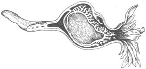
**Figure 1** Tubal Pregnancy with Late Abortion
(2) **Rupture of Tubal Pregnancy**: This most often occurs in the isthmus of the fallopian tube. Due to the narrow lumen, the trophoblast erodes through the muscular layer and serosa, eventually penetrating the tubal wall and causing rupture (Figure 2).
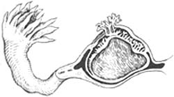
**Figure 2** Schematic of Ruptured Tubal Pregnancy with Late Abortion
In tubal pregnancy with late abortion, rupture occurs within the membrane without major vascular injury, causing only bleeding from the detached trophoblast. Thus, the progression is slow, with recurrent episodes but rarely life-threatening hemorrhage. In contrast, tubal pregnancy rupture involves injury to larger vessels in the tubal wall, leading to direct blood flow into the abdominal cavity, often causing severe, life-threatening bleeding. However, in some cases, only small venous branches or even larger arterial branches may be injured. Hypotension from internal bleeding may gradually reduce hemorrhage, with thrombus formation temporarily stopping the bleeding. Rupture in the isthmus can occur early, as soon as the first week after conception (the fertilized egg can implant 3–6 days after fertilization), so there may be no history of amenorrhea before clinical symptoms of ectopic pregnancy appear. In interstitial pregnancies, rupture may not occur until 3–4 months, presenting with symptoms similar to uterine rupture and causing extremely severe bleeding.In chronic cases, it is often difficult to distinguish between late abortion and rupture types, as they may overlap. Clinically, incomplete late abortion may be followed by tubal rupture due to persistent trophoblastic growth.
(3) **Secondary Abdominal Pregnancy**: When a tubal pregnancy ruptures or undergoes late abortion, the fetus may be expelled through the perforation or fimbriated end, while the placenta remains attached to the tubal wall or grows outward, implanting on the uterus, fallopian tube, broad ligament, pelvic wall, or other structures, forming a secondary abdominal pregnancy. If the rupture occurs between the two layers of the broad ligament, the embryo may continue developing into a broad ligament pregnancy or extraperitoneal pregnancy, another form of abdominal pregnancy.
(4) **Advanced Tubal Pregnancy**: In rare cases, a tubal pregnancy may progress to an advanced stage.
(5) Pelvic hematoma and infection: An abdominal mass in the hematoma of the uterus-rectal fossa can gradually be surrounded by a layer of connective tissue through the connective tissue reaction of the abdominal membrane and adhere to adjacent organs.
(6) Degeneration of the embryo or fetus: Some tubal pregnancies may resolve spontaneously due to degeneration, often occurring when the fertilized egg implants in the mucosal folds of the ampulla of the fallopian tube without invading the tubal wall. In some cases, even if the egg invades the muscular layer of the tubal wall, early embryonic death occurs due to nutritional disturbances, leading to spontaneous degeneration before obvious clinical symptoms appear. This condition is often discovered later during laparotomy for other reasons.
(7) Others: Occasionally, twin tubal pregnancies may be observed. The contralateral fallopian tube may also become filled with blood due to reflux from intrauterine hematometra. In rare cases, a tubal pregnancy may coexist with an intrauterine pregnancy.
II. Changes in the uterine endometrium During tubal pregnancy, the uterine muscles, influenced by endocrine factors, undergo hyperplasia and hypertrophy, causing the uterus to become larger than normal and softer, though smaller than the expected size for the amenorrhea period. A more notable change is the decidual transformation of the uterine endometrium shortly after fertilization. The presence of decidua is linked to the survival of the fertilized egg. In tubal pregnancies, the fetus often survives only for a short period. After fetal death, the uterine decidua may shed as a whole (triangular-shaped), known as a uterine cast (Photo 1), or as small fragments. In many cases, degenerative changes in the uterus cause the decidua to disintegrate before being expelled. It is believed that 50% of cases involve the expulsion of a true uterine cast.
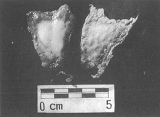
Photo 1: Uterine decidual cast
After the death of the fertilized egg, the uterine endometrium undergoes degenerative changes, so its appearance often depends on the timing of pregnancy termination.
Degenerative secretory pattern: After tubal pregnancy termination, some chorionic villi may survive for a period, with some deeply embedded in the muscular layer of the fallopian tube, closely connected to the maternal tissue. Therefore, the regression of the corpus luteum is slower than after normal pregnancy termination, delaying the maturation of new follicles. During the regression of the corpus luteum, the decidua, as sex hormone levels gradually decline, exhibits various degenerative processes or even atrophy. However, since the decidua represents the extreme development of the secretory phase of the uterine endometrium, it maintains secretory activity throughout the degeneration process.
Regenerative pattern: After a certain period, the endometrium in the degenerative secretory phase gradually regenerates. The stromal density becomes looser, glands appear round or oval, and glandular epithelial cells become columnar, with nuclei neatly arranged at the base or center of the cells, mostly resembling the proliferative phase endometrium. However, upon close examination, a small portion may still show degenerative secretory features. Thus, the endometrial patterns in ectopic pregnancy vary widely, depending on factors such as sex hormone concentration, the ratio of progesterone to estrogen, the duration of embryonic development, and the time elapsed since embryonic development ceased.
bubble_chart Clinical Manifestations
Before the fallopian tube pregnancy ruptures, there are generally no obvious symptoms. Some patients may experience early pregnancy reactions, such as loss of appetite, nausea and vomiting, or food aversions. Others may have intermittent dull pain on one side of the lower abdomen. During a bimanual examination, the uterus may not show significant enlargement or only slight enlargement, with a palpable mass on one side that is tender. Suspected fallopian tube pregnancy is then confirmed through further auxiliary examinations. Due to these characteristics—such as the absence of a clear history of amenorrhea, no history of infertility, and slight vaginal bleeding mistaken for side effects of an intrauterine device (IUD)—the misdiagnosis rate is high. Misdiagnosis further increases the danger of this condition. The key to early diagnosis lies in maintaining high vigilance among obstetricians, gynecologists, and women's healthcare workers. 1. After IUD placement, if irregular slight vaginal bleeding or lower abdominal pain occurs—regardless of whether there is a history of amenorrhea—ectopic pregnancy should be ruled out while treating IUD side effects. Necessary examinations should be conducted, and patients should be advised to self-monitor. For example, if severe abdominal pain or rectal tenesmus occurs, they should seek follow-up care. Any expelled tissue should be brought in for pathological examination to determine if it is decidual tissue. 2. In cases of pregnancy with an IUD where a late abortion procedure results in an empty aspiration, a pregnancy test and ultrasound should be repeated to confirm the diagnosis before rupture.
After a fallopian tube pregnancy undergoes late abortion or rupture, the clinical manifestations become evident.
I. Symptoms
(1) Abdominal pain: Most patients seek medical attention due to sudden abdominal pain, with an incidence rate exceeding 90%. The pain often begins as intense pain on the affected side, described as tearing, and may quickly spread to the entire abdomen. The severity and nature of the pain depend on the amount and speed of internal bleeding. In cases of rupture, rapid and substantial internal bleeding irritates the peritoneum, causing severe pain that may radiate throughout the abdomen. In cases of fallopian tube late abortion, bleeding is slower and less severe, with pain typically localized to the lower abdomen or one side. In rare cases, significant bleeding may reach the upper abdomen, irritating the diaphragm and causing upper abdominal or shoulder pain, often misdiagnosed as an acute upper abdominal condition. Repeated ruptures or late abortions can lead to recurrent internal bleeding. If a large single or multiple small internal hemorrhages are not treated promptly, blood may pool in the lowest part of the pelvis (the rectouterine pouch), causing severe rectal tenesmus.
(2) Amenorrhea: Fallopian tube pregnancy is often accompanied by amenorrhea. The duration of amenorrhea is usually related to the location of the pregnancy. For pregnancies in the isthmus or ampulla, amenorrhea typically lasts around 6 weeks before abdominal pain symptoms appear, rarely exceeding 2–3 months. In women with regular menstruation, if internal bleeding occurs a few days after a missed period, fallopian tube pregnancy should be considered. Interstitial fallopian tube pregnancy, due to the thicker surrounding muscle layer, often ruptures at 3–4 months of pregnancy, resulting in longer amenorrhea. When taking a medical history, detailed questions should be asked about the amount, quality, and duration of menstruation compared to past cycles, to avoid mistaking slight vaginal bleeding for a menstrual period. In rare cases, the chorionic gonadotropin produced by the trophoblast in fallopian tube pregnancy may be insufficient to induce an amenorrhea response in the uterine lining, resulting in no amenorrhea.
(3) Irregular vaginal bleeding: After the termination of a fallopian tube pregnancy, endocrine changes occur, leading to degenerative changes and necrosis of the uterine lining. The decidua may shed in fragments or entirely, causing uterine bleeding. The bleeding is often irregular, spotting, and dark brown, and it only stops completely after the lesion is removed (via surgery or medication). In rare cases, vaginal bleeding may be heavier, with some attributing the source to the fallopian tube rather than just the shedding uterine lining.
(4) Syncope and shock: Along with abdominal pain, patients often experience dizziness, blurred vision, cold sweats, palpitations, or even syncope. The severity of syncope and shock depends on the speed and volume of bleeding.
(5) History of infertility: Patients often have a history of primary or secondary infertility. In a Shanghai study of 2,822 cases, 66.28% had a history of infertility.
II. Signs
(1) General examination: Body temperature is usually normal, may be slightly lower in cases of shock, and can be mildly elevated (generally not exceeding 38°C) during absorption of internal bleeding. Blood pressure drops with internal hemorrhage, accompanied by rapid and weak pulse, and pale complexion.
(2) Abdominal examination: There is tenderness in the abdomen, with marked rebound tenderness, most pronounced on the affected side. The rigidity of the abdominal muscles is less severe than that seen in typical peritonitis, indicating that the bloody peritoneal irritation caused by internal hemorrhage differs from that of infectious peritonitis. When there is a significant amount of intraperitoneal bleeding, shifting dullness may be present. In cases of slow bleeding or delayed medical attention, a hematoma may form, presenting as a semi-solid, tender mass palpable in the abdomen.
(3) Vaginal examination: There is often slight vaginal bleeding originating from the uterine cavity. The posterior vaginal fornix is frequently full and tender. The cervix exhibits marked cervical motion tenderness, meaning the patient experiences severe pain when the cervix is gently moved upward or laterally. In cases of significant internal hemorrhage, the uterus may feel floating during examination. The uterus is normal in size or slightly enlarged and somewhat soft. A distended fallopian tube may be palpated on one side of the uterus. In patients who seek medical attention later, a semi-solid mass may be felt in the rectouterine pouch; the longer the delay, the more organized and hardened the hematoma becomes.
The patient's hemoglobin and red blood cell levels correlate with the extent of internal bleeding and the timing of the examination. At the onset of acute internal bleeding, hemoglobin levels may initially appear normal due to hemoconcentration. After 1–2 days, as hemodilution occurs, hemoglobin levels drop; or if bleeding continues, hemoglobin levels continue to decline. Therefore, in closely monitored patients, repeated hemoglobin measurements can be used for comparison. The white blood cell count is often elevated to 10×109/L.
bubble_chart Auxiliary Examination
1. B-mode ultrasound As an imaging diagnostic technique, ultrasound examination has the advantages of being simple to operate, highly intuitive, non-injurious to the human body, and repeatable. However, ultrasound images are complex, and the skill and experience of the examiner can vary greatly, with a misdiagnosis rate of up to 9.1%.
(1) Intrauterine images: There is no gestational sac, fetal bud, or primitive fetal heartbeat in the uterus. However, the incidence of pseudogestational sac sonograms is about 20%, caused by decidualization of the uterine membrane and a small amount of retained blood in the uterine cavity due to pregnancy. These sacs generally have unclear contours, incomplete layers, irregular edges, and do not enlarge with gestational age—sometimes even shrinking. Careful observation can differentiate them.
(2) Characteristics of adnexal masses and/or fluid in the uterorectal pouch: Adnexal masses are usually composed of gestational sacs, hematomas, and adherent bowel loops.
1. Before tubal pregnancy rupture, an irregular, poorly defined hypoechoic area can be seen beside the uterus. When the gestational sac develops to a certain extent, a round or oval hypoechoic gestational sac may be visible within this area. In rare cases, even the fetal bud and primitive fetal heartbeat can be observed, providing definitive evidence for diagnosing ectopic pregnancy. Reports indicate that the former is seen in 20% of cases, while the latter is observed in 12%.
2. During tubal pregnancy late abortion, blood flows from the fimbriated end of the fallopian tube, resulting in small amounts of free fluid in the adnexal mass and uterorectal pouch, appearing as anechoic or hypoechoic dark areas.
3. When tubal pregnancy rupture occurs, the gestational sac escapes through the ruptured fallopian tube. In the early stages, a hyperechoic mass may be seen beside the uterus due to hemorrhage, with chaotic internal echoes and enhanced echogenicity. The gestational sac is enveloped in the hematoma, and in very rare cases, the gestational sac echo may be visible, along with the fetal bud and primitive fetal heartbeat. If the rupture persists, repeated bleeding from the rupture site forms an enlarging adnexal mass with chaotic internal echoes, varying reflection intensities, and thickened edges—clinically presenting as an old ectopic pregnancy.
(3) Interstitial tubal pregnancy: Before the fertilized egg penetrates the muscle layer, the gestational sac may be seen surrounded by thickened muscle, with sonographic features similar to those of a rudimentary horn pregnancy. The two are difficult to differentiate.
2. Measurement of chorionic gonadotropin Techniques for measuring chorionic gonadotropin have significantly improved over the past decade. The hCG-β subunit radioimmunoassay can accurately detect early pregnancy and is a reliable method for diagnosing ectopic pregnancy. Syncytiotrophoblasts in the chorion secrete chorionic gonadotropin. Due to the thin mucosa and muscle layer of the fallopian tube, which cannot supply adequate nutrients to the trophoblasts, the plasma β-hCG concentration in ectopic pregnancy is lower. The β-hCG radioimmunoassay can detect the presence of a fertilized egg as early as the ninth day. In normal early pregnancy, β-hCG levels double every 1.2–2.2 days, whereas in 86.6% of ectopic pregnancies, the doubling time is prolonged, and the absolute β-hCG value is lower than in normal pregnancy.
3. Culdocentesis This is currently a widely used method for diagnosing ectopic pregnancy. If pus or serous fluid is aspirated, tubal pregnancy can be ruled out. However, failure to aspirate fluid does not exclude tubal pregnancy. If the mass is hard and difficult to aspirate, a small amount of saline can be injected first, followed by aspiration. If the aspirated saline appears reddish-brown and contains small blood clots, it confirms an old hematoma. If the blood is accidentally drawn from a vein, it will clot after a short time, whereas blood from a tubal pregnancy does not clot.
To further enhance the diagnostic value of posterior fornix puncture, the blood obtained from the posterior fornix puncture can be compared with peripheral venous blood through laboratory tests. The former shows slowed erythrocyte sedimentation rate (ESR), providing reliable evidence of thrombocytopenia. Whether it is a tubal pregnancy, late abortion, or rupture, and regardless of the duration of the onset, the ESR of the posterior fornix puncture blood is significantly slower, averaging 12.1 mm slower. Platelet counts are also markedly reduced, averaging 100,000 fewer. In contrast, the ESR and platelet counts of blood from accidental vascular puncture and peripheral vascular blood are almost identical.
IV. Laparoscopy For most ectopic pregnancies, the aforementioned examinations can confirm the diagnosis. However, for atypical cases, laparoscopy is highly valuable. It allows detailed observation of the ectopic pregnancy's location, its relationship with surrounding organs, and the state of adhesions. In some cases, surgical intervention can be performed simultaneously.
Laparoscopic Findings: The implantation site of the tubal pregnancy appears tumor-like, dark red, swollen, with proliferated and engorged surface blood vessels. If there is intra-abdominal bleeding, the visual field becomes obscured, and blood clots may adhere, making observation of the implantation site slightly difficult. In such cases, the abdominal cavity can be thoroughly irrigated with saline to clear the visual field, facilitating observation of the implantation site. Simultaneously, intra-abdominal blood and clots can be rapidly suctioned to ensure a clear view.
V. Diagnostic Curettage Diagnostic curettage is used to observe changes in the uterine membrane. If only decidual tissue is seen without chorionic villi, intrauterine pregnancy can be ruled out.
Additionally, in ectopic pregnancy, about 10–25% of cases show atypical hyperplasia of the uterine membrane resembling endometrial carcinoma. Features include highly convoluted glands, serrated edges, foamy cytoplasm, deeply stained nuclei, and irregularity. The Arias-Stella reaction, resembling an exaggerated secretory endometrium, also holds diagnostic significance. However, by the time patients seek medical attention, prolonged uterine bleeding may have already caused the endometrium to revert to a non-pregnant state. Thus, diagnostic curettage has significant limitations in diagnosing ectopic pregnancy.
VI. Hysterosalpingography (HSG) HSG has certain diagnostic value for tubal pregnancy before rupture. Before tubal rupture, HSG reveals the following characteristics:
(1) The uterine image appears relaxed and dilated, transforming from a triangular shape to a spherical one.
(2) The cervical canal image is not visible.
(3) After removing the HSG catheter, the contrast medium does not flow out.
These three points are similar to the HSG findings of intrauterine pregnancy, but the following features distinguish it from intrauterine pregnancy:
(1) No implantation site defect is seen in the uterine image.
(2) The uterine cavity edges appear irregular.
Typical cases present with acute abdominal pain, short-term amenorrhea, and irregular vaginal spotting, often with a history of primary or secondary infertility. Examination reveals tenderness and swelling of the affected fallopian tube. In cases of significant internal bleeding, hemorrhagic shock may occur. For cases where diagnosis remains uncertain, auxiliary diagnostic methods can be employed.
bubble_chart Treatment Measures
The treatment of tubal pregnancy has traditionally been primarily surgical. Over the past decade, the early diagnosis of ectopic pregnancy has significantly improved due to the development of highly sensitive radioimmunoassays for β-hCG, high-resolution ultrasound, and laparoscopy. Consequently, conservative surgery and medical treatment have been increasingly applied in clinical practice.
I. Surgical Therapy
(1) Salpingectomy: Whether it is a late abortion type or a ruptured tubal pregnancy, salpingectomy can promptly stop bleeding and save lives. For women who already have children and do not plan to conceive again, the contralateral tube may be ligated during the same seasonal epidemic. For women who wish to preserve fertility, if the tubal lesion is too large, the rupture is too extensive, the tubal mesentery and blood vessels are compromised, and/or vital signs are severely unstable, salpingectomy should also be performed. During conservative surgery, if tubal bleeding cannot be controlled, the tube should be immediately removed.
The surgery can be performed under acupuncture anesthesia or local anesthesia. After laparotomy, the first priority is to stop bleeding by clamping the bleeding point with forceps. In cases of shock, rapid blood transfusion should be administered, and salpingectomy should proceed step-by-step once the shock improves. If the ipsilateral ovary is normal, it should be preserved. If the contralateral tube is normal and the patient requests sterilization, tubal ligation should be performed. If the contralateral tube is diseased, management depends on the patient's condition, wishes, and the extent of the lesion. The principle is to minimize surgical time. Tubal implantation should not be considered during acute blood loss or in the presence of infection. If the free blood in the abdominal cavity shows no obvious infection, autotransfusion can be performed, especially when blood supply is scarce. Autotransfusion is an extremely effective measure for treating hemorrhagic shock. The blood should not be clotted, viscous, or foul-smelling, and red blood cell destruction under the microscope should not exceed 30%. Add 10 ml of 3.8% sodium citrate per 100 ml of blood. For autotransfusion exceeding 500 ml, administer 10–20 ml of 10% calcium gluconate to prevent citrate toxicity. Autotransfusion does not require cross-matching and can promptly restore blood volume, which is crucial for patients with severe internal bleeding and shock. It conserves banked blood, reduces financial burden, and provides fresh red blood cells with strong oxygen-carrying capacity, while also avoiding serum hepatitis and other pestilence diseases. In recent years, some have proposed autotransfusion without anticoagulants and applied it clinically. However, the degree of blood clotting varies among individuals. To fully leverage the advantages of autotransfusion, it is advisable to add sodium citrate or ACD solution as an anticoagulant.
(2) Conservative Surgery: The principle of conservative surgery is to remove the ectopic pregnancy while preserving the anatomy and function of the fallopian tube as much as possible, creating conditions for future intrauterine pregnancy.
Indications: Young women experiencing their first tubal pregnancy; women who have already lost one fallopian tube and have no children.
Surgical Methods: Salpingotomy with embryo removal involves making a 1–2 cm longitudinal incision along the antimesenteric border of the dilated segment of the affected tube. The pregnancy tissue is gently extruded, and the incision is sutured under a microscope with fine silk or 0/8 atraumatic sutures. Alternatively, a "window" technique can be used, where the incision edges are sutured intermittently for hemostasis without closing the incision, leaving a "window." For isthmic pregnancy, the affected segment is excised, and end-to-end anastomosis is performed. For isthmic pregnancy near the uterine cornu, tubal implantation into the uterine cornu can be performed.
Postoperative prevention of adhesions is one of the key measures to preserve fertility. This can be achieved by placing 250–300 ml of medium-molecular-weight dextran or 100 ml of 0.25% procaine, 250 mg of hydrocortisone, and 10 ml of glycerol in the abdominal cavity. Postoperative treatment is also critical for restoring fertility, such as timely tubal patency testing and invigorating blood and resolving stasis Chinese medicinals therapy.
(3) Laparoscopic Surgery: Under laparoscopy, first use an irrigator to flush and aspirate the accumulated blood in the pelvic cavity, and locate the implantation site of the gestational sac. If it is an ampullary pregnancy, the gestational products can be directly aspirated from the ampulla (via the fimbriated end) or removed with large spoon forceps. For isthmic or interstitial pregnancies, a salpingotomy is required. Inject 5% POR-8 (20–30 ml) into the mesosalpinx to induce local ischemia, which can prevent bleeding during the tubal incision. After electrocoagulating the convex area on the dorsal side of the tube, the tubal wall is incised until the gestational products are exposed. Using two atraumatic instruments, the tubal wall is separated, and the gestational products are slowly removed with large spoon forceps. Finally, the implantation site is flushed with an irrigator, and the serosal layer of the tube is sutured using an intra-abdominal knotting technique to close the wound.
II. Integration of Chinese and Western Medicine Treatment
According to the Eight Principles of Pattern Identification, tubal pregnancy belongs to the excess syndrome of blood stasis in the lower abdomen, where pain indicates obstruction. Therefore, the treatment principle should focus on invigorating blood and resolving stasis to relieve pain. This should be combined with pattern identification of cold, heat, deficiency, and excess, as well as clinical classification (shock type, stable type, mass type) for medication adjustment.
(1) Shock Type: Heavy internal bleeding. During the shock stage, attention should be paid to both deficiency and excess aspects, while also considering the patient's constitution in terms of cold or heat. Since tubal pregnancy itself is an excess syndrome, while internal bleeding, decreased blood pressure, pale complexion, cold sweating, and weak pulse are deficiency symptoms, the patient's condition must be analyzed at the time. If deficiency symptoms are more severe, use Ginseng to tonify qi to prevent blood deficiency, while also assisting with invigorating blood and resolving stasis to promote the absorption of internal bleeding.
Prescription: Salvia 30g, Red Peony Root 10g, Peach Kernel 10g, Frankincense 10g, Myrrh 10g. Decoct with 300ml of water and take in two doses. For severe deficiency, add Ginseng; for severe pain, add Corydalis Tuber. Simultaneously, manage shock with blood transfusion, intravenous fluids, oxygen, and warmth to maintain systolic blood pressure around 12kPa.
During treatment, closely monitor the patient's pulse, blood pressure, abdominal pain, and hemoglobin levels to determine if internal bleeding continues. The patient must rest absolutely in bed and avoid early movement, minimizing sudden changes in posture or factors that increase abdominal pressure. Treatment should be conducted under conditions where blood transfusion and emergency laparotomy are available. This type should ideally undergo immediate surgical treatment, though individual patients who have undergone multiple abdominal surgeries and refuse further surgery may be considered for conservative management.
(2) Stable Type: The condition is stable, blood pressure is steady, abdominal pain is alleviated, free blood in the abdominal cavity has initially formed a mass or partially absorbed, abdominal tenderness and rebound pain are reduced, and shifting dullness gradually disappears. Vaginal examination may reveal a palpable mass. Based on the main prescription, heat-clearing and detoxicating medicinals such as Skullcap Root, Honeysuckle, and Forsythia can be added to prevent infection. During this stage, bed rest remains primary, with gradual and appropriate activity.
(3) Mass Type: This type is characterized by blood stasis excess pattern in the lower abdomen with a mass. In addition to the main prescription for invigorating blood and resolving stasis, add medicinals to dissolve and break up masses, such as Sparganium Rhizome and Zedoary. However, prolonged use of strong dispersing medicinals like Sparganium Rhizome and Zedoary may lead to deficiency pattern. Therefore, based on symptoms and pulse manifestation, qi-tonifying medicinals like Tangshen and Astragalus Root should be added. To accelerate mass absorption, apply an external plaster: Chaulmoogra Tree Seed 15g, Cochinchina Momordica Seed 15g, Verdigris 15g, and 10 Chinese Dates (pitted), mixed and ground into fine powder, wrapped in gauze, and placed on the lower abdomen with a hot compress. Alternatively, use Musk 0.6g, Camphor 6g, Dragon's Blood Resin 9g, Rosin 9g, and Cinnabar 9g. Grind the latter four ingredients into fine powder, spread on cloth, melt with heat, and finally add Musk before applying to the lower abdominal mass.
If the pregnancy is not terminated and the pregnancy test remains positive, add Centipede (two pieces), Achyranthes Root 15g, Snakegourd Root 9–15g, and Gleditsia Sinensis 9g to eliminate the embryo and turn the pregnancy test negative. However, the effect is not entirely satisfactory. Treatment can also be monitored with β-hCG and ultrasound, and MTX may be added to accelerate pregnancy termination.
III. Drug Therapy
Methotrexate (MTX) is mainly used for unruptured tubal pregnancy, with intact tubal serosa, no active bleeding, tubal pregnancy product diameter <3–4cm, intraperitoneal blood <100ml, β-hCG <3000mIU/ml, stable vital signs, and young patients who desire fertility.
Administration method: ① Oral MTX 0.4mg/(kg·d) for 5 days as one course. The usual dosage is 25mg/d for 5 days, with no toxic reaction, but it is rarely used clinically; ② Intramuscular MTX 0.4mg/(kg·d) for 5 days as one course. Some authors reported 23 cases of ectopic pregnancy, with 95.7% absorption of the gestational sac after treatment, and hysterosalpingography or laparoscopy confirmed tubal patency in 10/19 (52.6%) patients; ③ MTX-CF regimen, citrovorum factor (CF), which can reverse the toxic effects of MTX, is currently the most commonly used method. When the MTX dosage is 1mg/kg, the plasma concentration reaches 10-8M, and CF rescue must be used to achieve high efficacy with low toxicity. The intravenous infusion time of MTX should be less than 4 hours, and CF is 1/10 of MTX, with an interval of 24 hours between the two; ④ Local injection of MTX, guided by ultrasound into the gestational sac; or direct injection into the fallopian tube under laparoscopy. During drug therapy, close monitoring of abdominal pain, vital signs, and drug toxic reactions is essential. β-hCG and ultrasound should be used to monitor the local condition of the fallopian tube.
If a ruptured tubal pregnancy is diagnosed in time, the treatment outcomes are generally good, whether through surgical intervention or Chinese medicine Chinese medicinals. For a ruptured interstitial pregnancy, timely diagnosis and emergency treatment also yield favorable results, with almost no fatalities currently reported.
Fertility after ectopic pregnancy: Most patients with ectopic pregnancy are eager to understand their future fertility prospects. For those whose ectopic pregnancy resulted from organic or functional sexually transmitted disease-related changes, the likelihood of future infertility increases, especially for women over 30 who have had pelvic inflammatory disease. The chances of a normal intrauterine pregnancy reaching full term are very low. For those whose first pregnancy was an ectopic pregnancy, fertility prospects are even poorer. The possibility of a subsequent intrauterine pregnancy is only about 1/10 of normal, and even if pregnancy is achieved again, the risk of another ectopic pregnancy can be as high as 50%.
Differential diagnosis must be made with the following diseases:
1. **Early pregnancy late abortion** In late abortion, abdominal pain is usually mild, located in the central lower abdomen, and intermittent. Vaginal bleeding is typically heavy, and the amount of bleeding corresponds to systemic blood loss symptoms. There is no or only slight abdominal tenderness, generally no rebound tenderness, and no shifting dullness. Vaginal examination reveals no cervical motion tenderness, the posterior fornix is not full, the uterus size matches the amenorrhea duration, and there are no masses beside the uterus. For patients who already have children or experience heavy bleeding, diagnostic curettage may be performed after discussion with the patient and family.
2. **Acute salpingitis** There is no history of amenorrhea or early pregnancy symptoms, and no signs of shock. Body temperature is elevated, with abdominal muscle tension and tenderness on both sides of the lower abdomen. Vaginal examination shows no fullness in the posterior fornix, a normal-sized uterus, and often thickened adnexa, masses, and tenderness on both sides (sometimes more pronounced on one side). Pus may sometimes be aspirated during posterior fornix puncture. Leukocyte and neutrophil counts are elevated, and pregnancy tests are negative. Particularly in hemorrhagic salpingitis, there may be lower abdominal tenderness and rebound tenderness, and sometimes shifting dullness. Fresh blood may be aspirated during posterior fornix puncture, making preoperative differentiation difficult, and diagnosis is often confirmed only during laparotomy. However, reports indicate that most patients have a recent history of induced late abortion, with negative hCG. If significant internal bleeding occurs (some reports mention volumes exceeding 1200 ml), exploratory laparotomy is necessary, revealing thickened, congested, and edematous fallopian tubes with fresh blood flowing from the fimbriated end. Pathology shows acute inflammation without chorionic villi.
3. **Acute appendicitis** There is no amenorrhea or early pregnancy symptoms, and no vaginal bleeding. Abdominal pain usually starts in the upper abdomen and later localizes to the right lower quadrant, often accompanied by nausea and vomiting, with no signs of internal bleeding. Examination reveals right lower abdominal muscle tension, tenderness, and rebound tenderness at McBurney's point, with no shifting dullness. Vaginal examination shows no cervical motion tenderness and a normal-sized uterus. If appendicitis spreads to involve the right fallopian tube or a broader area, there may be right adnexal tenderness or bilateral tenderness; otherwise, the adnexa show no significant abnormalities. Pregnancy tests are negative, with elevated body temperature and increased white blood cell count.
4. **Ovarian cyst torsion** There is a history of abdominal masses. If torsion resolves spontaneously, abdominal pain is transient; if intracystic hemorrhage occurs due to torsion, the pain becomes persistent, but tenderness and rebound tenderness are limited to the mass and its surroundings, with no shifting dullness. Vaginal examination reveals a tender cystic mass beside the uterus. There is no history of amenorrhea or early pregnancy symptoms, nor vaginal bleeding, but note that early pregnancy can sometimes trigger torsion of a preexisting ovarian cyst.
5. **Corpus luteum rupture** This often occurs in the premenstrual phase, frequently after intercourse, with no amenorrhea or early pregnancy symptoms and no vaginal bleeding. The nature of abdominal pain and signs resemble those of ruptured tubal pregnancy, but pregnancy tests are negative.
6. **Chocolate cyst rupture** This condition mostly occurs in young women and is prone to spontaneous rupture, causing acute abdominal pain, but there is no amenorrhea or early pregnancy symptoms, nor vaginal bleeding. The patient may have a history of progressive dysmenorrhea and pelvic masses.


