| disease | Pancreatic Trauma |
The pancreas is a gland with both endocrine and exocrine functions. Its deep location, protected anteriorly by the rib cage and posteriorly by the spine, makes it less prone to injury, often leading to misdiagnosis. It wasn't until 1952 that comprehensive reports on pancreatic injuries emerged. Pancreatic injuries occur in 0.4 per 100,000 people, accounting for 0.2% to 0.6% of abdominal trauma. In wartime, pancreatic injuries are mostly penetrating wounds, often accompanied by severe hemorrhage and high mortality. On average, they result from severe blunt abdominal trauma, sometimes due to surgical mishaps. The ratio of penetrating to blunt pancreatic injuries is approximately 3:1. In a study of 1,984 cases of pancreatic trauma, penetrating wounds accounted for 73%, while blunt injuries made up 27%.
bubble_chart Etiology
In traffic accidents, especially when a car is traveling at high speed and suddenly collides with an object, the strong inertia causes the driver's upper abdomen to hit the steering wheel, resulting in pancreatic injury. Sometimes, when a person falls from a height, the waist becomes excessively flexed, while both costal arches are severely retracted, and an instantaneous explosive force compresses the pancreas, causing varying degrees of pancreatic injury (Figure 1).
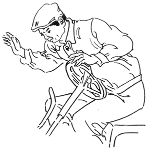
Figure 1 Mechanism of pancreatic injury
The location of pancreatic injury varies with the direction of the external force, with the head and body of the pancreas being the most commonly affected. When the force is applied to the upper right abdomen or the right side of the spine, the pancreatic head is prone to compression, often accompanied by injuries to the duodenum, bile duct, and liver. Such injuries have severe consequences, with a mortality rate as high as 70–80%. When the force is directly applied to the middle of the upper abdomen, the injury often involves partial or complete rupture of the pancreatic neck or body, along with injury to the superior mesenteric artery. If the force is applied to the left side of the spine, the pancreatic tail is frequently injured, often accompanied by splenic rupture.
bubble_chart Pathological ChangesThe pathological changes of pancreatic closed injury are progressive. Surgeons often pay great attention to pancreatic rupture and focus their efforts on managing it, while they tend to underestimate local pancreatic contusion. This is mainly due to insufficient understanding of its pathological characteristic—progression.
After pancreatic injury, general contusion marks initially appear at the local site. However, pancreatic fluid often leaks from the contusion line into the pancreatic interstitium, leading to self-digestion of the pancreas, which further digests the contusion area and results in "secondary rupture." The time from self-digestion to secondary pancreatic rupture varies, depending on the degree and extent of the pancreatic contusion. The process of self-digestion leading to secondary rupture is illustrated in the diagram (Figure 1).
(1) Contusion area
(2) Hematoma and digestion in the contusion area
(3) Rupture
Figure 1 The self-digestion process of pancreatic contusion can take anywhere from a few hours to several days.
Simple pancreatic contusion: The pancreatic capsule may remain intact or rupture. The former is a simple pancreatic injury, with grade I injury to the pancreatic interstitium, which is referred to as traumatic pancreatitis. The latter (ruptured pancreatic capsule) is more severe than the former, but there is no significant hematoma within the pancreas, nor is there pancreatic duct rupture. Contusion can occur in any part of the pancreas.
Deep laceration of the pancreas: Accompanied by hematoma and liquefaction within the pancreatic parenchyma, but without pancreatic duct injury.
Pancreatic rupture: The implications of pancreatic rupture include: ① Pancreatic rupture or fracture, exceeding more than half of the pancreatic diameter; ② Central penetrating injury of the pancreas; ③ Visible injury to the pancreatic duct; ④ Severe crushing and fragmentation injury of the pancreas.Contusion of the pancreatic head: Due to the uniqueness of its anatomical location, it should be classified independently, whether it is a simple contusion or a severe rupture. Duodenal injury refers to cases accompanied by traumatic rupture. Most duodenal injuries occur on the anteromedial wall, while a few patients may have rupture of the posterior wall of the second segment of the duodenum. Large ruptures on the posterior wall are easier to diagnose, but small ruptures are more prone to misdiagnosis. During surgery, if a hematoma is observed behind the lateral peritoneal membrane of the duodenum, sometimes accompanied by crepitus, the posterior peritoneal membrane should be incised along the duodenum, and the duodenum should be flipped to the left to carefully check for any fissures. Once misdiagnosed, it will lead to irreparable consequences.
The Japanese Society for the Study of Pancreatic Trauma has proposed the following classification for pancreatic injury, which is worthy of clinical reference.
Type I (contusion type): The pancreas exhibits punctate hemorrhage or hematoma, but the membrane remains intact, and there is no fistula disease fluid in the abdominal cavity.
Type II (laceration type): Various types of pancreatic injury without damage to the main pancreatic duct.
Type III (main pancreatic duct injury type).
a type: Main pancreatic duct injury in the body and tail of the pancreas.
b type: Main pancreatic duct injury in the head of the pancreas, accompanied by injury to the pancreatic duct and intra-pancreatic bile duct.The severity is ranked as: III b > III a > II > I.
bubble_chart Auxiliary Examination
1. Laboratory tests: Serum phospholipase A2 (SPLA2), C-reactive protein, α1-antitrypsin, α2-macroglobulin polycytidylic acid ribonuclease (poly-(c)-specific RNAase), serum methemoglobin, plasma fibrinogen, etc. These tests have good reference value but are not yet widely used.
2. B-ultrasound and CT examination: Small omental sac effusion, pancreatic edema, etc., can be observed. Since the pathological changes of pancreatic injury are progressive, imaging examinations should also be dynamically monitored. However, it can sometimes be easily confused with retroperitoneal hematoma.
4. Peritoneal lavage or abdominal puncture: In the early stages of pancreatic injury, there may be very little intraperitoneal fluid, and the puncture is often negative. Therefore, in addition to timing the abdominal puncture correctly, multiple punctures are necessary to achieve a clear diagnosis.
(1) The diagnosis of pancreatic trauma must first clarify several clinical issues to achieve a comprehensive and accurate diagnosis:
1. Isolated pancreatic injury often does not cause immediate death in the early stages. Early fatalities are usually due to combined injuries to other solid organs or massive hemorrhage from major vascular injuries.
2. In cases of simple pancreatic injury or Grade I combined injuries, early symptoms and specific signs are often absent, making diagnosis difficult. Delayed treatment increases the incidence of complications.
3. The digestive action of pancreatic enzymes leads to necrosis and hemorrhage in surrounding tissues, resulting in a complication rate of 30–50% post-injury.
4. Due to tissue necrosis, contamination, blood loss, shock, and weakened immunity, infections can easily spread, often leading to multiple organ failure and high mortality.
5. In the early stages of Grade II injury, combined with temporary suppression of pancreatic secretion or inactive enzyme release, symptoms are often atypical, leading to misdiagnosis. Only 50% of cases are correctly diagnosed preoperatively.
6. Pancreatic injury has a high incidence of combined injuries to other organs. - Open injuries combined with other organ injuries: liver injury (45–47%), gastrointestinal injury (47%), duodenal injury (24%), splenic injury (21–25%), renal injury (23%), small intestine injury (15%), colon injury (19%), vascular injury (30%). - Closed pancreatic injuries combined with other organ injuries: liver injury (18%), gastric injury (5%), duodenal injury (15%), splenic injury (15%), small intestine injury (8%), vascular injury (9%).
The number of combined organ injuries is directly proportional to mortality: - 1 organ injury: 4% mortality, - 2–3 organ injuries: ~15% mortality, - 4 or more organ injuries: >40% mortality. Therefore, when diagnosing pancreatic injury, a thorough examination of other abdominal organs is essential.
(2) Key points for diagnosing pancreatic injury are as follows:
1. Do not overlook upper abdominal contusion.
Any blunt contusion to the upper abdomen, regardless of the direction of force, should raise suspicion of pancreatic injury. If pancreatic rupture is accompanied by major vascular injury, obvious abdominal signs are usually present. However, minor or concealed pancreatic injuries may be overlooked early and only detected days or weeks later.
2. Accurately assess serum amylase levels.
A common misconception is that amylase levels must rise after pancreatic injury, ignoring the timing of elevation and the fact that severe pancreatic injuries may not show elevated amylase, leading to delayed diagnosis. - Serum amylase rises in most pancreatic injuries (~90%), but the degree of elevation correlates with injury severity. - In 179 cases of blunt pancreatic contusion, only 36 (20%) showed elevated serum amylase within 30 minutes post-injury. - In the initial stage of pancreatic injury, temporary suppression of enzyme secretion may prevent amylase elevation. Repeated dynamic measurements are necessary. - A single normal serum amylase reading should not rule out pancreatic injury. - Some suggest that measuring amylase in a 2-hour urine collection is more reliable than serum testing. - Peritoneal fluid amylase testing (via paracentesis or lavage) can aid diagnosis, as amylase rises rapidly in peritoneal fluid post-injury, with most cases showing positive results.
3. Fully understand the progression of pancreatic injury.
- Mild pancreatic injury manifests as contusion, while severe cases may involve rupture or transection, sometimes combined with duodenal injury. - Pancreatic contusion may initially present with subtle symptoms, becoming apparent only after pancreatic fluid leakage triggers autodigestion. - In severe contusions with intact pancreatic membranes, tissue swelling and the "constrictive" effect of the membrane can lead to progressive tissue damage and necrosis.
4. Pancreatic injury is often confused with injuries to other organs.
Due to the proximity of the pancreas to major blood vessels and organs, it is often complicated by injuries to other organs, which can confuse symptoms and make diagnosis difficult. Sometimes, attention is focused solely on major vascular injuries or injuries to other parenchymal organs, leading to a missed diagnosis of pancreatic injury or fistula disease.
5. Other Examinations
B-mode ultrasound and CT scan: These have certain diagnostic value for pancreatic injury, with a relatively high positive rate.
Endoscopic retrograde cholangiopancreatography (ERCP): This has a very high positive rate for diagnosing pancreatic injury, especially in determining whether there is pancreatic duct injury, which is particularly significant.
Peritoneal lavage or abdominal paracentesis: This method has great diagnostic value, with a positive rate of almost 100% (elevated amylase in the extracted peritoneal hemorrhagic fluid).
6. Key Points for Intraoperative Diagnosis
Severe pancreatic contusion or laceration can be clearly diagnosed upon laparotomy: intraperitoneal hemorrhage, retroperitoneal hematoma, or hematoma in the lesser omental sac usually make the diagnosis straightforward. However, minor injuries are prone to be missed. Therefore, when pancreatic injury is suspected, a comprehensive examination must be performed.
The laparotomy incision should be sufficiently large. Lift the transverse colon and push the small intestine downward to palpate the root of the mesocolon, the lower edge of the pancreas, and adjacent tissues. Incise the gastrocolic ligament, lift the stomach upward, and pull the colon downward. Then, incise the retroperitoneum lateral to the duodenum to mobilize the duodenum and explore the dorsal side of the pancreatic head, thereby assessing whether there is concomitant duodenal injury. Additionally, incise the retroperitoneum along the upper and lower edges of the pancreas and, if necessary, further mobilize the dorsal surface of the pancreas. During exploration, if a hematoma is found on the pancreas, it should be incised and examined—even small hematomas should not be overlooked, as injured pancreatic tissue often lies beneath them. Some have emphasized that any retroperitoneal hematoma in the upper abdomen should raise suspicion of pancreatic injury. In our treated cases, retroperitoneal hematomas were almost universally present. Grade I pancreatic injuries typically involve an intact capsule with only localized edema, ecchymosis around the pancreas, and varying degrees of hemorrhage.
To confirm whether the pancreatic duct is ruptured, some advocate resecting a small portion of the pancreatic tail for retrograde catheterization and contrast imaging. Alternatively, the duodenum can be incised for catheterization and contrast imaging via the duodenal papilla. This method is only used for severe and extensive pancreatic contusions where ductal rupture is difficult to confirm. For simple contusions, adequate drainage alone is usually sufficient for healing. Rashly resecting the pancreatic tail or performing duodenal catheterization and contrast imaging may exacerbate trauma, leading to pancreatic or duodenal fistulas and complicating treatment. Alternatively, methylene blue injection can be employed: mix 1 ml of methylene blue with 4 ml of water (saline) and inject it into normal pancreatic tissue distal to the injury. The methylene blue will then leak out through the injured main pancreatic duct.
bubble_chart Treatment Measures
1. Emergency Management of Pancreatic Injury
After pancreatic injury, the main manifestations are intra-abdominal hemorrhage and acute pancreatic peritonitis, followed by water, electrolyte, and acid-base imbalances. Therefore, immediate anti-shock measures must be taken, including aggressive fluid resuscitation and appropriate albumin infusion to reduce exudation. Regardless of whether blood pressure stabilizes, surgery should not be delayed and must be performed immediately under active anti-shock treatment. If the injury is severe with significant bleeding, surgery should proceed while continuing anti-shock measures, without waiting for blood pressure to recover.
(1) Pancreatic injury is challenging to treat, with high complication and mortality rates. The following principles are often overlooked during treatment, leading to failure:
1. Pancreatic injury accompanied by major vascular damage is life-threatening. Upon laparotomy, rapid exploration and appropriate management of these injured vessels should be prioritized. Hemorrhagic pancreatic tissue should not be clamped or sutured (especially deep sutures) to avoid damaging major pancreatic ducts.
2. Accurately assess the extent, range, and presence of pancreatic duct rupture.
3. Perform reasonable resection of the injured area to minimize impact on endocrine and exocrine functions.
4. Prevent activation of pancreatic enzymes due to pancreatic fluid leakage.
5. Apply internal and external drainage appropriately.
6. Prevent complications such as pancreatic fistula and pancreatic cyst formation.
The pancreas is deeply located, horizontally elongated, extending from the duodenum to the splenic hilum. An improper surgical incision can greatly hinder exploration, and poor exposure may lead to missed injury sites, resulting in fistula formation.
Various incisions can be used for pancreatic surgery. For exploration, a midline upper abdominal incision is preferable. For confirmed diagnoses, a pancreatic projection incision or an upper abdominal curved incision can fully expose the head, body, and tail of the pancreas. While these incisions provide excellent exposure, they cause significant abdominal wall damage and prolong surgery. In emergencies, a midline incision can suffice for complete pancreatic exploration.
(2) Emergency Management of Different Types of Pancreatic Trauma:
1. Pancreatic Contusion
Can be categorized into intact capsule and ruptured capsule types. The former is a simple pancreatic injury, often referred to as "traumatic pancreatitis." For pancreatic contusion with capsule rupture, cigarette drainage plus double-catheter drainage can be used. If no pancreatic fluid leaks from the drainage tube, it can be removed after a few days. Even minimal pancreatic fluid output warrants continued drainage. To reduce bile reflux into the pancreatic duct, biliary fistula creation may also be performed. For pancreatic injury with an intact capsule, omitting drainage is inadvisable, as minor capsule ruptures—especially on the dorsal side—may be missed even after meticulous exploration.
2. Pancreatic Fracture
Distal pancreatic fractures are typically managed by resecting the distal end and suturing the proximal stump. For fractures of the pancreatic neck or body, pancreatic duct anastomosis is not recommended due to the high risk of complications like fistula and stenosis. Instead, distal pancreatectomy is preferred. This approach reduces fistula risk, avoids endocrine insufficiency (as removal of the distal pancreas does not significantly impact insulin production), and prevents pancreatitis caused by enzyme activators introduced during intestinal anastomosis. Although the pancreatic tail has a higher density of islets than the head or body, resection of 80–90% of the pancreas generally does not cause endocrine insufficiency. However, resecting beyond this range (e.g., to the right of the mesenteric artery) may lead to pancreatic insufficiency. If excessive pancreatic tissue is removed, postoperative insulin therapy should be administered to prevent degeneration of the remaining islet cells due to overproduction.
After partial pancreatectomy, the regenerative capacity of the remaining pancreas differs from that of the liver, exhibiting limited spontaneous regeneration. Parekh reported the results of a group of rat experiments using a synthetic trypsin inhibitor (FOY-305), which stimulates the growth of normal rat pancreas by increasing the release mechanism of endogenous cholecystokinin (CCK). The experimental results showed that after pancreatic resection (66% distal resection), significant regenerative capacity could be observed in the pancreas when stimulated by FOY-305 via gavage. The regeneration process initially involved hypertrophy followed by hyperplasia as the treatment duration increased. The extent of pancreatic mass hyperplasia exceeded that of the normal unresected pancreatic mass only after 27 days of treatment. Although these findings are still in the research stage, they provide new insights into the treatment of pancreatic insufficiency following subtotal pancreatectomy or acute necrotizing pancreatitis.
3. Injury to the head of the pancreas
Injury to the head of the pancreas is difficult to manage. Simple drainage will fail, while resection of the fractured caudal segment will lead to pancreatic insufficiency, making both approaches inappropriate. The correct management principles are as follows: ① For contusion or laceration alone, anastomosis with the jejunum can be performed at the site; ② If the pancreas is fractured, the duodenal side of the fracture should be closed, and the distal pancreatic stump should be anastomosed to the jejunum to preserve pancreatic function. Alternatively, a segment of jejunum can be interposed between the two pancreatic stumps for double-end jejunal anastomosis to retain pancreatic function; ③ If the injury is very close to the duodenum or accompanied by duodenal rupture, the duodenum should be resected together, and the distal pancreatic stump should be anastomosed to the jejunum.
4. Combined injuries involving the pancreatic head
Injury to the pancreatic head is often combined with duodenal rupture and may also involve injuries to the inferior vena cava, portal vein, or superior mesenteric vessels. Cases with major vascular injuries often result in immediate death. The mortality rate for combined pancreatic head and duodenal injuries is very high.
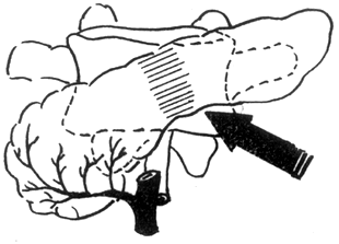
For contusion of the pancreatic head and duodenal rupture, the following approach can be adopted: partial resection of the gastric antrum, end-to-side gastrojejunostomy, duodenostomy, suturing of the duodenal rupture, vagotomy, and common bile ductostomy—effectively "diverticulizing" the duodenum (Figure 1)—along with latex tube drainage and double-catheter drainage. To prevent reflux, the gastrojejunostomy should be at least 60 cm away from the injured duodenum. However, some hold differing opinions, suggesting that simple repair of the injury site, jejunostomy, and high-calorie parenteral nutrition (TPN) may suffice.
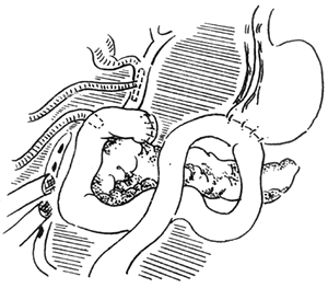
Figure 1 "Diverticulization" of the duodenum
Injury to the pancreatic head often involves bile duct injury, especially when the pancreatic duct is injured near the duodenum. Intraoperative cholangiography should be performed to assess the condition of the common bile duct. The junction of the common bile duct and duodenum requires careful examination to avoid missing a fistula.
Pancreaticoduodenectomy is a highly destructive procedure and should not be performed lightly. It is generally indicated in the following situations: ① Severe injury to the pancreatic head or pancreatic duct rupture that precludes anastomosis with the intestine; ② Severe duodenal contusion or laceration with irregular margins, extensive rupture, or involvement of the ampulla of Vater that makes repair difficult; ③ Pancreatic head injury combined with portal vein rupture; ④ Avulsion of the pancreas from the duodenum.
Regarding whether the residual pancreas after pancreaticoduodenectomy for contusion or laceration of the pancreatic head requires anastomosis of the pancreatic stump to the jejunum, some have compared it with the conventional Whipple procedure. The authors suggest that after pancreaticoduodenectomy, reconstruction with gastrojejunostomy and biliary-enteric anastomosis suffices, without the need for pancreaticojejunal anastomosis. The pancreatic duct at the stump can be ligated, and external drainage placed around it. Comparative studies of the two approaches showed no significant statistical difference in mortality or complications. Moreover, in severely traumatized patients, ligation of the pancreatic duct without pancreaticojejunal anastomosis simplifies the procedure and is better tolerated.
Clinically, cases of grade I contusion of the pancreatic head with predominant rupture of the second part of the duodenum are frequently encountered. Management must address both injuries. For pancreatic head contusion, simple drainage around the site suffices, while the focus should be on repairing the duodenal injury. If the duodenal rupture is simple, the following methods can be employed: ① Simple suturing with gastrostomy and high jejunostomy; ② Simple suturing with gastrojejunostomy; ③ Jejunal seromuscular patch repair; ④ Pedicled open ileum graft for duodenal rupture repair. The choice of technique depends on the extent of the duodenal rupture.
In recent years, some have reported good results using fibrin glue (Fibrin Glue) to seal pancreatic injury sites. In 15 cases treated with fibrin glue, no postoperative pancreatic fistula, pancreatic abscess, or pseudocyst formation was observed.
Fiber egg beautiful steetgum resin Although both pancreatic duct ligation and occlusion techniques have been reported, the number of cases is limited. Further discussion is needed before widespread clinical application can be considered.
After pancreatic injury or pancreatectomy, drainage around the pancreatic bed or the pancreas is crucial. Proper use of drainage not only reduces infection but, more importantly, diverts pancreatic enzymes away to prevent them from "digesting" surrounding blood vessels and organs. Cases of major vascular erosion and bleeding or skin digestion due to improper drainage after pancreatectomy have been frequently reported, necessitating high clinical vigilance. It can be said that the adequacy of drainage directly determines the success or failure of pancreatic trauma treatment and surgical outcomes. The principles of drainage are as follows: ① Ensure thorough drainage; ② Avoid retrograde infection; ③ Use drainage tubes with minimal irritation and appropriate softness/hardness; ④ Maintain suitable negative pressure in the drainage tube; ⑤ Remove the drainage tube at the right time using a gradual withdrawal method. This approach helps create a "dry" environment around the injured pancreas, minimizing complications (Figure 2).

Figure 2 Pancreatic Drainage
The placement of drainage material in pancreatic injury must be considered from a pathological perspective. For instance, in cases of contusion, the injury may appear "mild" during exploration, but over time, the contused area may undergo autodigestion by pancreatic enzymes. Therefore, drainage should be placed at the contusion site, with gentle mobilization of the pancreas's upper, lower, and posterior aspects to ensure minimal irritation and effective drainage.
Choice of drainage materials: Common options include cigarette drains, Penrose drains, plasma tube drains, double-lumen tube drains, and closed double-lumen tube drains. The drawbacks of cigarette drains and Penrose drains are well-known and will not be discussed here. Double-lumen tube drains significantly reduce complications and achieve thorough drainage, but their major flaw is the risk of retrograde infection due to the open lumen. Closed double-lumen tube drains eliminate this issue. Literature reports indicate no significant difference in drainage efficacy between the two, with the closed system overcoming the retrograde infection drawback.
Premature removal of drainage postoperatively is inappropriate. Generally, drainage tubes should remain in place longer, even if output is minimal. The typical drainage duration should be no less than 5–7 days. If the fluid from the pancreatic stump or bed has high amylase levels, drainage should be extended until no fluid is discharged, with gradual withdrawal rather than immediate removal.
After pancreaticoduodenectomy, the stent drainage tube in the pancreaticojejunal anastomosis typically drains little pancreatic fluid in the first 1–3 days, increasing to around 50ml daily by days 3–5. If no pancreatic fluid is observed, adjustments are needed. Ensuring pancreatic fluid drainage is critical for anastomotic healing and must not be overlooked.
Post-pancreatic injury, significant fluid loss occurs due to intraoperative fluid depletion, gastrointestinal decompression, exudation from the pancreatic bed or stump, and pancreatic duct fluid loss. Daily infusion of 5000–7000ml may still struggle to maintain normal blood volume. Thus, for the first 3–5 days, fluid administration should be monitored via CVP, with regular measurements of urine output and specific gravity. Electrolyte replacement should be based on lab results. To reduce pancreatic secretion, continuous gastrointestinal decompression, total parenteral nutrition, and pancreatic secretion inhibitors are essential. During fasting, adequate daily protein, vitamins, and trace elements must be provided.
II. Treatment of Common Complications of Pancreatic Injury
Complications such as major bleeding, pancreatic abscess, pseudopancreatic cyst, and pancreatic fistula may arise days, months, or even years after pancreatic injury. Therefore, these issues must not be neglected during treatment.
1. Massive hemorrhage: Massive hemorrhage often occurs when pancreatic fluid, which has leaked due to pancreatic injury, fails to be drained out of the body in time. The pancreatic enzymes then erode and dissolve the surrounding major blood vessels, leading to ulceration of the vessel walls and resulting in severe bleeding that is often difficult to manage. Surgical hemostasis is also highly challenging because the entire peripancreatic area is in a state of "digestive necrosis," making ligation difficult. Even if temporary suturing and ligation stop the bleeding, if the pancreatic fluid is not completely drained, the erosion and bleeding will persist. The only effective solution is prevention—enhancing drainage to maintain a "dry" environment around the pancreas.
2. Pancreatic Abscess: The preventive measure remains to enhance effective drainage, directing necrotic tissue out of the body. Pancreatic abscess is a result of pancreatic contusion. In some cases, postoperative abdominal symptoms persist, accompanied by varying degrees of fever. At this point, attention should be paid to observing whether localized necrotic abscesses have formed in the pancreas. Dynamic Pancreatography can be used to predict pancreatic necrosis. The method involves intravenous administration of a contrast agent, measuring the density of the contrast agent within the pancreas, and simultaneously determining the density in each main stirred pulse image as a reference for pancreatic contrast. In the absence of pancreatic necrosis, the average contrast density in the head, body, and tail sections of the pancreas is essentially consistent, with a density >50Hu. When pancreatic necrosis is present, the density is uniformly <50Hu. Additionally, after contrast injection, the main stirred pulse density increases threefold, while the pancreas only increases twofold, with the necrotic area being particularly low, resulting in a ratio of less than 30% between the two.
3. Pancreatic Fistula: The treatment methods can be divided into local and systemic therapies. Local treatment primarily involves enhancing drainage. Systemic treatment includes replenishing water, electrolytes, and various nutrients while reducing pancreatic secretion through fluid pathways.
Total Parenteral Nutrition (TPN) provides the calories and nutrients required for metabolism in fasting patients with external fistulas, maintaining internal balance. The hypertonic glucose in TPN can inhibit pancreatic exocrine secretion by increasing plasma osmotic pressure. Within 30 minutes of amino acid infusion, pancreatic protein and HCO3- concentrations significantly decrease, and pancreatic fluid volume can be reduced by 60%. Previously, fat emulsion infusion was thought to promote pancreatic exocrine secretion, but recent studies have found no effect on pancreatic exocrine function. During TPN administration, the gastrointestinal tract remains in a "resting" state, reducing the stimulatory effect of intestinal diet on pancreatic exocrine secretion.
Octreotide (Sandostatin) is a peptide hormone widely distributed in the central nervous system, gastrointestinal tract, and neuroendocrine organs, possessing multiple inhibitory functions. Octreotide can significantly reduce pancreatic exocrine secretion. The mechanism may involve direct (or indirect) inhibition of pancreatic exocrine secretion. Studies have found that pancreatic cell membranes contain somatostatin receptors, which have a strong affinity for octreotide. Their direct binding inhibits adenylate cyclase activity, reduces intracellular cAMP synthesis, and decreases pancreatic exocrine secretion. Octreotide can also inhibit pancreatic exocrine secretion by suppressing secretin and cholecystokinin. Additionally, octreotide lowers vagal nerve activity, reduces acetylcholine release, and subsequently inhibits neurogenic pancreatic exocrine secretion.
Feedback Effect of Pancreatic Enzymes: Oral pancreatic enzyme therapy for pancreatic external fistulas has been successfully reported. Garcia et al. reported five cases where the use of pancreatic enzyme mixtures rapidly reduced pancreatic fluid volume and trypsin concentration, with pancreatic fluid cessation and sinus healing occurring within 1–12 days of treatment.
Most pancreatic external fistulas can heal through TPN, octreotide, pancreatic enzyme feedback, and enhanced local drainage. For persistent fistulas unresponsive to treatment, imaging may reveal that the fistula originates from the pancreatic duct, with significant proximal stenosis or obstruction. After 3–4 months of palliative treatment, once surrounding edema and inflammation subside, surgical intervention may be considered. The surgical approach should be determined based on the specific circumstances.
After pancreatic injury, although reasonable treatment is administered, the mortality rate remains high. Deaths caused by associated major vascular or surrounding organ injuries often exceed those caused by the pancreatic injury itself. Among survivors, more than 30% develop complications such as massive hemorrhage, pancreatic abscess, pseudopancreatic cyst, pancreatic fistula, etc.
1. Massive hemorrhage: This is one of the most dangerous complications following pancreatic injury, often leading to death due to the difficulty in treatment.
2. Pancreatic abscess: This is relatively rare and usually occurs secondary to severe contusion in the pancreatic area, where the contused pancreatic tissue becomes necrotic and further develops into an abscess.
3. Pancreatic fistula: This is the most common complication of pancreatic trauma, with an incidence rate as high as 20–40%. It occurs most frequently in cases of pancreatic head contusion.


