| disease | White Line Hernia |
A hernia protruding through the linea alba is called a linea alba hernia, also known as an epigastric hernia. The linea alba is formed by the interlacing of the sheaths of the rectus abdominis muscles on both sides at the midline of the abdomen. The linea alba above the umbilicus is wider, while below the umbilicus it is narrow and firm. Therefore, linea alba hernias commonly occur above the umbilicus, often due to poor development or the presence of gaps in the linea alba.
bubble_chart Clinical Manifestations
The main clinical manifestation is the appearance of a mass along the midline above the umbilicus. When lying flat with the rectus abdominis relaxed, a defect in the linea alba (hernial ring) can be palpated after the hernia is reduced. Protrusion of the omentum or intestine may cause dull pain and a pulling sensation. A few linea alba hernias may become incarcerated, leading to severe pain accompanied by nausea and vomiting. Smaller linea alba hernias may lack a hernial sac, with only extraperitoneal fat protruding through the weakened or defective linea alba. These small masses require careful examination to be detected.
bubble_chart Treatment Measures
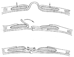
Small and asymptomatic linea alba hernias do not require surgery, while all others should be treated surgically. A midline abdominal incision is made at the site of the linea alba hernia, the hernial sac is incised, the hernial contents are reduced, the neck of the hernial sac is ligated high, and the hernial ring is sutured closed. If there are multiple defects in the linea alba, the Berman procedure (Figure 1) can be used. This involves repairing the transversus abdominis membrane and then making equal vertical incisions in the anterior sheaths of both rectus abdominis muscles, overlapping and suturing the inner layers of the anterior sheaths to reinforce the weakened or defective linea alba.

Figure 1 Berman procedure (cross-sectional view)
Top: Equal vertical incisions are made in the anterior sheaths of both rectus abdominis muscles
Middle: Repair of the transversus abdominis membrane, with the inner layers of the anterior sheaths of both rectus abdominis muscles flipped over
Bottom: Overlapping suturing of the inner layers


