| disease | Primary Mediastinal Tumor |
| alias | Primary Mediastinal Tumor |
Primary mediastinal tumors are not uncommon. According to a report from Shanghai Chest Hospital involving 1,228 cases of mediastinal tumors, thymomas were the most frequent, followed by neurogenic tumors and teratomas. Other types such as cysts, intrathoracic thyroid, and bronchogenic cysts were relatively rare. Most of these tumors are benign but have the potential for malignant transformation.
bubble_chart Pathological Changes
Pathology and Classification:
The mediastinum is located in the center of the thorax. It extends from the thoracic inlet superiorly to the diaphragm inferiorly, bounded laterally by the mediastinal pleura, and anteriorly and posteriorly by the sternum and thoracic vertebrae, respectively. The region above the level of the sternal angle is called the superior mediastinum. The area anterior to the pericardium is termed the anterior mediastinum, the area containing the pericardium is the middle mediastinum, and the space between the pericardium and the spine is the posterior mediastinum (Figure 1). Common mediastinal tumors have their preferred locations (Figure 2), which is clinically significant for diagnosis.
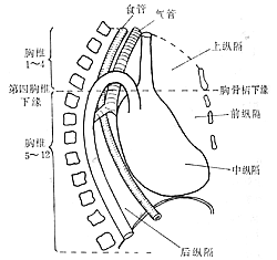
Figure 1: Division of the Mediastinum
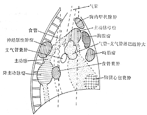
Figure 2: Primary Locations of Mediastinal Tumors
(1) Tumors of the Superior Mediastinum: The most common are thymomas and intrathoracic thyroid tumors.
1. Thymoma: Most are located in the anterior superior or anterior middle mediastinum, accounting for about 1/4 to 1/5 of primary mediastinal tumors, with equal incidence in males and females. 30% are malignant, 30% are benign, and 40% are potentially or low-grade malignant. Benign thymomas are often asymptomatic and occasionally discovered during X-ray examinations. If the tumor is small and faint in density, closely adherent to the posterior sternum, it may be difficult to detect on X-ray. Thymomas are often adjacent to the ascending aorta, so significant transmitted pulsations may be observed. Histologically, they can be classified into lymphocyte-predominant, epithelial-reticular cell, and mixed epithelial-lymphocyte types. Benign thymomas with predominant epithelial and lymphocyte components may recur or metastasize if not completely resected. Zhongshan Hospital in Shanghai reported 12 cases of thymoma, 5 of which showed obvious malignancy during surgery, suggesting that thymomas should be considered low-grade malignant tumors requiring postoperative radiotherapy. Malignant thymomas tend to invade surrounding tissues, causing varying degrees of retrosternal pain and dyspnea. Advanced-stage patients may develop symptoms due to vascular or nerve compression, such as superior vena cava syndrome, diaphragmatic paralysis, or hoarseness. About 10–75% of thymoma patients may exhibit symptoms of myasthenia gravis, but only 15–20% of myasthenia gravis patients have thymic lesions. After tumor resection, approximately two-thirds of patients show improvement in myasthenia gravis symptoms. A few patients may develop aplastic anemia, Cushing’s syndrome, lupus erythematosus, γ-globulin deficiency, or idiopathic granulomatous myocarditis. On X-ray, a round or oval mass is seen in the anterior superior mediastinum; benign tumors have clear, smooth contours with intact capsules and often cystic changes, while malignant tumors show irregular, rough contours and may exhibit pleural reactions. Surgical resection of thymomas yields good outcomes. Legg’s analysis of 51 thymoma cases showed a 5-year survival rate of 23% for locally invasive tumors and 80% for non-invasive tumors. Shanghai Chest Hospital reported 207 cases with a postoperative 5-year survival rate of 59.7% and a 10-year survival rate of 43.4%.
2. Intrathoracic Goiter This includes congenital aberrant thyroid and acquired retrosternal thyroid. The former is rare, consisting of thyroid tissue remnants in the mediastinum from embryonic development that grow into thyroid adenomas, entirely located within the thorax without a fixed position. The latter refers to cervical thyroid tissue extending retrosternally into the anterior superior mediastinum (Figures 3A, B), mostly situated beside and anterior to the trachea, with a minority located posterior to the trachea. Most intrathoracic goiters are benign, though rare cases may be adenocarcinoma. The mass may pull or compress the trachea, leading to symptoms such as irritating cough and dyspnea. These symptoms may worsen when lying supine or turning the head and neck to the side. Compression of the sternum or spine can cause chest tightness or back pain, and occasionally symptoms of hyperthyroidism may appear. If severe cough, hemoptysis, or hoarseness occurs, malignant goiter should be considered. Approximately half of the patients may have a nodular goiter palpable in the neck. On X-ray examination, a shadow of the anterior superior mediastinal mass can be seen, appearing oval or spindle-shaped with clear margins, often deviating to one side of the mediastinum or bulging bilaterally. The presence of calcification in the tumor on plain films is diagnostically significant. In most cases, signs of tracheal compression and displacement, as well as upward movement of the tumor shadow during swallowing, can be observed.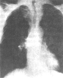
Figure 3A Retrosternal Thyroid
The tumor is located in the upper left mediastinum, with a wider upper end and a narrower lower end. The trachea is slightly compressed (anteroposterior view).
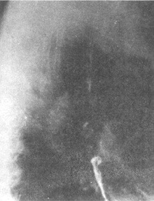
Figure 3B Retrosternal Thyroid
The tumor extends downward and forward from the thoracic inlet into the retrosternal space (right lateral view).
(II) Anterior Mediastinal Tumors Tumors growing in the anterior mediastinum are most commonly teratoid tumors. They can occur at any age, but symptoms appear in half of the cases between the ages of 20 and 40. Histologically, they are all abnormalities or malformations of embryonic development. Teratoid tumors can be divided into two types:
1. Dermoid Cyst This is a fluid-filled cyst containing skin, hair, teeth, and other tissues originating from the ectoderm. It is often unilocular but can also be bilocular or multilocular. The cyst wall is composed of fibrous tissue, and the inner wall is lined with stratified squamous epithelium.
2. Teratoma This is a solid mixed tumor composed of tissues from the ectoderm, mesoderm, and endoderm, including cartilage, smooth muscle, bronchi, intestinal mucosa, neurovascular components, etc. Teratomas have a greater tendency to become malignant than dermoid cysts, often transforming into epidermoid carcinoma or adenocarcinoma. Literature reports 386 cases of teratoma, of which 14.2% showed malignant transformation. Among 10 cases of teratoma at Zhongshan Hospital in Shanghai, 2 were malignant. Small tumors often have no symptoms and are mostly discovered during X-ray examinations. If the tumor enlarges and compresses adjacent organs, corresponding compressive symptoms may occur, such as superior vena cava syndrome if the superior vena cava is compressed; hoarseness if the recurrent laryngeal nerve is compressed; and dyspnea if the trachea is compressed, which worsens when the patient lies supine. If the cyst ruptures into a bronchus, the patient may cough up gelatinous fluid containing hair and sebum. If this fluid is aspirated into the lungs, it can cause lipoid pneumonia and lipoid granuloma. If the cyst becomes secondarily infected, fever and systemic toxic symptoms may appear. If the cyst enlarges rapidly in a short period, the possibility of malignant transformation, secondary infection, or hemorrhage within the tumor should be considered. If a purulent cyst ruptures into the pleural cavity or pericardium, it can cause empyema or pericardial effusion.
X-ray Examination The cyst is located in the anterior mediastinum, at the junction of the heart and aortic arch. A few are positioned higher, near the anterior superior mediastinum, or in the anterior inferior mediastinum. Most protrude to one side of the mediastinum, while a few may bulge to both sides. Large tumors may protrude into the posterior mediastinum or even occupy one side of the thoracic cavity. They are mostly round or oval with clear margins, and calcification of the cyst wall is common. Sometimes, characteristic shadows of teeth and bone fragments can be seen (Figure 4).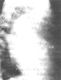
Figure 4 Teratoid Tumor Containing Bone Fragments, Teeth, and Hair
(III) Middle Mediastinal Tumors The vast majority are tumors of the lymphatic system. Common types include Hodgkin's disease, reticulum cell sarcoma, and lymphosarcoma. Most are characterized by enlarged lymph nodes in the middle mediastinum, but they can also invade lung tissue, forming infiltrative lesions. The disease has a short course, with rapid progression of symptoms, often accompanied by systemic lymphadenopathy, irregular fever, hepatosplenomegaly, anemia, etc. X-ray examination shows enlarged lymph nodes on both sides of the trachea and at the hila of the lungs. Markedly enlarged lymph nodes may fuse into a mass, with uniform density and large lobulations but no calcification. The bronchi are often compressed and narrowed.
(4) Posterior Mediastinal Tumors Almost all are neurogenic tumors. They can originate from spinal nerves, intercostal nerves, sympathetic ganglia, and vagus nerves, and may be benign or malignant. Benign types include schwannomas, neurofibromas, and ganglioneuromas; malignant types include malignant schwannomas and neurofibrosarcomas. Electron microscopy reveals that the ultrastructure of schwannomas and neurofibrosarcomas is similar, but their collagen content differs. The vast majority of neurogenic tumors are located in the paravertebral sulcus of the posterior mediastinum (Figures 5A, B), though they may occasionally be found in the superior mediastinum, often with a capsule. Radiographic features show a smooth, round, solitary mass. Large masses may widen the intercostal spaces or enlarge the intervertebral foramina. Sometimes, the tumor extends into the intervertebral foramen in a dumbbell shape, invading the spinal canal and causing spinal cord compression symptoms. Neurofibromas are more common in young adults and are usually asymptomatic. Larger tumors may produce compression symptoms, such as interscapular or posterior back pain, dyspnea, etc.
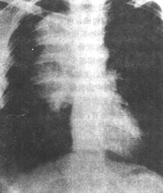
Figure 5A Mediastinal schwannoma
The tumor is located in the right paravertebral region, presenting a "bottle gourd peel" appearance (posteroanterior view)

Figure 5B Mediastinal schwannoma
The tumor is located in the posterior mediastinum, overlapping the thoracic vertebrae, showing a "bottle gourd peel" appearance (right lateral view)
(5) Bronchogenic cyst Can occur in any part of the mediastinum, mostly located near the trachea, bronchi, or carina. Bronchogenic cysts are mostly congenital, originating from tracheal aberrant buds, and are commonly seen in children under 10 years old. Usually asymptomatic, but if connected to the bronchus or pleura, a fistula may form. Secondary infection can lead to cough, hemoptysis, purulent sputum, or even empyema. X-ray examination reveals a round or oval, uniformly dense, well-defined mass shadow in the upper-middle mediastinum near the trachea or major bronchi, without lobulation or calcification. If the cyst communicates with the bronchus, an air-fluid level may be observed.
Mediastinal tumors are sometimes difficult to distinguish morphologically from primary or secondary lung tumors, enlarged lymph nodes, hemangiomas, etc. Common examination methods are as follows.
(1) X-ray examination: If fluoroscopy reveals pulsation in the tumor, it should first be determined whether it is expansile or transmitted pulsation. If it is the former, a stirred pulse tumor may be preliminarily suspected, and X-ray kymography or angiography can be used for confirmation. If an upper mediastinal tumor moves upward with swallowing during X-ray fluoroscopy, it can be preliminarily diagnosed as a thyroid tumor. X-ray plain films in frontal, lateral, and oblique views, tomograms, or high-kilovoltage radiographs can clarify the tumor's location, shape, density, and the presence of calcification or ossification, thereby providing a preliminary judgment of the tumor type. A barium swallow examination can assess whether the esophagus or adjacent organs are compressed.
(2) Fiberoptic bronchoscopy or esophagoscopy: Helps clarify the extent and degree of bronchial compression and whether the tumor has invaded the bronchus or esophagus, thus estimating the feasibility of surgical resection.
(3) Diagnostic pneumothorax: Can determine whether the tumor originates from the chest wall or lung, or whether it is intra- or extrapulmonary. Diagnostic pneumoperitoneum can differentiate subdiaphragmatic factors, such as diaphragm hernia.
(4) Mediastinal pneumography: Quite helpful in displaying the morphology of anterior mediastinal tumors and confirming the presence of mediastinal lymph node metastasis.
(5) Mediastinoscopy: Useful for identifying enlarged lymph nodes near the trachea or under the carina and for obtaining biopsy samples to clarify disease cause diagnosis.
(6) Computed tomography (CT): CT is more reliable than any other X-ray method for examining anterior mediastinal tumors, enlarged lymph nodes, and lesions in mediastinal fat tissue (e.g., lipomas). The accuracy of CT in diagnosing mediastinal tumors and lymph node enlargement exceeds 90%.
(7) Magnetic resonance imaging (MRI): Offers the following advantages: multiple imaging parameters; high soft tissue resolution; flexible slice orientation; absence of bony artifacts; safety and reliability, with no ionizing radiation injury. It has unique advantages in diagnosing mediastinal tumors.
(8) Cervical lymph node biopsy: Bronchial lymph subcutaneous nodes and lymphomas often involve surrounding and cervical lymph nodes, and biopsy can aid in diagnosis.
(9) Radionuclide examination: For suspected intrathoracic goiter, radionuclide 131I scanning can be performed, which is very helpful in diagnosing ectopic thyroid goiter and thyroid adenoma.
(10) Diagnostic radiotherapy: For suspected cervical malignancy with cachexia that cannot be confirmed by other examinations, radiotherapy may be attempted. Cervical malignancy with cachexia is relatively radiosensitive, and the tumor shrinks rapidly after irradiation with 20–30 Gy (2000–3000 rad).
(11) Thoracotomy: If the nature of the tumor cannot be determined through various examinations but cervical malignancy with cachexia has been ruled out, thoracotomy may be performed if the patient's overall condition permits.
bubble_chart Treatment Measures
For localized cervical malignancy with cachexia, radiation therapy can be performed. For extensive lesions, chemotherapy may be considered.
The primary treatment for other mediastinal tumors is surgical resection. Some mediastinal tumors, such as teratomas, neurofibromas, and thymomas, have the potential for malignant transformation, and postoperative adjuvant radiotherapy or chemotherapy should be administered.
The following diseases need to be differentiated from mediastinal tumors:
1. Central lung cancer presents with respiratory symptoms such as cough and expectoration. X-ray findings show hilar masses, which appear semicircular or lobulated. Tumors are often visible during bronchoscopy, and tumor cells can be detected in sputum.
2. Mediastinal lymph subcutaneous nodes are more common in children or adolescents and often have no clinical symptoms. A few may present with low-grade fever, night sweats, and other grade I toxic symptoms. Round or lobulated masses can be seen at the hilum, often accompanied by pulmonary subcutaneous node lesions. Calcification spots may sometimes be observed in the lymph nodes. If differentiation is difficult, a subcutaneous node bacillus test or short-term anti-subcutaneous node drug therapy can be administered.
3. Main stirred pulse tumors are more common in older patients. Vascular murmurs can be heard during physical examination, and expansile pulsations are visible under fluoroscopy. Retrograde main stirred pulse angiography can confirm the diagnosis.


