| disease | Kidney Injury |
The kidneys are deeply embedded in the renal fossa and are well-protected by surrounding structures: behind the kidneys are the ribs, spine, and the long muscles of the back; in front are the abdominal wall and the contents of the abdominal cavity; and above, they are covered by the diaphragm. Normal kidneys have a mobility range of 1 to 2 cm, making them less susceptible to injury. However, from another perspective, the bony structures behind can also cause kidney injury, such as the fractured end of a lower rib penetrating the renal parenchyma; the kidney can also be compressed between the spine and its transverse processes, leading to injury.
bubble_chart Epidemiology
The incidence of renal injury is not high. Renal injury accounts for 0.03% to 0.063% of the total number of hospitalized patients. Renal injury is often part of severe multiple injuries. In a group of 326 autopsies of accidental casualties, 36 cases of renal injury (11%) were found. Domestic reports indicate that renal injury accounts for 14.1% of abdominal injury cases; in cases of penetrating abdominal injuries, renal injury accounts for 7.5%. However, the actual incidence of renal injury is higher than these figures suggest. This is because severe multiple injury cases often overlook renal injury, while minor renal injuries are frequently diagnosed as fistula disease due to the absence of severe symptoms.
Renal injury is most commonly seen in males aged 20 to 40. This is related to engaging in intense physical labor and sports activities. The ratio of male to female patients is approximately 4:1. However, renal injury is relatively common in infants and young children. This is related to anatomical characteristics: ① The kidneys of infants and young children are relatively large and positioned lower; ② They have less protective perirenal fat and underdeveloped muscles; ③ The perirenal fascia, which acts as a buffer, is underdeveloped, and the kidneys directly rely on a rather tense abdominal membrane; ④ Some patients have congenital conditions such as hydronephrosis or nephroblastoma, making them more susceptible to injury. It has been statistically noted that for every 2000 hospitalized children, there is 1 case of renal injury, with children under 15 years old accounting for 20% of all renal injury cases. In infants and young children, the influence of gender on the mechanism of disease in renal injury is not significant.
Most renal injuries are closed injuries, accounting for 60% to 70%. They can be caused by direct violence (such as impact, falls, or compression) or indirect violence (such as contrecoup injuries). Open injuries are more common during wartime and in accidents. Whether caused by cold weapons or firearms, they are often accompanied by injuries to other organs and have serious consequences. Occasionally, renal injury can also occur during medical procedures such as renal puncture, endourological examinations, or treatments.Kidney injury can occur under the following circumstances:
(1) Direct violence: The kidney area is directly struck, the injured person falls onto a hard object, or is squeezed between two external forces.
(2) Indirect violence: When a person falls from a height and lands on their feet or buttocks, the intense shock can injure the kidneys.
(3) Puncture wounds: Often penetrating wounds, which can injure the entire kidney or a part of it, usually accompanied by injury to other internal organs in the abdominal or thoracic cavity.
(4) Spontaneous rupture: The kidney can also rupture spontaneously without obvious external violence. Such "spontaneous" kidney ruptures are often caused by pre-existing kidney conditions such as hydronephrosis, tumors, stones, and chronic inflammation.
bubble_chart Pathological Changes
Renal injuries can be divided into two categories: closed injuries (such as renal contusion and renal laceration) and penetrating injuries (such as gunshot wounds and stab wounds).
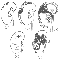
According to the severity of renal injury, it can be classified as follows: (see Figure 4).


Figure 4 Types of Renal Injuries
(1) Grade I renal contusion injury is limited to part of the renal parenchyma, forming intraparenchymal ecchymosis, hematoma, or small subcapsular hematoma, and may also involve the renal collecting system with slight hematuria. Due to the reduced urine secretion function of the injured renal parenchyma, there is rarely any urine extravasation. Symptoms are generally mild and healing is rapid.
(2) Renal contusion and laceration involve the renal parenchyma. If accompanied by rupture of the renal capsule, it can lead to perirenal hematoma. If the renal pelvis and calyces mucosal membrane are ruptured, significant hematuria may be observed. However, it generally does not cause severe urine extravasation. Most cases can heal on their own with medical treatment.
(3) Full-thickness renal laceration involves severe contusion of the renal parenchyma extending to the renal capsule and reaching the mucosal membrane of the renal pelvis and calyces, often accompanied by perirenal hematoma and urine extravasation. If the perirenal fascia is ruptured, extravasated hematuria can spread along the retroperitoneal space. If the hematoma ruptures into the collecting system, it can cause severe hematuria. Sometimes, one pole of the kidney may be completely avulsed, or the kidney may be severely lacerated into a shattered state—shattered kidney. These types of renal injuries have obvious symptoms and serious consequences, and all require surgical treatment.
(5) Pathological renal rupture can occur with grade I force in kidneys with pathological changes, such as renal tumors, hydronephrosis, renal cysts, or pyonephrosis. Sometimes, the force may not even be noticed, and it is referred to as "spontaneous" renal rupture.
Severe renal trauma, especially penetrating injuries, is often accompanied by injuries to other abdominal and thoracic organs. Hematuria can seep into the thoracic or abdominal cavity. Patients often die due to massive blood loss and lack of medical treatment.
In addition to bleeding and urine extravasation, infection is a serious complication. Its occurrence is later than bleeding, and the kidney and surrounding tissues are prone to bacterial invasion and proliferation due to hematoma and urine extravasation. During the healing process, fibrous changes and adhesions may form in the surrounding tissues of the kidney.
bubble_chart Clinical Manifestations
The clinical manifestations of kidney injury are quite inconsistent. When other organs are injured simultaneously, the symptoms of kidney injury may not be easily noticeable. The main symptoms include shock, hemorrhage, hematuria, pain, rigidity of the abdominal wall on the injured side, and swelling in the lumbar region.
(1) Shock Early shock may be caused by severe pain, but later it is related to significant blood loss. The degree of shock depends on the severity of the injury and the amount of blood loss. Apart from hematuria and blood loss, when the renal fascia is intact, the hematoma is confined to the renal fascia; if the renal fascia is ruptured, blood may seep outside the fascia, forming a large retroperitoneal hematoma; if the peritoneum is ruptured, a large amount of blood may flow into the peritoneal cavity, rapidly worsening the condition. Shock that occurs rapidly within a short period or cannot be corrected after rapid transfusion of 2 units of blood often indicates severe internal hemorrhage.
Advanced stage secondary hemorrhage commonly occurs 2-3 weeks after the injury, and occasionally may occur even 2 months later.
(2) Hematuria More than 90% of patients with kidney injury have hematuria, with mild cases showing microscopic hematuria. However, gross hematuria is more common. In severe cases, the hematuria is very concentrated, may be accompanied by strip-like or cast blood clots and renal colicky pain, with significant blood loss. In most cases, hematuria is transient. It starts with a large amount of hematuria, which gradually subsides after a few days. Getting up and moving, exertion, and secondary infections are common triggers for secondary hematuria, often seen 2-3 weeks after the injury. In some cases, hematuria can persist for a long time, even several months. Comparing the color of urine collected hourly in test tubes arranged in order on a test tube rack can help understand the progression of the condition. The absence of hematuria does not rule out the presence of kidney injury, and the amount of blood in the urine cannot determine the extent and severity of the injury. In cases of extensive injury to the renal pelvis, injury to the renal vessels (renal artery thrombosis, renal pedicle avulsion), complete obstruction of the ureter by blood clots or renal tissue fragments, blood flowing into the abdominal cavity, and simultaneous extravasation of blood and urine into the perirenal tissues, despite the severity of the injury, hematuria may not be obvious. If the urine sample is obtained by catheterization, it needs to be differentiated from traumatic hemorrhage caused by the catheterization itself.
(3) Pain and Abdominal Wall Rigidity There is pain, tenderness, and rigidity in the renal area on the injured side. The pain worsens with body movement, but the severity varies. This pain is caused by injury to the renal parenchyma and expansion of the renal capsule. Although abdominal wall rigidity may affect accurate palpation, in some cases, a mass formed by renal hemorrhage can still be felt in the lumbar region. The pain may be localized to the lumbar region or upper abdomen, or may spread to the entire abdomen, radiating to the back, shoulders, hip region, or lumbosacral area. If accompanied by peritoneal rupture with a large amount of urine and blood flowing into the abdominal cavity, it can cause generalized abdominal tenderness and muscle guarding, signs of peritoneal irritation. This situation is more likely to occur in young children.
When blood clots pass through the ureter, severe renal colicky pain may occur.
Penetrating injuries to the abdomen or lumbar region often cause extensive abdominal wall rigidity, which may be caused by injury to the abdominal or thoracic organs, but can also be due to a renal hematoma or intra-abdominal hemorrhage.
(4) Lumbar Swelling Extravasation of blood or urine from a ruptured kidney can form an irregular, diffuse mass in the lumbar region. If the renal fascia is intact, the mass is localized; otherwise, it can cause extensive swelling in the retroperitoneal space. Later, subcutaneous ecchymosis may appear. This swelling can often be palpated even when the abdominal muscles are rigid. The progression of the swelling can help estimate the severity of the kidney injury. To alleviate lumbar pain, the patient's spine often shows lateral curvature. Sometimes, it is necessary to differentiate from masses formed by subcapsular hemorrhage of the spleen or liver.
The diagnosis of kidney injury can be determined based on medical history, symptoms and signs, urinalysis, and X-ray urography. In most cases, the diagnosis of kidney injury can be confirmed through the aforementioned steps or solely based on clinical manifestations and hematuria. Kidney injury is often accompanied by severe injuries to the brain, thoracic and abdominal organs, and fractures. Due to the severity of these injuries, the manifestations of kidney injury are often overlooked. However, as long as there is vigilance for the possibility of kidney injury, while promptly addressing these injuries and treating shock, a detailed inquiry into the circumstances of the injury, the nature of the trauma, the direction of penetrating wounds, and careful examination of signs and routine urinalysis can lead to a definitive diagnosis in most patients. Rapid deterioration of the condition indicates severe injury and requires active resuscitation. To choose between conservative or surgical treatment, it is often necessary to rely on some auxiliary examinations to understand the true condition of the injured kidney.
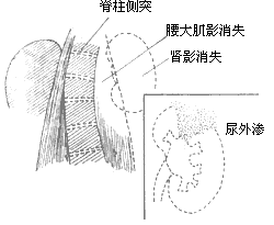
X-ray examination is extremely important for the diagnosis of kidney injury. It should be performed as early as possible; otherwise, the outline of the kidney shadow may be obscured due to abdominal distension. On a plain X-ray of the abdomen, an enlarged kidney shadow suggests a subcapsular hematoma, while an expanded kidney shadow indicates perirenal hemorrhage. The disappearance of the psoas muscle shadow, curvature of the spine toward the injured side, blurred or enlarged kidney shadow, restricted kidney movement, and elevated diaphragm with reduced movement on the injured side can further indicate significant blood or urine extravasation in the perirenal tissues. Due to intestinal paralysis, obvious intestinal gas may be seen. Additionally, evidence of severe injuries such as free intra-abdominal gas, air-fluid levels, displacement of abdominal contents, pneumothorax, fractures, and foreign bodies may be found (see Figure 1). Excretory urography can determine the extent and severity of kidney injury. Grade I kidney injury may show no signs or only slight compression deformation of individual calyces or localized cystic shadows outside the calyces. Blood clots in the renal pelvis or calyces appear as filling defects. On tomographic images, negative shadows may be seen in the renal parenchyma. In cases of extensive kidney injury, a diffuse irregular shadow may extend to part of the renal parenchyma or the perirenal area, with delayed excretion of contrast. Extravasation of contrast may be seen if there is a laceration in the collecting system. The ureter may be displaced toward the spine due to extravasation of hematuria, with upward displacement of the ureteropelvic junction and narrowing of the calyces. Excretory urography can also reflect the function of both kidneys. Although congenital solitary kidney is rare, this possibility should be considered. Shock, vascular spasm, severe kidney injury, intravascular thrombosis, reflex anuria, or obstruction of the renal pelvis or ureter by blood clots can lead to non-visualization of the kidney. Therefore, shock must first be corrected, and systolic blood pressure should be raised above 12 kPa (90 mmHg) before performing excretory urography. High-dose excretory urography (50% diatrizoate meglumine 2.2 ml/kg + 150 ml saline rapid intravenous infusion) can yield better results than standard doses and avoids pain caused by abdominal compression. Tomography can reduce interference from intestinal contents, making the imaging clearer. To avoid the impact of intestinal gas on the clarity of X-ray images, excretory urography should be performed as soon as possible after the injury. Cystoscopic retrograde urography can achieve the same objectives as excretory urography, except for assessing kidney function. However, due to the risk of retrograde urinary tract infection and the fact that trauma patients often cannot tolerate this procedure, it is generally avoided. Main renal artery and selective renal artery angiography should be performed more than 2 hours after the injury to avoid the effects of early vascular spasm caused by trauma. In Grade I kidney injury, renal artery angiography may appear completely normal. In cases of renal parenchymal laceration, typical splitting of the renal parenchymal edge may be seen, sometimes requiring differentiation from embryonic lobulated kidneys. Based on the elongation or displacement of capsular artery and renal pelvic artery, smaller perirenal hematomas can be diagnosed. Typical intrarenal hematomas are characterized by displacement or distortion of interlobar arteries and reduced local parenchymal contrast enhancement. If the surrounding area shows uniform normal enhancement, it indicates good blood supply, while patchy or reduced enhancement suggests traumatic vascular embolism or severe and persistent vascular spasm in the surrounding renal tissue. These patients are prone to delayed bleeding or retroperitoneal urinoma formation. If the avascular area is limited to a small part of the renal parenchyma, the injury is mild, and the prognosis is good. Renal artery thrombosis appears as a blind end of the main renal artery or its branches, with a cut-off phenomenon, often accompanied by spherical dilation of the proximal artery and poor enhancement of the corresponding renal parenchyma; the renal vein is not visualized during the venous phase. Traumatic renal arteriovenous fistula is characterized by premature visualization of the renal vein, with a cystic structure connecting the artery and vein. When the arteriovenous fistula is large, hemodynamic changes and the siphon effect of the fistula can cause ischemia in the corresponding renal parenchyma, leading to reduced enhancement. Renal artery angiography can also reveal the presence of collateral circulation after renal cortical infarction. If other visceral injuries are present, selective angiography of the corresponding organs can also be performed.Computed tomography (CT) can also diagnose some small renal lacerations and other visceral injuries.

Figure 1: Manifestation of renal trauma on X-ray plain film
B-mode ultrasound can be used to follow up the size and progression of hematoma, and it can also be used to differentiate subcapsular hematoma of the liver and spleen. In radionuclide renal scanning, the injured area appears as a "cold area" with low radionuclide concentration, and the renal contour is irregular. This method is safe, simple, and not interfered by intestinal contents, especially suitable when excretory urography shows poor imaging.
Serum alkaline phosphatase often increases after renal injury. It generally begins to rise 4 hours after injury, peaks at 16-24 hours, and then gradually decreases. Therefore, it is advisable to conduct the examination 16-24 hours after injury.
bubble_chart Treatment Measures
The treatment of kidney injury is determined based on the general condition of the patient, the extent and severity of the kidney injury, and the presence of severe injuries to other organs. Therefore, the following considerations should be taken into account during management: ① Treatment of shock; ② Treatment of injuries to other organs; ③ Management of kidney injury: supportive treatment or surgical intervention; ④ Timing and method of surgery. Choosing the correct initial stage [first stage] treatment method is often a critical factor in determining the prognosis.
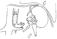
For patients with severe shock, emergency resuscitation should be prioritized, including bed rest, sedation, pain relief, maintaining warmth, blood transfusion (or plasma), and fluid infusion. Many cases show improvement in general condition after shock is corrected. If shock is caused by massive bleeding or diffuse peritonitis, an early and relatively safe period should be chosen for exploratory surgery. For extensive injuries requiring surgical exploration, a flank incision is typically used due to its simplicity and lower risk. If necessary, the lower angle of the incision can be extended transversely to open the peritoneum and explore the abdominal cavity. In cases of concomitant intra-abdominal organ injury, emergency laparotomy is required. The abdominal incision can be used for exploration. Before opening the retroperitoneum to explore the injured kidney, isolating and blocking the renal vessels can prevent unexpected massive bleeding and avoid unnecessary nephrectomy (Figure 2).

Figure 2: Controlling the renal pedicle via the abdominal approach before opening the peritoneum to explore both kidneys can prevent unexpected massive bleeding.
For simple kidney injuries without severe bleeding or shock, supportive treatment is generally adopted. This includes: ① Absolute bed rest for at least 2 weeks, allowing the patient to get up only after the urine clears. However, small lacerations require 4–6 weeks to heal, so strenuous activities should be avoided until at least 1 month after symptoms completely disappear. ② Sedation, pain relief, and antispasmodics; ③ Appropriate antibiotics for prevention and infection control; ④ Hemostatic medications; ⑤ Regular monitoring of blood pressure, pulse, blood tests, lumbar and abdominal signs, and the progression of hematuria. Local cold compresses can be applied, and blood transfusion may be necessary to replenish blood volume; ⑥ Re-evaluation with excretory urography after 3–5 weeks, paying attention to the possibility of hypertension.
The principles of debridement, hemostasis, and initial stage [first stage] suturing in the surgical field also apply to kidney injuries. Immediate initial stage [first stage] repair of kidney lacerations yields better outcomes than intermediate stage [second stage] surgery performed after infection or scar adhesion formation. In severe kidney contusions and lacerations, rupture of the collecting system, urinary extravasation, and infection are the main causes of complications. In such cases, reoperation often necessitates nephrectomy. Renal pedicle injuries have a higher likelihood of surgical repair. Therefore, surgery should be performed as early as possible in these situations.
The following are common surgical methods for kidney injuries:
(1) Renal drainage: Early surgery in patients with kidney injuries often achieves complete repair, with drainage being only a part of the overall procedure. However, in cases of urinary extravasation with infection, perirenal hematoma with secondary infection, or critical conditions where the contralateral kidney's status is unknown, drainage alone may be performed. If peritoneal rupture is found, blood and urine in the abdominal cavity should be aspirated, and the peritoneal tear should be repaired, with drainage placed outside the peritoneum. Drainage must be thorough, as incomplete drainage often leads to uncontrolled perirenal infection and extensive fibrous scar formation. Silicone negative-pressure ball drainage is the most effective. Postoperative drainage should remain in place for at least 7 days and can only be removed after the daily drainage volume is less than 10ml for 3 consecutive days. If the kidney injury is severe and the patient is in critical condition, packing should be used for hemostasis (major bleeding points should be ligated); nephrectomy can be performed once the patient's condition improves.
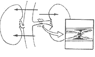
(2) Kidney repair or partial nephrectomy (see Figure 3). Lacerations of the kidney parenchyma can be sutured with silk thread. Absorbable sutures should be used to repair lacerations of the collecting system, and fat or muscle pads can be inserted to prevent suture cutting. Devitalized and fragmented tissue should be debrided. If there is no obvious infection, internal stents or fistulas are generally not necessary. The wound should be thoroughly drained. In cases of closed kidney injuries in peacetime, the outcomes of these methods are generally good. However, in wartime penetrating injuries with infection, the results are often unsatisfactory. Due to kidney parenchyma infection, necrosis, and advanced stage bleeding, a second surgery is often required, or even a total nephrectomy may be forced.

Figure 3 Kidney Root of nose Severe contusion Partial nephrectomy performed
(3) Nephrectomy Every effort should be made to preserve the injured kidney. However, nephrectomy is simpler than repair surgery, as it can eliminate the source of bleeding and infection, and also avoid the need for reoperation and the consequences of advanced stage disability. When the condition is critical and nephrectomy is required, it must be confirmed that the contralateral kidney is functioning well before proceeding. At least, the abdominal membrane should be opened to check the condition of the contralateral kidney. Nephrectomy is indicated for ① uncontrollable massive hemorrhage; ② extensive renal lacerations, especially penetrating wounds during wartime; ③ severe injury to the renal pedicle that cannot be repaired; ④ pre-existing pathological changes in the injured kidney that cannot be repaired, such as renal tumors, renal abscesses, large stones, and hydronephrosis. Renal angiomyolipomas are prone to rupture and bleeding, but they are benign. Moreover, the tumors are often multiple and may involve both kidneys, so partial nephrectomy should be attempted as much as possible.
(4) Renal Vascular Repair Surgery The renal stirred pulse is a terminal branch, and ligation of any stirred pulse can lead to infarction of the corresponding kidney excess tissue. However, there are extensive anastomoses between the branches of the renal vein, and as long as one of the thicker branches remains patent, renal function will not be affected. The left renal vein also drains through branches such as the spermatic vein (or ovarian vein) and the adrenal vein. Therefore, the main trunk of the renal vein can be ligated near the vena cava without affecting renal blood circulation. Thus, the left kidney has more chances of being saved in case of renal vein injury. Once renal stirred pulse thrombosis caused by contrecoup injury is confirmed by stirred pulse angiography, thrombectomy should be performed. There are reports in the literature of successful thrombectomy even 9 days after injury, so active efforts should be made. Arteriovenous fistulas and main stirred pulse aneurysms should be repaired, and if located within the kidney excess tissue, partial nephrectomy can be performed.
(5) Renal Stirred Pulse Embolization Therapy Satisfactory hemostasis can be achieved by injecting embolic agents through selective stirred pulse angiography. Commonly used embolic agents include absorbable autologous blood clots and gelatin sponge fragments. If a small amount of norepinephrine solution is injected first to constrict the normal renal vessels, the embolic agent can be more concentrated at the injured site.
Currently, it is possible to perfuse the kidney with cold kidney preservation solution and preserve it frozen for 72 hours without affecting the recovery of renal function, both domestically and internationally. Therefore, it is possible to carefully repair the injured kidney on the workbench, freeze and preserve it, and then implant it into the iliac fossa after the patient's condition stabilizes.
Direct deaths from kidney injury are uncommon. Most fatalities result from injuries to other vital organs (liver, spleen, pancreas, duodenum, etc.).
The complications of severe injuries are mostly caused by blood or urine extravasation and secondary infections. These mainly include perinephric abscess, urinary fistula, pyelonephritis and pyonephrosis, ureteral stricture, hydronephrosis, pseudocyst, stones, loss of renal function, arteriovenous fistula, hypertension, and hematoma calcification. In some cases, the injured kidney may exhibit persistent morphological changes such as calyceal diverticula, calyceal deformation, and partial renal parenchymal atrophy, but without any accompanying symptoms.


