| disease | Lung Abscess |
| alias | Lung Abscess |
Lung abscess is a suppurative lesion of lung tissue caused by various disease factors. In the early stage, it manifests as suppurative inflammation, followed by necrosis leading to abscess formation. The clinical features include high fever, cough, and expectoration of large amounts of foul-smelling purulent sputum. It predominantly occurs in middle-aged adults, with a higher incidence in males than females. Since the widespread use of antibiotics, the incidence of lung abscess has significantly decreased.
bubble_chart Etiology
The infectious bacteria of acute lung abscess are the common resident bacteria of the upper respiratory tract and oral cavity. It is often a mixed infection, including both aerobic and anaerobic gram-positive and gram-negative cocci and bacilli. The most common pathogens include staphylococci, streptococci, pneumococci, fusiform bacteria, and spirochetes. The importance of anaerobic bacteria in pulmonary suppurative infections has only been recognized in recent years due to improvements in culture techniques. Gorbach and Bartlett reported in 1974 that anaerobic infections accounted for approximately 85–90% of aspiration pneumonia and lung abscess cases. Bartlett et al. reported data from 45 cases of acute lung abscess, isolating 114 strains of anaerobic bacteria, with purely anaerobic infections accounting for 58% and mixed aerobic and anaerobic infections accounting for 42%. The more significant anaerobic bacteria include peptostreptococci, peptococci, Fusobacterium nucleatum, Bacteroides species, Veillonella, and spirochetes. In addition to these anaerobic bacteria, aerobic or facultative anaerobic bacteria are also present. In recent years, foreign reports indicate that pneumonia caused by Legionella pneumophila forms abscesses in about 25% of cases.
The pathogenesis of lung abscess is closely related to its disease causes, which can be categorized as follows.(1) Aspiration lung abscess: Pathogens are inhaled through the mouth or nasopharynx, which is the primary cause of lung abscess. Purulent secretions from tonsillitis, sinusitis, alveolar pyorrhea, or dental caries; blood clots after oral, nasal, or pharyngeal surgery; dental plaque or vomitus can be inhaled into the lungs under conditions such as unconsciousness or general anesthesia, leading to bronchiolar obstruction and subsequent bacterial proliferation and disease. Additionally, some patients show no obvious predisposing factors, with domestic and foreign case reports at 29.3% and 23%, respectively. Possible triggers include cold exposure or extreme fatigue, which reduce systemic immunity and respiratory defense functions, leading to inhalation of contaminated oral secretions during deep sleep. This type is often solitary and related to anatomical structure and body position. Because the right main bronchus is straighter and wider, aspirated secretions are more likely to enter the right lung, making right lung involvement more common than the left. In the supine position, the posterior segment of the upper lobe or the dorsal segment of the lower lobe is most affected; in the sitting position, the posterior basal segment of the lower lobe is more likely to be involved. In the right lateral position, the anterior and posterior segments of the right upper lobe, forming the axillary subsegment, are commonly affected.
(2) Hematogenous lung abscess: Septicemia and pyemia caused by skin trauma, infections, boils, carbuncles, osteomyelitis, postpartum pelvic infections, or subacute bacterial endocarditis can lead to pathogen (often Staphylococcus aureus) or septic emboli entering the lungs via the pulmonary circulation, causing small vessel embolism, pulmonary inflammation, and necrosis, resulting in abscess formation. Lesions are often multiple and distributed randomly, typically occurring at the edges of both lungs.

(4) Amebic lung abscess: This is often secondary to amebic liver abscess. Since liver abscesses commonly occur in the dome of the right lobe of the liver, they can easily penetrate the diaphragm into the right lower lobe of the lung, forming an amebic lung abscess.
bubble_chart Pathological Changes
Early bronchiolar obstruction leads to inflammation of the lung tissue, small vessel thrombosis, and suppuration and necrosis of the lung tissue, eventually forming an abscess. The lesion can spread to surrounding areas, even crossing the interlobar fissure to invade adjacent lung segments. Bacterial emboli cause local tissue ischemia, promoting anaerobic infection and exacerbating tissue necrosis. The liquefied pus and abdominal mass increase tension within the abscess cavity, eventually rupturing into the bronchus and resulting in the expectoration of large amounts of purulent sputum. If air enters the abscess cavity, a fluid level appears within the abscess. Sometimes, inflammation spreads to surrounding lung tissue, forming one or several abscess cavities. If the abscess is near the pleura, localized fibrinous pleuritis may occur, leading to pleural adhesions. A tension abscess located at the edge of the lung, if rupturing into the pleural cavity, can form pyopneumothorax. If bronchial drainage is inadequate, necrotic tissue remains in the abscess cavity, and inflammation persists, the condition may progress to chronic lung abscess. Fibrous tissue proliferates around the abscess cavity, the abscess wall thickens, and surrounding bronchioles become involved, leading to deformation or dilation.

Acute aspiration lung abscess has a sudden onset, with patients experiencing {|###|}fear of cold{|###|} and fever, with body temperature potentially rising to 39–40°C. It is accompanied by {|###|}cough{|###|} and the expectoration of mucoid or mucopurulent sputum. When inflammation spreads to the local pleural {|###|}membrane{|###|}, it may cause {|###|}chest pain{|###|}. If the affected area is extensive, shortness of breath may occur. Additionally, patients may exhibit lethargy, {|###|}lack of strength{|###|}, and poor appetite. After about 7–10 days, the {|###|}cough{|###|} worsens, and the abscess ruptures into the bronchus, leading to the expectoration of a large amount of foul-smelling purulent sputum, which can reach 300–500ml per day, followed by a rapid drop in body temperature. Since the causative pathogens are often anaerobic bacteria, the sputum tends to have a fishy, foul odor. Sometimes, the sputum may contain blood or moderate amounts of {|###|}hemoptysis{|###|}.
Patients with chronic lung abscess typically present with chronic {|###|}cough{|###|}, purulent sputum production, recurrent {|###|}hemoptysis{|###|}, secondary infections, and irregular fever. They often appear anemic, emaciated, and in a chronic wasting state.
Hematogenous lung abscess usually begins with systemic septicemia symptoms such as {|###|}fear of cold{|###|} and high fever caused by the primary infection. Pulmonary symptoms, such as {|###|}cough{|###|} and sputum production, may only appear after several days to two weeks. Generally, the amount of sputum is not excessive, and {|###|}hemoptysis{|###|} is rare.
{|###|}Sign{|###|}: The clinical manifestations depend on the size and location of the abscess. If the lesion is small or located deep in the lung, there may be no abnormal {|###|}sign{|###|}. For larger lesions with significant surrounding inflammation, percussion may reveal dullness or flatness, and auscultation may show diminished breath sounds, sometimes accompanied by moist rales. Most hematogenous lung abscess cases show negative {|###|}sign{|###|} findings. In chronic lung abscess, the affected side of the chest may appear slightly collapsed, with dullness on percussion and reduced breath sounds. Clubbing of the fingers (toes) may also be present.
bubble_chart Auxiliary Examination
(1) Peripheral Blood Picture The white blood cell count and neutrophil count in the blood are significantly increased, with the total count reaching 20,000 to 30,000/mm3, and neutrophils accounting for more than 80–90%. In patients with chronic lung abscess, the white blood cell count shows no significant change, but grade I anemia may be present.
(2) Pathogen Examination of Sputum and Blood Gram staining of sputum smears, sputum culture (including anaerobic culture), and bacterial drug sensitivity tests help identify the pathogen and select effective antibiotic treatment. Blood cultures in patients with hematogenous lung abscess may reveal the causative bacteria.
X-ray Examination:
The X-ray manifestations of lung abscess vary depending on the type, stage, patency of bronchial drainage, and the presence of pleural complications.
In the early suppurative inflammatory stage of aspiration lung abscess, the typical X-ray finding is a large, dense, and poorly defined inflammatory infiltrate shadow, distributed in one or several lung segments, resembling bacterial pneumonia. After abscess formation, a round lucent area with an air-fluid level appears within the dense inflammatory shadow (Figures 1 and 2). During the resolution phase, the surrounding inflammation gradually subsides, the abscess cavity shrinks and eventually disappears, leaving behind minor fibrous streak shadows. In chronic lung abscess, the abscess wall thickens, the inner wall becomes irregular, the surrounding inflammation partially resolves but incompletely, accompanied by significant fibrous tissue proliferation and varying degrees of lung lobe contraction, as well as pleural thickening. The mediastinum shifts toward the affected side, and compensatory lung emphysema occurs in the unaffected lung.
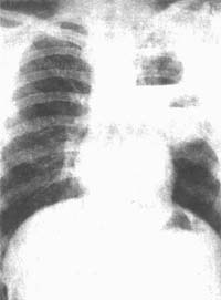
Figure 1: Abscess in the Left Upper Lobe (Posteroanterior View)
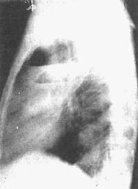
Figure 2: Abscess in the Left Upper Lobe (Lateral View)
Hematogenous lung abscess presents with multiple scattered small patchy inflammatory shadows or spherical lesions with relatively smooth edges in one or both lungs, within which abscess cavities and air-fluid levels may be seen. After inflammation resolves, localized fibrosis or small air cysts may remain.
In cases complicated by empyema, the affected side of the chest shows a large, dense shadow; if pneumothorax is present, an air-fluid level may be visible.
Lateral X-ray examination can clarify the location and extent of the abscess in the lung, aiding in postural drainage or surgical treatment.
Chest CT scans often reveal a thick-walled, round-shaped abscess cavity, possibly with an air-fluid level. The inner wall of the cavity is typically irregular, surrounded by hazy inflammatory shadows.
Fiberoptic Bronchoscopy
This helps identify the disease cause. If a bronchial tumor is present, a biopsy can be performed. Foreign bodies, if seen, can be removed to restore drainage. Fiberoptic bronchoscopy can also be used to collect samples with a protected brush for bacterial culture, aspirate pus, and inject antibiotics into the lesion to promote bronchial drainage and abscess healing.
Based on the history of oral surgery, unconsciousness vomiting, foreign body aspiration, acute onset of fear of cold, high fever, cough, and expectoration of large amounts of foul-smelling purulent sputum, combined with a significant increase in total white blood cell count and neutrophils, as well as X-ray findings of dense inflammatory shadows in the lung fields with abscess cavities and fluid levels, a diagnosis can be made. Blood and sputum cultures, including anaerobic cultures, to isolate bacteria, aid in determining the pathogenic diagnosis. The presence of skin wound infections, boils, carbuncles, or other purulent sexually transmitted disease foci, persistent fever accompanied by symptoms such as cough and expectoration, and chest X-ray findings showing multiple small abscesses in both lungs can lead to a diagnosis of hematogenous lung abscess.
bubble_chart Treatment Measures
Infections in the upper respiratory tract and oral cavity must be eradicated. During oral surgery, secretions should be aspirated as much as possible. For patients with unconsciousness or under general anesthesia, enhanced care is necessary to prevent pulmonary infections. Early and thorough treatment is the key to curing lung abscess.
The treatment principles are anti-inflammatory therapy and drainage.
(1) Antibiotic Treatment: Most anaerobic bacteria causing acute lung abscess, including the vast majority, are sensitive to penicillin, which is highly effective and thus most commonly used. The dosage depends on the condition; severe cases may require intravenous infusion of 2.4–10 million units per day, while a general dose of 1.6–2.4 million units can be administered intramuscularly in 2–3 divided doses daily. With effective antibiotic treatment, fever typically subsides to normal within 3–10 days. Most cases of acute lung abscess can be cured with penicillin. For penicillin-resistant Bacteroides fragilis, oral lincomycin 0.5g 3–4 times daily or intramuscular injection of 0.6g 2–3 times daily may be used. Severe cases may require 1.8g added to 500ml of 5% glucose solution for intravenous infusion once daily. Alternatively, clindamycin 0.15–0.3g can be taken orally four times daily, or metronidazole 0.4g three times daily. For lung abscess caused by Legionella pneumophila, erythromycin is highly effective. The antibiotic course generally lasts 8–12 weeks or until clinical symptoms completely disappear, and X-ray shows resolution of the abscess cavity and inflammatory lesions, leaving only fibrous streaks. In addition to systemic medication, local treatments such as cricothyroid membrane puncture, nasal catheter intratracheal instillation, or fiberoptic bronchoscopic instillation can be used. Typically, 800,000 units of penicillin (diluted in 2–5ml) is instilled, followed by positioning the patient based on the abscess location and maintaining supine rest for 1 hour.
Hematogenous lung abscess is a complication of sepsis and should be treated as such.
(2) Sputum Drainage: Expectorants such as ammonium chloride 0.3g, ambroxol 30mg, resolving phlegm tablets 500mg, or expectorant syrup 10ml taken orally three times daily can facilitate sputum expectoration. For thick sputum, airway humidification methods like steam inhalation or ultrasonic nebulization can aid drainage. Patients in relatively good condition with low fever may benefit from postural drainage to expel pus. Positioning the abscess area upward and gently percussing the affected area 2–3 times daily for 10–15 minutes each session can help. In cases of significant sputum obstruction, fiberoptic bronchoscopic lavage and suction may be performed.
(3) Surgical Treatment: Surgical intervention is required for suspected bronchial obstruction due to bronchial carcinoma; chronic lung abscess unresponsive to medical treatment for three months, with persistent abscess cavity and uncontrolled infection; complications such as bronchiectasis, empyema, or bronchopleural fistula; or life-threatening massive hemoptysis.
Lung abscess should be differentiated from the following diseases.
(1) Bacterial pneumonia Early-stage lung abscess is very similar to bacterial pneumonia in terms of symptoms and X-ray findings. Among bacterial pneumonias, pneumococcal pneumonia is the most common, often presenting with herpes labialis and rusty sputum but without large amounts of yellow purulent sputum. Chest X-ray shows lobar or segmental consolidation or patchy, faint inflammatory changes with blurred margins but no cavity formation. Other types with a tendency to suppuration include staphylococcal and Klebsiella pneumonia. Bacterial isolation from sputum or blood can aid in differentiation.
(2) Cavitary pulmonary tuberculosis The onset is slow, with a prolonged course, often accompanied by tuberculous toxic symptoms such as afternoon low-grade fever, weakness, night sweats, chronic cough, and hemoptysis. Chest X-ray reveals a thick-walled cavity surrounded by tuberculous infiltrates or accompanied by speckled or nodular lesions. There is usually no air-fluid level in the cavity, and sometimes ipsilateral or contralateral tuberculous dissemination may be present. Mycobacterium tuberculosis can be found in the sputum. In cases of secondary infection, large amounts of yellow purulent sputum may also be present. A confirmed diagnosis can be made by combining past medical history and repeated sputum examinations while treating the secondary infection.
(3) Bronchogenic lung cancer Tumor obstruction of the bronchus leads to distal obstructive pneumonia, distributed in lobes or segments. Necrosis and liquefaction of the cancerous lesion may form a cancerous cavity. The onset is slow, often with no or only mild toxic symptoms. Chest X-ray typically shows an eccentric, thick-walled cavity with irregular inner walls, usually without an air-fluid level, and no inflammatory reaction around the cavity. Since lung cancer often metastasizes, enlarged hilar lymph nodes are frequently observed. Diagnosis can be confirmed through X-ray tomography, chest CT scans, sputum cytology, and fiberoptic bronchoscopy.
(4) Secondary infection of pulmonary cysts Pulmonary cysts appear round, with thin and smooth walls, often accompanied by an air-fluid level and no surrounding inflammatory reaction. Patients usually have no significant toxic symptoms or cough. Comparison with pre-infection X-rays makes differentiation easier.


