| disease | Chronic Maxillary Sinusitis |
Chronic maxillary sinusitis is a common disease. It can occur alone, but is often seen with multiple sinus involvement.
bubble_chart Etiology
1. Systemic resistance weakening: Anemia, hypoproteinemia, hypogammaglobulinemia, diabetes, malnutrition, etc. After the maxillary sinus is infected by bacteria, it is difficult to cure and often develops into chronic maxillary sinusitis. Many cases of maxillary sinusitis show no history of an acute phase and begin directly as chronic.
2. Sinus ostium drainage obstruction: The natural opening of the maxillary sinus has many variations in position within the middle meatus, making it prone to obstruction. Structures such as the uncinate process, hypertrophic middle turbinate, concha bullosa, high deviation of the nasal septum, and nasal polyps can hinder the maxillary sinus opening, affecting its ventilation, drainage, and mucociliary clearance function, leading to chronic inflammation.
3. Chronic infection of the ethmoid sinus: The lower air cells of the anterior ethmoid sinus extend to the superomedial angle of the maxillary sinus, where the bony wall is very thin, allowing infection to easily spread to the maxillary sinus. Additionally, purulent discharge from ethmoid sinusitis flowing into the maxillary sinus via the middle meatus is another common cause.
4. Nasal allergy: Due to maxillary sinus mucosal edema and impaired ciliary clearance function, the sinus ostium may experience poor ventilation and drainage, leading to chronic inflammation, where allergy and inflammation coexist.
5. Odontogenic infection.
bubble_chart Pathological Changes
The duration of chronic maxillary sinusitis varies, and the pathological changes are also inconsistent. It can be divided into five types: polyp, papillomatous, follicular, glandular, and fibrous. These inflammatory types often mix or transform into one another. The details are as follows:
1. Polypoid type: Also known as the hypertrophic or edematous type, it is often associated with allergic reactions. The mucous membrane exhibits varying degrees of edema, with infiltration of lymphocytes, plasma cells, and eosinophils. In severe cases, polypoid changes and cystic degeneration occur, and prolonged conditions may lead to loosening of the smooth wall.2. Papillomatous hyperplasia type: The mucous membrane undergoes a transformation from pseudostratified columnar epithelium to stratified squamous epithelium, with thickened and protruding surfaces forming papillae. This type is related to viral infections and bacterial invasion.
3. Follicular type: The mucous membrane contains large aggregations of lymphocytes, forming follicular structures.
4. Glandular type: There is hyperplasia of mucous and serous glands, and glandular duct obstruction may lead to cyst formation.
5. Fibrous type: Also known as the sclerotic or atrophic type. It often involves small vessel endarteritis and perivascular inflammation, leading to vessel obstruction, insufficient blood supply to the mucous membrane, glandular degeneration, reduced and thickened secretions, and may even cause mucosal atrophy, loss of cilia, and crust formation.
bubble_chart Clinical Manifestations
The main symptoms include nasal discharge from the affected or both sinuses, either anterior or posterior nasal drip. Sometimes, nasal secretions flow out with changes in head position. Patients often complain of excessive and foul-smelling phlegm, with secretions being mucopurulent or purulent. They frequently experience dizziness or purulent discharge, headaches, memory decline, and difficulty concentrating. However, some patients may forget their symptoms, and chronic maxillary sinusitis is only discovered upon nasal examination.
bubble_chart Diagnosis1. Medical history inquiry: Pay attention to the history and treatment of acute rhinitis and acute sinusitis, and inquire about any history of nasal allergies.
2. Nasal endoscopy: Observe whether the middle turbinate is hypertrophic or has polyps, whether the middle nasal meatus is obstructed or has purulent secretions, and whether the nasal septum is deviated. Then, use a 1% ephedrine cotton swab to shrink the nasal mucosa, followed by a head-position test, positioning the affected maxillary sinus upward. After a few minutes, observe whether there is pus discharge from the middle nasal meatus on the affected side.
3. X-ray imaging: Take a naso-chin (Water's) view to compare the density of the bilateral maxillary sinuses with that of the orbits. If the density is higher than that of the orbits, it indicates shadow blurring, suggesting possible mucosal thickening or purulent secretions in the sinus, warranting further examination.
4. Maxillary sinus contrast imaging: After maxillary sinus irrigation, inject 2 ml of iodized oil into the sinus, change the head position, and take another X-ray to observe mucosal thickening, polyps, sinus tumors, cysts, and other sinus conditions. Mucosal thickness exceeding 3 mm is considered thickening.
5. Mucosal clearance function test: Perform another X-ray on the 4th day after iodized oil contrast imaging. If the mucosal clearance function is normal, the iodized oil should have been cleared. If iodized oil remains in the maxillary sinus, it indicates impaired mucosal clearance function.
6. Maxillary sinus ostium resistance measurement: Perform maxillary sinus puncture, inject water into the sinus, and measure the water column pressure in the manometric tube when the fluid flows steadily. If the ostium resistance remains at 6 kPa after 3–4 rounds of irrigation and medication, surgical treatment is required.
bubble_chart Treatment Measures
1. Maxillary Sinus Puncture and Irrigation Maxillary sinus puncture and irrigation can be used for both diagnosis and treatment. It was first introduced by Mikulicz in 1887.
1. Indications ① History of purulent nasal discharge with X-ray showing opacity in the maxillary sinus region. ② For subacute and chronic maxillary sinusitis, irrigation can drain accumulated pus, promote the recovery of mucosal ciliary function, and allow for the injection of medications into the sinus cavity via the puncture needle. ③ Through the puncture hole, various angled maxillary sinus endoscopes can be inserted for procedures such as biopsy, imaging, and video recording.
2. Contraindications ① Not suitable for children under 7 years old due to underdeveloped sinus cavities and lack of cooperation. ② Contraindicated for patients with blood disorders such as hemophilia and leukemia.
3. Procedure
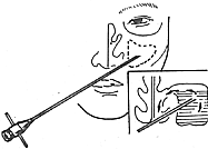
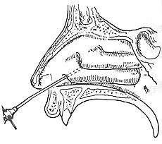
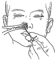
(1) Irrigation via the natural orifice After mucosal surface anesthesia, insert a curved-tip maxillary sinus irrigation tube into the middle nasal meatus, reaching about halfway in depth. Rotate the tip outward and downward, then slowly pull forward to pass through the uncinate process and enter the natural orifice. The orifice diameter is 5–7 cm, and its length is 8–10 mm. After insertion, irrigate with saline. This method is difficult for patients with high septal deviation, hypertrophic middle turbinate, or enlarged ethmoid bulla and uncinate process (Figure 1).


Figure 1: Maxillary sinus puncture and irrigation via the inferior nasal meatus
(1) Frontal view (2) Lateral view (3) Schematic diagram of puncture and irrigation
(2) Puncture and irrigation via the medial wall of the middle nasal meatus Following the above method, direct the tip of the maxillary sinus irrigation tube toward the lateral wall near the upper edge of the inferior turbinate. When a soft sensation is felt, puncture into the sinus cavity for irrigation. The advantage of this method is that it avoids injury to the nasolacrimal duct and the branches of the greater palatine artery, thus preventing bleeding. The puncture hole here is less likely to close.
(3) Puncture and irrigation via the inferior nasal meatus Anesthetize the anterior mucosal area of the inferior nasal meatus, or use 1% procaine for submucosal infiltration anesthesia. The patient is best seated. The operator stabilizes the patient's head with one hand and holds the puncture needle with the other, placing it near the attachment of the inferior turbinate in the inferior nasal meatus, about 1 cm behind the anterior end of the inferior turbinate. Puncture at a 45° angle toward the outer canthus of the eye. A sudden loss of resistance indicates entry into the sinus cavity. If the bone wall is not penetrated, the puncture point can be adjusted posteriorly or rotated to advance. After entering the sinus cavity, remove the needle core and instruct the patient to lower their head, holding a basin with both hands and raising their elbows to prevent irrigation fluid from flowing into the sleeves. Attach a syringe and attempt aspiration. If air or pus is aspirated, it confirms entry into the sinus cavity, and saline can be injected for irrigation. The patient must breathe through the mouth during this process until the outflow is clear. Depending on the condition, residual fluid is drained, and appropriate antibiotic or metronidazole solution is injected. After irrigation, remove the puncture needle and pack the inferior nasal meatus with 1% ephedrine-soaked cotton for hemostasis, removing it after 10 minutes. The advantage of this method is its high success rate, ensuring the needle tip is within the maxillary sinus cavity. The drawback is that it is not entirely painless, carries a risk of injuring the nasolacrimal duct, and is unsuitable for children.
(4) Puncture via the canine fossa method: The patient lies supine, and the area above the labiogingival groove is disinfected. Then, 5 ml of 1% lidocaine with epinephrine is injected, reaching deep to the periosteum. The maxillary sinus puncture needle is then inserted into the maxillary sinus 1 cm below the infraorbital margin. After successful puncture, the patient is instructed to sit up for irrigation. The advantages of this method are that it is well-tolerated by patients, suitable for pediatric cases, and avoids syncope due to nervousness. The disadvantage is that the anterior wall of the maxillary sinus is thicker than the lateral wall of the inferior nasal meatus, requiring greater force for puncture and sometimes the use of a bone chisel.
(5) Indwelling irrigation with a plastic tube in the sinus cavity. A thicker puncture needle is used to penetrate the sinus cavity, and a suitable thin polyethylene or silicone tube, 10–15 cm in length, is inserted through the needle hole into the sinus cavity. The external end of the tube is secured with adhesive tape to the upper lip or nasal wing. The advantage is that it eliminates the pain of multiple punctures, allows for daily multiple irrigations, shortens the treatment time, and enables the collection of sinus secretions as needed for cytological and bacteriological studies.
4. Irrigation solutions. To enhance efficacy, the following types of medications can be added to physiological saline as needed:
(1) Vasoconstrictors. These can shrink the mucosal blood vessels and reduce swelling, facilitating ventilation and drainage. Among them, 0.5% Aramine (metaraminol) is the most effective, with mild secondary vasodilation and no inhibitory effect on mucosal cilia.
(2) Antibiotics. Various antibiotics can be used. Since pathogens have different resistance profiles to different antibiotics, bacterial culture and sensitivity testing of secretions should be performed before use. If sensitivity testing is not feasible, a broad-spectrum antibiotic with high efficacy can be added. For chronic maxillary sinusitis, especially odontogenic cases, which are often caused by anaerobic bacteria, metronidazole and chloramphenicol must be added to the irrigation solution to achieve therapeutic goals.
(3) Adrenocortical hormones. Aqueous solutions are preferred, such as hydrocortisone acetate, while alcohol solutions should be avoided to prevent mucosal irritation. These medications can reduce mucosal swelling and assist antibiotics in exerting their anti-inflammatory effects.
(4) Enzymes. These can liquefy thick purulent material in the sinus, facilitating its expulsion. Empirical evidence shows no contraindication to combining antibiotics with enzymes. Commonly used enzymes include streptokinase (500–5000 U/ml), streptodornase (1000 U/ml), and deoxyribonuclease (50,000–100,000 U/ml).
5. Errors in maxillary sinus puncture. According to domestic data, the error rate in 321 puncture cases was 4.1%. ① Puncture outside the maxillary sinus, such as into the orbit, soft tissues of the cheek, pterygopalatine fossa, or submucosa of the inferior nasal meatus. ② Puncture into the submucosa of the medial or lateral wall of the maxillary sinus. These errors are caused by lack of technical proficiency or excessive force. After puncture, a syringe should be used to aspirate; if no air is withdrawn, an error should be suspected, and water should not be forcibly injected to avoid complications.
6. Common complications
(1) Syncope. This is a transient loss of consciousness caused by reflexive dysfunction of the vasomotor center due to neuropsychiatric factors, leading to cerebral anemia. It is more likely to occur in cases of excessive nervousness, pain, weakness, hunger, fatigue, excessive indoor steam, or poor air circulation. The author believes that harsh language or behavior from medical staff, causing patients to lose trust, also plays a role. Therefore, detailed explanations should be given before puncture, and patients should be frequently asked about their sensations. Early symptoms of syncope include weakness, chest tightness, nausea, tinnitus, blurred vision, vertigo, and unsteadiness while sitting upright, but patients may collapse and lose consciousness before they can report these symptoms. Examination may reveal pale complexion, sweating, shallow breathing, slow pulse, and slightly low blood pressure; in severe cases, there may be no response to stimuli and dilated pupils. This process is brief, lasting seconds to minutes, after which consciousness gradually returns. The patient should be placed in a supine or head-down position, with airways kept clear. Acupuncture at the philtrum point, oxygen inhalation, and a cup of warm water may help, but further puncture should be avoided.
(2) Collapse is a manifestation of acute systemic vascular tension reduction and heart failure. It is prone to occur in individuals with chronic wasting diseases, inadequate stress responses, and low adrenal cortical hormone secretion, with pain and mental tension as precipitating factors. The symptoms are more severe than syncope, presenting as pale skin, cyanosis, weak and rapid pulse, shallow breathing, lowered blood pressure, decreased body temperature, clouded consciousness, and inability to recover quickly. Collapse is generally reversible, but if not promptly treated, it can be life-threatening. When performing maxillary sinus puncture on long-term bedridden patients, adequate preparation is necessary, such as fluid infusion, correction of electrolyte imbalances, and administration of hormones. The puncture should be performed in a supine position. For patients who have already experienced collapse, attention should be paid to blood pressure, pulse, and respiration, and 40–60 ml of 10% glucose solution can be immediately administered intravenously.
(3) Air Embolism This complication is rare but can be fatal. It occurs when the needle punctures a vein in the maxillary sinus mucosa during the procedure, and after irrigation, air is forcefully injected into the sinus to expel residual fluid. The air may travel through the facial vein, internal jugular vein, and into the right heart, or bubbles may ascend to the brainstem, embolizing the respiratory center and causing death. During air injection, the patient may feel a bubbling sensation in the neck on the affected side, followed by cyanosis, collapse, loss of consciousness, and rapid cessation of breathing and heartbeat leading to death. Emergency treatment involves immediately placing the patient in a head-down position and lying on the left side to prevent more bubbles from entering the brain, left heart system, and coronary circulation. Artificial respiration and oxygen inhalation should be administered. If ineffective, cardiac massage and cardiac puncture to aspirate air from the heart may be necessary.
(4) Allergic Reaction to Topical Anesthetic Although rare, this reaction can be fatal. Symptoms manifest as initial excitation followed by paralysis of the central nervous system, progressing downward. Signs include spasms, convulsions, irregular breathing progressing to apnea, hypotension, excitement transitioning to unconsciousness, and pupils dilating from constriction. Emergency treatment involves administering antispasmodics, artificial respiration, and cardiac pacing if needed.
2. Intranasal Antrostomy Also known as maxillary sinus fenestration, this technique was introduced by Mikulicz in 1886. The procedure is similar to maxillary sinus puncture and irrigation via the inferior meatus, with the key difference being the creation of a window in the inferior meatus to allow repeated catheter insertion for irrigation. This method also facilitates sinus ventilation and restores mucociliary transport function. The purpose of fenestration is not drainage. Through this window, a maxillary sinus endoscope can be inserted to examine pathological changes.
1. Procedure First, topical anesthesia with 1% tetracaine (containing epinephrine) is applied to the inferior meatus. Then, infiltration anesthesia with 1% procaine is administered to the lateral wall of the inferior meatus, approximately 2 cm from the inferior turbinate. A bone chisel is used to create a mucoperiosteal flap with its pedicle posteriorly, which is then turned into the sinus to prevent window closure. If necessary, the window can be enlarged using a bone file in the superior, inferior, and anterior directions. Part of the inferior turbinate covering the window may also be excised to prevent obstruction.
2. Causes of Treatment Failure Similar to those in maxillary sinus puncture and irrigation.
3. Transalabial Fold Antrostomy This method was introduced by Xu Weixin in 1965. First, topical anesthesia is applied to the inferior meatus and inferior turbinate, followed by infiltration anesthesia with 1% procaine to the labiogingival groove, paranasal soft tissues, and canine fossa. A gauze pad is placed between the teeth and cheek to absorb oozing blood. A horizontal incision is made 5–6 mm above the free gingival margin, extending from the first canine to the midline, cutting through the mucosa and periosteum while avoiding injury to the labial frenulum. The tissue is dissected to expose the piriform crest, and the incision is extended upward to the mucoperiosteum above the attachment of the inferior turbinate, approximately 1.5 cm above the nasal floor, where the inferior meatus fenestration is performed. A dissector is used to separate the mucoperiosteum of the inferior meatus bone wall up to about 3 cm from the piriform crest, from the inferior turbinate attachment down to the nasal floor, advancing slowly. The mucoperiosteum of the inferior meatus is retracted medially, and a bone chisel is used to enter the maxillary sinus below the inferior turbinate. If necessary, the piriform crest may also be removed until the anteromedial angle of the maxillary sinus is visible. The anterior wall of the maxillary sinus does not need to be disrupted. The window should be enlarged as much as possible to minimize the risk of closure. Through the window, the sinus can be inspected, and the sinus mucosa can be treated. The maxillary sinus mucosa is dissected along the lower edge of the window to reach the sinus floor, and the bone wall is removed down to the nasal floor, which is on average 5 mm higher than the maxillary sinus floor. The bony ridge between the maxillary sinus and nasal cavity must be completely chiseled away to ensure proper drainage. The maxillary sinus mucosal flap is then turned toward the nasal cavity to cover the bone surface, with its pedicle placed at the anterior edge of the window and secured under pressure. The maxillary sinus is packed with iodoform gauze for five days, and the incision is sutured with silk thread, with stitches removed on the sixth day.
IV. Radical Maxillary Sinus Surgery This procedure was first performed in 1893 by George Galter Caldwell and Henry Paul Luc, hence it is called the Caldwell-Luc operation (Ke-Lu surgery).
1. Indications for Surgery
(1) Chronic suppurative maxillary sinusitis, with persistent purulent discharge after one month of continuous puncture and irrigation, or half a month of intrasinus medication.
(2) Pathologically confirmed subcutaneous nodular inflammation or fungal infection in the maxillary sinus.
(3) Imaging-confirmed polyps, cysts, or benign tumors in the maxillary sinus.
(4) Foreign bodies in the maxillary sinus.
(5) Odontogenic maxillary sinusitis and maxillary sinus-oral fistula.
(6) Other procedures performed via the maxillary sinus, such as exploration of the posterior nasal aperture, sphenoid sinus, or sella turcica; vidian neurectomy; ligation of the internal maxillary artery; orbital decompression; reduction of blow-out fractures; parotid gland transplantation for atrophic rhinitis; drainage before radiotherapy for maxillary sinus cancer; removal of foreign bodies from the pterygopalatine fossa; medial displacement of the lateral nasal wall; and ethmoid sinus opening.
2. Surgical Procedure
(1) Anesthesia: Local infiltration anesthesia combined with nasal and sinus mucosal anesthesia. Key nerves anesthetized include the infraorbital nerve, alveolar nerve, and sphenopalatine ganglion.
(2) Incision: A transverse incision is made from the canine eminence to the second premolar at the junction of the upper lip and gingival mucosa, down to the bone. The periosteum is elevated to expose the canine fossa.
(3) Opening the Anterior Wall: The medial part of the canine fossa (anterior maxillary sinus wall) is opened using a round chisel or electric drill, then enlarged to a 1–1.5 cm diameter bone window with rongeurs. Bleeding is controlled with bone wax or compression by chiseling adjacent areas.
(4) Removal of Pathologic Tissue: The classic Caldwell-Luc procedure involves complete removal of the sinus mucosa for radical cure. However, postoperative fibrosis often leads to reinfection and unsatisfactory outcomes. Current approaches preserve reversible mucosa (e.g., sparing non-odontogenic areas in odontogenic sinusitis). Irreversible changes (necrosis, abscesses, granulomas, cysts, polyps) require excision, confirmed preoperatively via endoscopic biopsy.
(5) Creating an Antrostomy to the Inferior Meatus: A bony window is chiseled in the anteroinferior medial sinus wall and enlarged (≥1.5 cm anteroposteriorly, ≥1.0 cm vertically), aligning its inferior edge with the nasal floor. Mucosal flaps from the inferior meatus are rotated into the sinus to promote epithelialization. Hypertrophic inferior turbinate tips may require resection.
(6) Packing and Suturing: The sinus is packed with iodoform gauze (if oozing persists), with the tail exiting the antrostomy for removal. The gingival incision is sutured, and cheek pressure dressing applied to reduce swelling and static blood.
3. Surgical Refinements The key to long-term success is maintaining antrostomy patency (achieved in ~60% of cases). Failures often result from antrostomy closure. Improvements focus on preventing closure and promoting mucosal regeneration:
(1) Antrostomy Stent: A plastic ring is placed at the antrostomy during packing to prevent stenosis. Grooved edges prevent dislodgement while allowing postoperative irrigation.
(2) Maxillary Sinus Nasal Anastomosis In 1964, Zhang Xiyi pioneered this technique. The method involves removing the sinus mucosa during the procedure while preserving the medial wall mucosa. The nasal mucosa of the inferior meatal antrostomy is divided into upper, lower, and posterior sections, which are then inverted into the sinus and sutured to the sinus mucosa. If the longitudinal and transverse diameters of the anastomosis can be maintained at 1 cm or more, the long-term patency of the antrostomy can be achieved.
(3) Mouth-held self-retaining hook This device was developed by the author in 1953. Its purpose is to replace the assistant dedicated to retraction, saving manpower during busy procedures. Additionally, it helps avoid excessive pulling force and prevents postoperative cheek swelling. The masseter muscle's strength is 45kg, while the force required for retraction is less than 4.5kg, so patients using the mouth-held self-retaining hook do not experience fatigue. This instrument is suitable for patients with temporomandibular joint dysfunction or those who have lost or have loose lower teeth.
(4) Maxillary sinus radical surgery with enlarged natural ostium In 1993, Xiao Bijun et al. adopted the method of enlarging the natural ostium of the maxillary sinus during radical surgery to improve sinus drainage, achieving better results compared to traditional procedures.
1. Common Complications of Maxillary Sinus Puncture and Irrigation:
1. Syncope: Caused by neuropsychiatric factors leading to reflexive dysfunction of the vasomotor center, resulting in temporary loss of consciousness due to cerebral anemia. It is prone to occur under conditions such as excessive mental stress, pain, physical weakness, hunger, fatigue, excessive indoor steam, or poor air circulation. The author believes that harsh language or behavior from medical staff, which erodes patient trust, also plays a role. Therefore, detailed explanations should be provided to the patient before the puncture, and their sensations should be frequently inquired about. Early symptoms of syncope include lack of strength, chest tightness, nausea, tinnitus, blurred vision, vertigo, and unsteadiness while sitting upright. However, patients often fail to report these symptoms to the doctor before fainting and losing consciousness. Examination may reveal pale complexion, sweating, shallow breathing, slow pulse, slightly low blood pressure, and in severe cases, unresponsiveness to stimuli and dilated pupils. This process is brief, lasting from a few seconds to minutes, with gradual recovery of consciousness. The patient should be placed in a supine or head-down position to maintain airway patency. Acupuncture at the philtrum point, oxygen inhalation, and drinking a cup of warm water may help, but further puncture should be avoided.
2. Collapse: A manifestation of acute systemic vascular tension reduction and heart failure. It is more likely to occur in individuals with chronic wasting diseases, inadequate stress responses, or low adrenal cortical hormone secretion, with pain and mental stress as triggers. Symptoms are more severe than syncope, including pale or cyanotic skin, weak and rapid pulse, shallow breathing, lowered blood pressure and body temperature, and clouded consciousness without quick recovery. Collapse is generally reversible, but failure to provide timely treatment can be life-threatening. For bedridden patients undergoing maxillary sinus puncture, thorough preparation is necessary, such as intravenous fluids, electrolyte correction, and hormone administration. The puncture should be performed in a supine position. For patients experiencing collapse, close monitoring of blood pressure, pulse, and respiration is essential. Immediate intravenous injection of 10% glucose solution (40–60 ml) may be administered.
3. Air Embolism: This complication is rare but potentially fatal. It occurs when the needle punctures a vein in the maxillary sinus mucosa during irrigation, and forceful air injection is subsequently used to expel residual fluid. Air may travel through facial veins, the internal jugular vein, and into the right heart, or bubbles may ascend to the brainstem, embolizing the respiratory center and causing death. During air injection, the patient may feel a bubbling sensation in the neck on the affected side, followed by cyanosis, collapse, loss of consciousness, and rapid cessation of breathing and heartbeat. Emergency measures include immediately placing the patient in a head-down position and lying on the left side to prevent further bubbles from entering the brain, left heart system, or coronary arteries. Artificial respiration and oxygen inhalation should be administered. If ineffective, cardiac massage and puncture to aspirate air from the heart may be necessary.
4. Allergic Reaction to Topical Anesthetics: Although uncommon, this can be fatal. Symptoms involve the central nervous system, initially exhibiting excitation followed by paralysis, such as spasms or convulsions. Breathing becomes irregular and then stops, blood pressure drops, consciousness shifts from excitation to loss, and pupils dilate. Treatment includes antispasmodics, artificial respiration, and cardiac pacemakers if needed. 2. Common Complications of Maxillary Sinus Fistulization:
1. Nasolacrimal Duct Injury: Persistent tearing on the affected side post-surgery, caused by an overly anterior fistula position. Recently, some advocate placing the fistula in the middle of the inferior nasal meatus to avoid this.
2. Epistaxis: Occurs if the fistula is positioned too posteriorly, injuring the nasal branch of the greater palatine artery, or too anteriorly, damaging the nasal branch of the superior labial artery.
3. Adhesion Between Inferior Turbinate and Nasal Septum: Caused by improper post-operative management, leading to adhesion between the inferior turbinate and the lateral nasal wall. 3. Common Complications of Radical Maxillary Sinus Surgery:
1. Postoperative bleeding According to domestic statistics, the incidence rate is 2.4-7%. Most cases occur within 24 hours after surgery. Minor bleeding from the edges of the anterior wall window of the maxillary sinus or the counter-opening may be caused by injury to the inferior turbinate and can be stopped by compression; subsequent bleeding is secondary bleeding, often caused by infection of the residual mucous membrane in the sinus. If the bleeding is significant, the maxillary sinus can be explored through the original incision, the bleeding mucous membrane can be removed, and packing can be performed again to stop the bleeding.
2. Facial Swelling This condition is a postoperative reaction, often caused by the injection of a large amount of high-concentration local anesthetic into the buccal submucosa, excessive force from retractors, or prolonged surgery. Treatment involves early removal of nasal sinus packing, applying warm compresses to the face, and using antibiotics to prevent infection.
3. Numbness of the Upper Lip and Upper Teeth This is usually due to surgical incision injury to the infraorbital nerve or because the incision is close to the midline, injuring the maxillary incisor nerve. Recovery may take several months to a year.


