| disease | Traumatic Spondylolisthesis of the Axis |
| alias | Hanged-manfracture, Hangmanfracture |
Fractures and dislocations of the axis were first discovered and described by Haughton in 1866 in a criminal executed by hanging. In 1931, Wood-Jones observed that placing the knot of the noose beneath the chin during hanging consistently resulted in the same fatal fracture and dislocation of the axis (bilateral pedicle fractures). In 1965, Schneider et al. identified the same injury in car accidents or other sudden deceleration incidents (such as hitting the bottom of a pool headfirst while diving) and first proposed the term "Hangman fracture" to describe this injury, which was gradually adopted by many authors. However, some have objected to this term. For instance, Nijima argued that it is inaccurate because "hang-man" refers to "a person who hangs another" (i.e., the executioner). According to Garfin and Rothman, this injury (Hangman fracture) represents the outcome every executioner strives to achieve, and thus they suggested renaming it "hanged-man fracture." In reality, this injury often manifests as anterior dislocation of the axis, making "traumatic spondylolisthesis of the axis" a more appropriate term, as the trauma results in fractures of the posterior structures of the axis. It is defined as: bilateral pedicle fractures of the axis, with or without anterior slippage. Recently, some authors have reported cases of axis fractures and dislocations involving fracture lines extending into the vertebral body under the title of "atypical Hangman fractures." Strictly speaking, these are vertebral body fractures of the axis, not Hangman fractures.
bubble_chart Pathological Changes
Anatomical and Biomechanical Characteristics
The axis (C2) serves as the connecting segment between the occipitocervical complex and the lower cervical spine, playing a crucial role in spinal biomechanics. The upper part of its anterior column consists of the odontoid process, which forms the atlantoaxial joint with the anterior arch of the atlas, the transverse ligament, and other accessory structures. The lower part connects to the C3
vertebral body via the intervertebral disc, anterior longitudinal ligament, and posterior longitudinal ligament. The posterior column features broad and robust laminae and spinous processes, with the spinous process being notably longer and bifid at its caudal end, distinguishing it morphologically from other cervical vertebrae. This makes it a key anatomical landmark in posterior cervical surgery. The middle column is relatively weak, with the superior articular process positioned anteriorly and the inferior articular process posteriorly. The narrow bony connection between these two articular processes is termed the pars interarticularis (isthmus), traversed by the vertebral artery foramen, rendering it an anatomically vulnerable area.From a biomechanical perspective, an axial load descending from above converges into a single force line at the level of the axis, passing through the pars interarticularis (Figure 1). A hyperextension force acting on the odontoid process creates a focal point of stress, inducing rotation around the X-axis in the sagittal plane. This force is balanced by two opposing mechanisms: tension exerted by the anterior longitudinal ligament, intervertebral disc, and posterior longitudinal ligament, and compression acting on the C2–3 facet joints. These equal and opposite forces establish an equilibrium point at the pars interarticularis of the axis (Figure 2), coinciding with its anatomical weakness. When stress exceeds its tolerance limit, a fracture may occur.
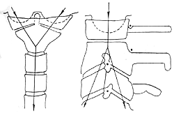
Figure 1: A vertically directed force concentrates at the axis pars interarticularis.
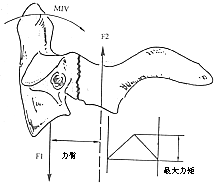
Figure 2: Hyperextension force on the odontoid process generates maximal torque at the axis pars interarticularis.

Several major injury mechanisms: (1) The hyperextension force mentioned above is a primary injury mechanism for axis isthmus fractures; (2) Similar to the mechanism of using a submental knot in hanging. Numerous studies have identified this injury, termed Hangman's fracture, which may occur at the anterior-most part of the lateral mass or extend into the pedicle, accompanied by rupture of the anterior longitudinal ligament, intervertebral disc, and posterior longitudinal ligament. The injury mechanism involves hyperextension combined with sudden and violent distraction force, causing craniocervical dissociation—where the axis body and cranioatlantal structures separate upward as a unit while the posterior axis structures remain intact with C3 connections (Figure 3). This often results in spinal cord transection and immediate death. However, some reports describe survival after such injury, even with transient neurological symptoms. This difference is attributed to variations in load direction, magnitude, and duration of force application. For executed prisoners, "suspension by the neck until death" ensures prolonged force application until critical soft tissues reach failure load, leading to craniocervical dissociation and death; (3) In car accidents or diving incidents, the injury mechanism involves hyperextension with axial compression. Extension occurs when the torso lunges forward, striking the forehead against an inclined windshield or pool bottom, while axial pressure—and possibly rotational components—are also involved. Rogers observed numerous C3 vertebral compression fractures associated with axis fractures, along with other injuries unexplainable by simple extension mechanisms. One of his cases involved C7
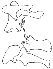
Figure 3 Craniocervical separation caused by hanging
In fact, there is a large group of cases involving fractures of the axis pedicle, where the combination of injuries depends on the specific vector of the applied force, including its magnitude, direction, point of application, and duration. Overall, the position of the spinal structures at the moment the force is applied, as well as the unique biomechanical characteristics of the patient's spinal anatomy, determine the specific injury, the structural components damaged, and the degree of displacement. When a physician observes a traumatic spondylolisthesis of the axis, flexion along the X-axis is the primary component of the injurious force, with hyperextension being the most likely mechanism involved.
bubble_chart Clinical Manifestations
Table 1 Francis Classification of Hangman Fracture
| Grade | Displacement | Angulation (degrees) |
| Ⅰ | <3.5mm | <11 |
| Ⅱ | <3.5mm | >11 |
| Ⅲ | >3.5mm or<0.5椎體寬度 | <11 |
| Ⅳ | >3.5mm or >0.5 vertebral body width | >11 |
| Ⅴ | Disc rupture |
Classification
It was not until 1981 that a standardized classification for Hangman fracture emerged. Francis et al. first categorized Hangman fractures into five grades based on displacement, angulation, and ligamentous instability (Table 1). Displacement is measured on lateral radiographs by drawing vertical lines along the posterior edges of the C2 and C3 vertebral bodies and measuring the distance between these lines; angulation is determined by drawing lines along the posterior edges of the C2 and C3 vertebral bodies and measuring the angle formed by their intersection (Figure 1).
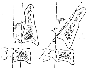
Figure 1 Measurement of horizontal displacement and angulation in Hangman fracture
Grade I fractures are considered stable, while Grades II–IV are unstable. Grade V fractures indicate displacement exceeding half the sagittal diameter of the C3 vertebral body or angulation deformity causing at least one C2–3 interspace to exceed the height of a normal cervical disc.
In the same year, Effendi et al. classified these fractures into three types based on stability: Type I is a stable fracture where the fracture line may involve any part of the vertebral arch, and the C23 intervertebral structures remain normal. Type II is an unstable fracture, with the axis showing flexion or extension angulation or significant anterior slippage, and injury to the C23 intervertebral structures. Type III is a displaced fracture, where the axis is anteriorly displaced with flexion, and the C23 facet joints are dislocated or locked (Figure 2).
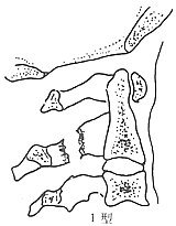

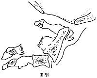
Figure 2 Effendi Classification of Hangman Fracture
In 1985, Levine and Edwards classified 52 patients with traumatic spondylolisthesis of the axis into four types based on fracture morphology and stability combined with injury mechanisms. Type I fractures exhibited minimal displacement with slight ligament injury, representing stable fractures and accounting for 28.8%. The injury mechanism involved hyperextension combined with axial loading, causing the pars interarticularis of the axis to fracture in an extended position. Type II fractures showed anterior displacement exceeding 2 mm and significant angulation, representing unstable fractures and accounting for 55.8%. The injury mechanism involved hyperextension and axial loading, resulting in a near-vertical fracture of the pars interarticularis, followed by sudden flexion leading to stretching of the posterior fibers of the intervertebral disc, anterior displacement, and angulation of the vertebral body. The C23 intervertebral disc could rupture due to the sudden flexion component involved in this injury mechanism. Type IIA fractures were a variant of Type II, displaying severe angulation and Grade I anterior displacement at C23. The fracture line was typically not vertical but obliquely traversed the pars interarticularis of the axis from posterosuperior to anteroinferior, accounting for 5.8%. The injury mechanism primarily involved flexion with a traction component. Type III fractures were bilateral pedicle fractures with posterior facet joint injuries, often accompanied by severe displacement and angulation of the pars interarticularis fracture, as well as unilateral or bilateral facet joint dislocation, accounting for 9.6%. The injury mechanism involved flexion force combined with axial compression (Figure 3).
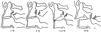
Figure 3 Levine-Edwards classification of Hangman fracture
The author believes that the classification method proposed by Levine and Edwards combines fracture morphology and injury mechanism, providing guidance for treatment selection.
Although traumatic spondylolisthesis of the axis is anatomically a highly dangerous injury, the incidence of neurological damage is relatively low, sometimes even难以置信. For example, among Levine's 52 cases, only 4 were accompanied by cervical spinal cord injury, while unrelated neurological injuries such as closed head trauma occurred in 11 cases. Among Brashear's 29 patients with this type of fracture, initial stage [first stage] symptoms included 1 case of left upper limb paralysis, which recovered after 6 hours; 1 case of transient全身麻木; 1 case of central cord syndrome; after 5 weeks, only left upper limb weakness remained; and another case of quadriplegia, with full recovery after 25 days. There are also reports of relatively higher neurological injury rates. In Tan's group of 31 patients, 20 were asymptomatic, 7 had incomplete quadriplegia (3 with central cord syndrome), 2 had incomplete paraplegia, 2 had Brown-Sequard syndrome, and 2 had complete bladder dysfunction. Among Marar's 15 cases, 11 were complicated by varying degrees of neurological damage, with 6 recovering within 24 hours and the remaining 5 recovering fully within 3 days to 3 months. The lower incidence and severity of neurological damage in this type of injury may be due to the anterior fracture fragment displacing forward, creating a defect in the middle arch and effectively enlarging the spinal canal, allowing the spinal cord to shift forward and avoid compression by the posterior arch of the atlas. However, when the fracture line involves the axis vertebral body, the posterior bone fragment of the axis vertebral body to be decocted later remains in place, posing a risk of spinal cord compression (Figure 4).
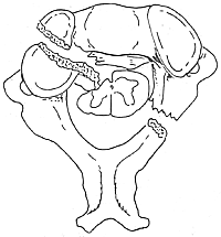
Figure 4 Fracture line involving the axis vertebral body, with the posterior fracture fragment compressing the spinal cord
The most common complaints are neck pain and stiffness, followed by numbness and weakness. A clear history of trauma, often from car accidents or falls, is present. Another clinical feature is the frequent association with head and maxillofacial injuries, typically involving contusions to the forehead or chin. Occasionally, fractures of other vertebrae or long bones may occur. For example, among Tan's 31 cases, 18 had forehead soft tissue injuries, and 15 had fractures of other vertebrae (5 cases) or long bones (10 cases). Among Levine's 52 cases, 13 also had fractures in other locations. Additionally, Okuchi reported one case complicated by a right vertebral arteriovenous fistula.
In the entire fracture of cervical vertebrae dislocation, traumatic anterior spondylolisthesis of the axis accounts for 4–7%. If there is a lack of accurate trauma history or insufficient understanding of the characteristics of this injury, it may lead to misdiagnosis of fistula disease. Sometimes the injury is more complex, accompanied by multiple injuries, especially when there are obvious life-threatening non-cervical injuries, which can divert the physician's attention and result in the cervical spine injury being overlooked. The importance of routine cervical spine films for patients with neck pain after trauma is emphasized once again. For patients with suspected diagnoses, do not let them go; repeatedly examine them until the diagnosis is confirmed or ruled out. Through a detailed history and physical examination, understand the point and direction of the force, combine it with imaging studies to determine the injury mechanism, and guide the selection of treatment options. The diagnosis should include: (1) classification of the fracture; (2) presence or absence of nerve injury; (3) presence or absence of associated injuries; (4) whether it is a multiple injury.
Imaging Studies
Conventional X-ray examinations include routine cervical spine films and tomograms. The diagnosis of traumatic anterior spondylolisthesis of the axis mainly relies on lateral films. Lateral films can clearly show the fracture line and the degree of displacement and angulation. Based on this, an imaging diagnosis of the fracture type can be made. Under the guidance and protection of a physician, cautious flexion and extension films of the cervical spine can provide further information on the stability of the fracture. Sometimes tomography is required to clearly visualize the fracture line. The typical X-ray findings are bilateral fractures of the axis pedicles, with the fracture line being vertical or oblique, and the axis vertebral body may show varying degrees of displacement and angulation. Additionally, attention should be paid to possible associated fractures in the atlas or lower cervical spine. For infants and young children, the possibility of congenital defects or cartilaginous connections in the axis pedicles should also be considered.
Examination of other injury sites can help determine the presence of multiple injuries.
CT scans can clearly show the fracture line, displacement, and its relationship with the spinal canal. Three-dimensional CT reconstruction aids in a comprehensive understanding of the fracture morphology. MRI can assess the condition of the spinal cord and surrounding soft tissues, provide a comprehensive evaluation of the entire injury, and serve as a basis for selecting the surgical approach.
bubble_chart Treatment Measures
The choice of treatment depends on the stability of the fracture. Most patients with traumatic spondylolisthesis of the axis can achieve solid bony union with minimal deformity through close non-surgical management, with a low incidence of nonunion.
Non-surgical treatment
Non-surgical treatment includes head-neck-thorax plaster casts, plaster cervical collars, Halo braces, and traction.
For stable fractures (Levine-Edwards Type I), direct plaster fixation for 12 weeks can be used, followed by a cervical collar for 6 weeks after radiographic confirmation of bony fusion. For unstable fractures (Levine-Edwards Type II), traction reduction can be performed. Bedside radiographs should be taken upon admission to check for displacement during transfer. Traction can begin with a small weight, starting at 2 kg and gradually increasing to 4–5 kg. The direction of traction and neck position should be selected based on the injury mechanism, displacement, and angulation. Close radiographic follow-up is necessary to monitor traction effectiveness. If increased displacement or over-traction is observed, immediate adjustments such as reducing weight or changing the traction direction are required. Once reduction is achieved, neutral traction with 2 kg should be maintained for 3–6 weeks to immobilize and maintain the reduction. The patient can then ambulate with a Halo brace. Note that in the initial stage of the fracture, the Halo plaster cannot achieve or maintain reduction, and early ambulation with a Halo brace may cause re-displacement. After 3 months, the fracture often heals, even with an initial gap, and C23 often fuses spontaneously.
Identifying Levine-Edwards Type IIA fractures is crucial. Traction in these patients can worsen C23 separation and displacement. The recommended treatment is Halo brace immobilization with Grade I compression under radiographic monitoring to achieve and maintain anatomical reduction. After radiographic confirmation of anatomical reduction, Halo brace immobilization should continue for 12 weeks, followed by a plastic cervical collar for 6 weeks once fracture healing is observed.
Some physicians strongly oppose traction, especially when imaging suggests rupture of the C23 annulus fibrosus and ligaments, as traction may cause significant over-traction. However, there are reports of cases with initial radiographs showing significant C23 separation where traction achieved anatomical reduction with contact. Clearly, cautious, light-weight traction can be used before external fixation or surgery to improve reduction, relieve muscular rigidity, and facilitate soft tissue repair, but it must be closely monitored. Over-traction must be stopped immediately if detected.
Surgical treatment
Clearly, Levine-Edwards type III fracture is the only Hangman fracture that requires surgical treatment, as the posterior facet joint fracture and dislocation, if not reduced, can lead to persistent neck pain. Posterior surgical reduction and "∞" shaped wire fixation with bone graft fusion can be performed, followed by Halo brace immobilization to achieve bone graft fusion and fracture healing. C23Rupture of the anterior ligament and intervertebral disc can result in extreme instability of the segment. Sometimes, traction fails to maintain reduction, necessitating surgical fixation. Surgical options include posterior pedicle screw internal fixation, C23slot bone graft fusion, and anterior plate internal fixation. Postoperative effective external fixation immobilization should be provided as protection until radiographic evidence of bony fusion is observed. The goals of surgery are decompression, reduction, and stabilization. Matsumoto et al. reported a case involving a patient with a C2 pedicle fracture affecting the vertebral body. MRI indicated spinal cord compression originating posteriorly—from the foramen magnum of the occipital bone and the posterior arch of the atlas. Initial treatment involved skull traction, but follow-up imaging days later showed no reduction, and neurological symptoms worsened. Decompression of the foramen magnum, resection of the posterior arch of the atlas for decompression, and occipitocervical fusion were performed, followed by Halo brace immobilization. Neurological symptoms improved within days postoperatively, and radiographic evidence of solid fusion was observed at 12 weeks, after which a cervical collar was used for protection. Follow-up MRI at this time confirmed decompression of the high cervical spinal cord, with normal subarachnoid space.
Regarding the prevention of traumatic spondylolisthesis of the axis, the use of seat belts in car accidents can significantly reduce this injury. Of course, compliance with traffic regulations is the most beneficial.


