| disease | Duodenal Stasis Syndrome |
| alias | Duodenal Stasis |
Duodenal stasis refers to a clinical syndrome caused by various factors leading to obstruction of the duodenum, resulting in proximal dilation of the obstructed segment and accumulation of chyme.
bubble_chart Etiology
There are many causes of this condition, with compression of the duodenum by the superior mesenteric artery leading to stasis being the most common (accounting for 50%). This condition is also known as superior mesenteric artery syndrome. Other causes include: ① Congenital anomalies: such as compression or traction by congenital peritoneal bands obstructing the duodenum; congenital stenosis or atresia of the distal duodenum; annular pancreas compressing the descending duodenum; megaduodenum due to duodenal dysplasia; and severe duodenal ptosis caused by congenital variations, which can kink the duodenojejunal angle and close it, resulting in stasis. ② Tumors: benign or malignant tumors of the duodenum; retroperitoneal tumors such as renal tumors, pancreatic cancer, or lymphoma; metastatic cancer to the duodenum; adjacent enlarged lymph nodes (due to cancer metastasis); mesenteric cysts; or compression by an abdominal aortic aneurysm. ③ Infiltrative or inflammatory diseases of the distal duodenum or proximal jejunum, such as progressive systemic sclerosis, Crohn’s disease, or strictures caused by diverticulitis-related adhesions or compression. ④ Postoperative adhesions from gallbladder or gastric surgery pulling on the duodenum; adhesions, ulcers, strictures, or afferent loop syndrome after gastrojejunostomy. ⑤ Other congenital malformations: duodenal inversion; duodenal obstruction caused by gallbladder-duodenal or intestinal bands; anterior portal vein; or abnormal positioning of the ampulla of Vater (where the common bile duct opens into the third part of the duodenum).
bubble_chart PathogenesisThe transverse part of the duodenum is located behind the abdominal membrane, crossing horizontally from right to left over the third lumbar vertebra and the abdominal main stirred pulse. Its anterior aspect is traversed by the superior mesenteric vascular and nerve bundle within the root of the mesenteric membrane, as shown in Figure 1. If the angle between the two is too small, it can compress the duodenum. The superior mesenteric stirred pulse usually branches off at the level of the first lumbar vertebra, forming an angle of 30° to 42° with the main stirred pulse. Additionally, the following five factors can also cause mechanical obstruction: ① The superior mesenteric stirred pulse is too long or too short; ② Variations in the superior mesenteric stirred pulse, such as an abnormally low branching point from the abdominal main stirred pulse or a narrow branching angle; ③ An abnormally large vein compressing the anterior aspect of the duodenum; ④ Kyphosis deformity reducing the space occupied by the duodenum; ⑤ In slender individuals or those with visceral ptosis, the weight of the intestinal tract pulls on the root of the mesenteric membrane.
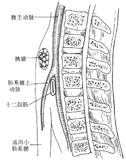
Figure 1 Schematic diagram of the anatomical position of the mesenteric membrane vessels.
bubble_chart Clinical Manifestations
Acute duodenal obstruction often occurs when the trunk is immobilized by a plaster cast or traction, leading to signs of acute gastric dilation. Chronic obstruction is the most common clinical type, with hiccups, nausea, and vomiting being frequent symptoms, typically appearing after meals. The vomitus may contain bile, and symptoms can be alleviated by changes in body position, such as lying on the side, prone, or in the knee-chest position. If not relieved, prolonged episodes can result in weight loss, dehydration, and general malnutrition.
bubble_chart Diagnosis①Typical symptoms are important diagnostic criteria. ②Features of X-ray barium meal examination: Interruption of the barium column is observed at the horizontal part of the duodenum (sudden vertical cut-off); pendulum movement formed by strong forward peristalsis and reverse peristalsis in the obstructed proximal intestinal segment; barium passes smoothly in the prone position, and reverse peristalsis disappears. ③If necessary, selective mesenteric angiography can be performed to reveal the anatomical relationship with the duodenum.
bubble_chart Treatment Measures
Patients without obvious symptoms may not require treatment. During the acute stage of an attack, intravenous nutrition including fat emulsions, nasogastric tube decompression, and antispasmodic medications should be administered to address acute gastric dilation. It is advisable to eat small, frequent meals, maintain a knee-chest position for half an hour after meals, and strengthen abdominal muscle exercises. If conservative medical treatment proves ineffective, surgical intervention may be considered. Surgical options include: ① Mobilization of the duodenal ligament; ② Duodenojejunostomy; ③ Duodenal repositioning.
Symptoms of indigestion need to be differentiated from peptic ulcers, and sometimes the two conditions may coexist. Duodenal stasis caused by tumors outside the duodenum, such as pancreatic head cancer or large pancreatic cysts, can be distinguished through endoscopic examination or retrograde cholangiopancreatography. Occasionally, this condition may also result from compression of the duodenum by an abdominal aortic aneurysm. The disease should also be distinguished from duodenal obstruction caused by stones, bezoars, ascaris masses, or foreign bodies within the duodenum.


