| disease | Urethral Injury |
The male urethra is a muscular mucous membrane tube, approximately 20 cm in length, and can be divided into anterior and posterior segments, demarcated by the urogenital diaphragm. The anterior urethra consists of the spongy part, including the glans, penile, and bulbous sections, totaling 15 cm in length. The posterior urethra includes the membranous and prostatic parts, measuring about 5 cm in length. The male urethra has two curvatures: the subpubic and prepubic curves. The subpubic curvature is relatively fixed, while the prepubic curvature disappears when the penis is pressed against the lower abdomen. The dorsal side of the urethra is shorter than the ventral side and is fixed. When the penis is in a relaxed state, the ventral side of the urethra has numerous folds. The mucous membrane of the urethra is rich in glands and is inherently soft; the submucosal tissue is well-supplied with blood. Due to its anatomical characteristics, the male urethra is prone to injury, and male urethral injury is a common urological emergency, which can lead to complications such as urinary extravasation, infection, urethral stricture, and fistula. The female urethra is short and rarely injured. However, during difficult delivery, pressure from the fetal head or the use of forceps can cause injury, resulting in a urethrovaginal fistula.
bubble_chart Etiology
(1) Urethral injuries are mostly caused by the use of transurethral instruments or the removal of foreign bodies (such as stones). A small number of injuries are caused by the insertion of foreign objects like hairpins, wires, or glass by individuals with sexual perversions, intoxication, or mental illness. Accidental injection of certain chemicals such as silver nitrate, copper sulfate, or phenol can cause chemical burns. Urethral electrocautery injuries can occur during transurethral resection.
(2) External violent injuries to the urethra are more common than internal urethral injuries. These can be either penetrating or blunt injuries. The former mainly occurs on the battlefield, where the urethra is punctured by firearms or sharp objects. The injury sites are mostly in the bulbous and membranous parts, while injuries to the cavernous and prostatic parts are rare. Closed urethral injuries can be seen both in wartime and peacetime. Injuries to the bulbous and membranous parts of the urethra are common in perineal straddle injuries or kicks, while injuries to the prostatic part of the urethra often accompany pelvic fractures.bubble_chart Pathological Changes
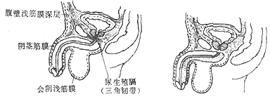
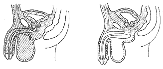
Urethral injury may only damage the mucous membrane or result in a contusion of the urethral wall, but most injuries involve the full thickness, leading to urethral rupture. This rupture can be longitudinal or transverse, partial or complete, causing the severed ends to retract, leaving a gap and misalignment between the two ends. After a full-thickness urethral rupture, hematuria may leak out. The extent of hematuria extravasation varies depending on the location and severity of the urethral injury. Familiarity with the anatomy of the perineum greatly aids in understanding the extent of hematuria extravasation. Clinically, there are three types of urinary extravasation following urethral trauma (Figure 3):


Figure 3 Urinary extravasation after urethral injury
(1) Normal anatomy of the penis, scrotum, and fascial membrane (2) Extravasation from penile urethral injury (with intact penile fascial membrane) (3) Extravasation from bulbar urethral injury (with ruptured penile fascial membrane) (4) Extravasation from prostatic urethral injury
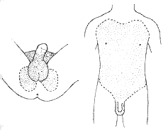
(2) In cases of anterior urethral injury, if the intrinsic fascial membrane of the penis is also ruptured, urine extravasates along the penis, scrotum, and superficial fascia of the abdominal wall to the scrotum, penis, superficial perineum, and abdomen. Since the superficial fascia of the abdominal wall is fixed at the inguinal ligament, urine does not extravasate to the thighs. This situation is the most common (Figure 4).

Figure 4 Extent of urinary extravasation from bulbar urethral injury
(3) When the urethral rupture occurs in the posterior urethra, either between the two layers of the urogenital diaphragm or posterior to it, urine extravasates along the prostatic area into the retropubic space and around the bladder. The bladder is primarily fixed to the urogenital diaphragm by the membranous urethra. If the urethra is completely severed, the bladder is often pushed upward by the extravasated blood and urine, creating a large gap between the two severed ends. If not promptly reduced and fixed during emergency treatment, it will inevitably complicate late-stage [third-stage] repair.
Urethral rupture can be complicated by perineal abscess and urinary fistula. In advanced stages, due to the formation of fibrous scars, urethral stricture may develop.
bubble_chart Clinical Manifestations
The symptoms of urethral injury depend on the disease cause of the injury, the extent and range of the urethral injury, and the accompanying injuries to other organs. Common symptoms include:
(2) Pain, with pain at the site of injury, sometimes radiating to the external urethral orifice. The pain is particularly severe during urination.
(3) Urethral bleeding. If the injury is distal to the membranous part of the urethra, blood may drip from the external urethral orifice even without urination. If the injury is in the posterior urethra, bleeding is more common during urination, with a small amount of blood dripping before or after urination.
(4) Difficulty in urination and urinary retention. Patients with complete urethral rupture experience urinary retention. In cases of urethral contusion and laceration, pain can cause sphincter spasm, leading to difficulty in urination and urinary retention.
(5) Local swelling and ecchymosis. Swelling and static blood appear in the injured tissue. In straddle injuries of the urethra, swelling and obvious ecchymosis can be seen in the perineum and scrotum.
(6) Urinary extravasation and urinary fistula. After a full-thickness urethral laceration, when the patient strains to urinate, urine can extravasate from the laceration into the surrounding tissues. Once secondary infection leads to cellulitis, sepsis can occur. If not treated promptly, it can lead to death. In the case of open injuries, urine can flow from skin wounds, intestinal or vaginal fistulas, eventually forming a urinary fistula.
Based on medical history, symptoms, and signs, the diagnosis of urethral injury is not difficult. The signs of anterior urethral injury are generally more obvious, making diagnosis easier. Diagnosis of posterior urethral injury is more challenging. Catheterization is a good method to check the integrity of the urethral continuity. Under sterile conditions, if a catheter can be smoothly inserted, it indicates that the urethral continuity is intact. If the catheter is smoothly inserted into the bladder, and upon examination the bladder wall is intact but the patient shows signs of urinary extravasation, urethral injury should be considered. However, catheterization must be performed under strict sterile conditions and satisfactory anesthesia, preferably in an operating room. If insertion is difficult on the first attempt, repeated probing should not be forced to avoid aggravating the trauma and causing infection. Immediate surgical exploration is necessary. Emergency high-dose intravenous urography, followed by voiding cystourethrography and retrograde urethrocystography after the contrast agent accumulates in the bladder, can also help confirm the diagnosis of urethral injury. Normally, digital rectal examination can palpate the membranous part of the urethra between the apex of the prostate and the anal sphincter. If this segment of the urethra cannot be palpated and the posterior edge of the pubic bone is directly felt, the membranous part of the urethra is completely ruptured. During digital rectal examination, if the prostate is pushed upward and it is fixed, it indicates that the posterior urethra is not completely transected. Conversely, if the prostate can be pushed upward or is suspended in the pelvic cavity due to loss of support and pushed upward by extravasated hematuria, it indicates that both the urethra and the puboprostatic ligament are ruptured. Therefore, digital rectal examination is also important in diagnosing posterior urethral injury.
bubble_chart Treatment Measures
First, shock should be corrected, and then the urethral injury should be addressed. The basic principles of treating urethral injury are to drain urine and reconnect the severed ends of the urethra.
(1) Drainage of Urine Under strict aseptic conditions and satisfactory anesthesia, if a catheter can be smoothly inserted, it indicates that the continuity of the urethra is still intact. If the hematoma and urinary extravasation are not severe, the catheter should be retained for 10 to 14 days to drain urine and support the urethra, waiting for the injury to heal. If catheterization fails, surgical exploration should be performed immediately. If the patient's condition is too severe to allow major surgery, a simple suprapubic bladder ostomy can be performed. A bladder ostomy can prevent urinary extravasation, reduce local irritation and infection, promote the absorption of inflammation, hematoma, and fibrous tissue, thereby reducing the degree of possible urethral stricture and surrounding scar formation, and providing convenience for intermediate stage [second stage] repair. A bladder ostomy can also be completed by puncture. It is suitable for cases of posterior urethral injury. Due to its simplicity, it is especially suitable for primary medical units.
(2) Urethral Repair
1. Perineal Urethral Repair Suitable for bulbar urethral injuries caused by straddle injuries, etc. Through a perineal incision, the bulbar urethra is exposed. If the urethra is not completely severed, a catheter is inserted from the external urethral orifice into the bladder under direct vision and finger palpation and retained. The laceration is sutured along the catheter. Generally, transverse lacerations are more likely to cause postoperative strictures than longitudinal lacerations. In cases of severe contusion or complete transection of the urethra, a catheter can be inserted from the external urethral orifice to locate the distal end, and abdominal pressure can be applied to observe urine outflow. Alternatively, a catheter can be inserted through the internal urethral orifice from a suprapubic bladder incision to locate the proximal urethral end. Necrotic tissue and hematoma are thoroughly removed, and the two ends are then anastomosed with absorbable sutures in an interrupted everted manner. The anastomosis should avoid tension. According to anatomical relationships, extravasated urine is thoroughly drained, and multiple skin incisions are made in the area of urinary extravasation to drain the extravasated urine. The incisions should reach below the superficial fascia. Postoperatively, the catheter should be retained for at least 3 to 4 weeks. After catheter removal, if urination is smooth, the suprapubic bladder fistula tube can be removed. To prevent postoperative urethral stricture, regular urethral dilation can be performed postoperatively. Alternatively, the urethra can be irrigated with 10 ml of urethral irrigation solution once or twice daily as a soft dilation (irrigation solution formula: dexamethasone 0.15 g, neomycin 25 g, procaine 10 g, 5% nipagin 10 ml, glycerol 400 ml, Tween-80 5 ml, add double-distilled water to 1000 ml). Audio frequency physiotherapy can also be used to prevent stricture.
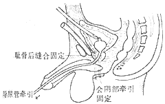
2. Transurethral Rendezvous Procedure In cases of posterior urethral injury, due to the frequent combination of severe injuries to other organs and critical conditions, the patient may not tolerate major surgery. In such cases, a transurethral rendezvous procedure can be performed through a suprapubic incision via the bladder (Figure 1). A male and female sound are inserted from the external urethral orifice and the internal urethral orifice via the bladder, respectively. After rendezvous, a balloon catheter is introduced, and the balloon is inflated to pull the catheter, bringing the two ends together. If male and female sounds are not available, a finger can be inserted from the bladder neck into the posterior urethra to rendezvous with a metal sound inserted from the external urethral orifice. If the tension is high, a nylon suture can be placed on each side of the prostatic end and brought out through the perineum with a straight needle, tied over a small gauze pad to aid in traction and fixation (Figure 2). The sutures are removed after 2 weeks. Although there is still a possibility of urethral stricture postoperatively, the approximation of the two ends and alignment of the axis facilitate intermediate stage [second stage] repair.


Figure 1 Rendezvous Procedure for Posterior Urethral Injury
(1) Rendezvous of male and female sounds (2) Introduction of a catheter through a suprapubic bladder incision (3) Introduction of a balloon catheter (4) Completion of the surgery (5) Self-made male and female sounds from a metal catheter (6) Schematic diagram of the rendezvous of male and female sounds.

Figure 2 Perineal Traction Fixation of the Prostate for Posterior Urethral Rupture
3. Suprapubic Approach Initial Stage [First Stage] Urethral Repair for Rupture Due to the frequent association of posterior urethral rupture with pelvic fracture, patients are often in a state of shock, with significant bleeding around the pubic bone and bladder. Performing a repair surgery requires the removal of hematomas and bone fragments, which could potentially lead to more severe bleeding, thus presenting certain difficulties. However, if the patient's condition permits and there is an ample blood supply, experienced surgeons may opt for this approach and achieve favorable outcomes.
The prognosis of urethral injury critically depends on the correctness of emergency management. It is crucial to avoid repeated attempts at catheterization, which can exacerbate the injury, potentially turning a partial urethral tear into a complete rupture. The choice of surgical method should be based on the patient's overall condition, the location and severity of the urethral injury, associated injuries, the surgeon's experience, and the available medical resources, and should not be generalized.
Regardless of the method used for urethral injury repair, there is a possibility of scar contraction leading to urethral stricture postoperatively. Regular postoperative urethral dilation may not always be effective. Additionally, infection and urinary fistula are common complications.


