| disease | Congenital Supravalvular Aortic Stenosis |
Supravalvular aortic stenosis is the rarest form of congenital aortic outlet stenosis, accounting for approximately 5-10% of cases. The incidence is similar in males and females. Patients with supravalvular stenosis may exhibit intellectual developmental delays. The stenotic lesion is located above the coronary artery ostia. The localized type is the most common form of supravalvular aortic stenosis, representing about 90% of cases.
bubble_chart Pathological Changes
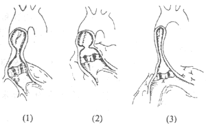
The most common lesion is a membranous stenosis above the coronary sinus. There is a small hole in the center of the membrane, and sometimes the membrane connects to the left coronary cusp, obstructing left coronary blood flow. The aortic valve leaflets may thicken. The ascending aorta appears normal without post-stenotic dilation. Another type of localized supravalvular stenosis presents with a narrowed external diameter at the stenotic site, resembling an hourglass or "8" shape. The aortic wall at this location shows fibrotic thickening, and the intima is also thickened. Histological examination reveals lesions similar to aortic coarctation. Diffuse supravalvular aortic stenosis is rare, with the stenosis extending from above the coronary sinus along the ascending aorta to the origin of the innominate artery, and even involving the aortic arch. Cases of supravalvular aortic stenosis often exhibit tortuous dilation of the coronary arteries and enlargement of the coronary sinus (Figure 3). They may also involve multiple peripheral pulmonary stenoses, such as pulmonary valve stenosis, hypoplasia of the main pulmonary artery, branch stenosis of the aortic arch, aortic coarctation, or ventricular septal defects.

Figure 3 Types of supravalvular aortic stenosis
(1) Annular constriction at the root of the ascending aorta
(2) Membranous supravalvular aortic stenosis
(3) Hypoplasia of the long segment of the ascending aorta
bubble_chart Clinical Manifestations
Most cases present with symptoms of aortic outlet stenosis during childhood. Due to the early onset of coronary atherosclerosis, colicky pain is relatively common. Some patients have a family history.
The signs are similar to other types of aortic outlet stenosis, but no systolic click is heard. The location of the heart murmur and tremor is higher than in valvular stenosis, and aortic diastolic murmurs are rare. Some patients exhibit poor growth and development, short stature, low intelligence, excessive talking, and distinctive facial features: retrognathia, upturned nostrils, low nasal bridge, thick lips, broad forehead, wide-set eyes, and malocclusion. Approximately 5% of patients have elevated blood calcium levels.
X-ray and electrocardiogram findings are similar to those of other types of aortic outlet stenosis.
Cardiac catheterization: Left heart catheterization with continuous pressure recording may reveal changes in the pressure waveform above the aortic valve.
Selective left ventriculography can demonstrate the location, length, and severity of supravalvular stenosis, while also assessing the morphology and function of the aortic valve, as well as the condition of the coronary sinuses and coronary arteries. Right heart angiography can reveal whether the pulmonary artery and its branches are also affected.
Cross-sectional echocardiography: Can directly visualize the location and length of supravalvular stenosis.
bubble_chart Treatment Measures
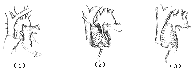
Localized supravalvular stenosis: After establishing extracorporeal circulation and implementing myocardial protection measures, the ascending aorta is clamped. A longitudinal incision is made from above the narrowed area of the ascending aortic root to the non-coronary aortic sinus, and the lesion is carefully examined. If the supravalvular membrane is adherent to the aortic valve, meticulous dissection is required. The membrane is then excised, or the thickened intimal membrane and fibrous tissue of the aortic wall are peeled and removed, with attention paid to relieving coronary artery obstruction. A diamond-shaped Dacron patch is used to suture and repair the aortic incision, enlarging the aortic lumen to normal size (Figure 1). Alternatively, an inverted "Y" incision is made in the ascending aorta, and after removing the thickened intimal membrane and fibrous tissue, a patch is used to suture and enlarge the ascending aorta (Figure 2).

Figure 1: Surgery for localized supravalvular aortic stenosis
⑴ Clamping the ascending aorta ⑵ Excision of the membrane or thickened intimal membrane of the aortic wall ⑶ Repairing the incision with a patch

Figure 32: Enlargement surgery for localized supravalvular aortic stenosis
⑴ Inverted "Y" incision of the ascending aorta ⑵ After removing the thickened intimal membrane and fibrous tissue, the ascending aorta is enlarged with a patch ⑶ Completion of suturing
Surgical outcomes: The operative mortality rate for localized supravalvular aortic stenosis is low, with good long-term results and disappearance of the systolic pressure gradient postoperatively. For diffuse supravalvular stenosis, if the obstructive lesions are not completely removed, the postoperative mortality rate is slightly higher.


