| disease | Intraocular Foreign Body |
| alias | Intraocular Foreign Bodies |
Intraocular foreign bodies represent a specific type of ocular trauma, posing greater harm than typical penetrating injuries near the eyeball. When a foreign object enters the eye, in addition to the mechanical injury caused at the time of trauma, the retained foreign body further increases the risk to the eye. Generally, intraocular foreign bodies require prompt diagnosis and timely surgical intervention to protect the eyeball and preserve vision. Types of intraocular foreign bodies: They are categorized into magnetic and non-magnetic. Magnetic foreign bodies can be extracted using a magnet during surgery, while non-magnetic foreign bodies include other metals, alloys, and non-metallic materials. The removal of non-magnetic foreign bodies is often more challenging. In terms of location within the eyeball, approximately 20% of foreign bodies are found in the anterior segment, while about 80% are located in the posterior segment, with 10% of these embedded in the eyeball wall. The left eye is more frequently affected than the right, and bilateral intraocular foreign bodies occur in about 1% of cases.
bubble_chart Auxiliary Examination
Localization of intraocular foreign bodies can be achieved through the following methods:
1. Ophthalmoscopic localization
(1) Contrast localization: Under the ophthalmoscope, the diameter of the optic disc (average 1.5 mm) is used as a measurement scale to determine the distance of the foreign body from the fovea centralis in terms of disc diameters. For foreign bodies in the peripheral region, the distance from the ora serrata is measured, and the meridian of the foreign body is identified, expressed in clock-hour notation. Peripheral foreign bodies require indirect ophthalmoscopy or slit-lamp three-mirror examination for localization.
(2) Perimeter localization: Using a handheld perimeter and direct ophthalmoscopy, the meridian and latitude of the foreign body are measured, and its position is calculated. The longitude does not require special calculation; the meridian is simply converted to clock-hour notation. The relationship between the meridian and clock-hour notation (same for both eyes) is as follows: starting from 3 o'clock and moving counterclockwise, 3 o'clock is 0°; 12 o'clock is 90°; 9 o'clock is 180°; and 6 o'clock is 270°.There are multiple methods for converting latitude, but the most accurate and practical is the conversion table developed by Dr. Yang Peilin. This table allows the determination of the arc distance, chord distance, vertical distance, and axial distance of the foreign body from the cornea based on the meridian and latitude degrees. It can also be used to create a conversion record chart for intraocular foreign bodies, serving both calculation and documentation purposes.
(3) Localization of floating foreign bodies
① Measuring the distance between the foreign body and the eyeball wall: Indirect ophthalmoscopy utilizes stereopsis to estimate the distance between the foreign body and the eyeball wall. Direct ophthalmoscopy can measure the distance in millimeters by observing the retina near the foreign body and the surface of the foreign body separately, using the weakest concave or strongest convex lens that provides clear visualization. The difference in lens power required to see both is used to calculate the distance, with every 3D difference equivalent to 1 mm.
② Assessing the range of mobility of the foreign body: After visualizing the foreign body, the eye is turned to one side and then rapidly returned to the original position to observe the extent of movement of the foreign body.
(4) Magnetic test: Any foreign body visible under direct ophthalmoscopy can undergo a magnetic test to determine its magnetic properties and fixation status. During the test, the foreign body is first visualized, and a handheld electromagnet is directed toward it, starting from 10 cm away and gradually approaching while repeatedly turning the magnet on and off to observe synchronized movement. If the foreign body remains immobile even with the magnet in contact with the eye, the result is considered negative. A giant electromagnet may also be used for further testing. A negative result indicates the foreign body is non-magnetic or firmly fixed to the eyeball wall, making extraction difficult. In such cases, surgical removal methods for non-magnetic foreign bodies are typically employed.
2. X-ray localization: X-ray localization is a primary method for intraocular foreign body localization, providing accurate and reliable results unaffected by opacities in the refractive media of the eye. It is the most commonly used clinical method.
(1) Direct localization: This involves directly measuring the foreign body's position from anteroposterior and lateral X-ray films. The specific method is as follows:
Limbus marking: The simplest method to mark the limbus is to suture a metal ring at the limbus, but the best option is a locator with an indicator rod (Figure 6). The latter is a plastic corneoscleral contact lens. At the position corresponding to the limbus, there is a metal ring with an inner diameter of 11mm; at the center of its front side, there is a metal indicator rod measuring 20mm in length and 2mm in diameter. This locator is fixed to the surface of the eyeball by suturing or adsorption.

Figure 6 Contact lens locator with indicator rod
Method for taking an anteroposterior radiograph: The patient lies prone with the head slightly extended, forming a 45° angle between the canthus line and the examination table (or film cassette). The eye gazes downward, and the X-ray tube projects vertically from above, aligning the central X-ray beam with the sagittal axis of the eyeball. Adjust the tube height so that the target-film distance to eye-film distance ratio is 10:1. The radiograph is taken under standard skull anteroposterior conditions, resulting in an image magnification of 1.1:1 compared to the actual size.
Method for taking a lateral radiograph: The patient lies on their side with the affected side close to the table (or film cassette). The sagittal plane of the skull is parallel to the table, and both eyes gaze horizontally. The X-ray tube projects vertically from above, aligning the central beam with the corneal plane. The target-film distance to eye-film distance ratio is set at 10:1. The radiograph is taken under standard skull lateral conditions, producing an image magnification of 1.1:1.
Film interpretation method: On the anteroposterior radiograph (Figure 7), the meridian of the foreign body and its distance from the sagittal axis can be measured. On the lateral radiograph (Figure 8), the vertical distance between the foreign body and the corneal plane can be determined. Both anteroposterior and lateral radiographs can be measured using an intraocular foreign body measuring device (Figure 9). This device is also scaled at 1.1:1, eliminating the need for magnification conversion during measurement.
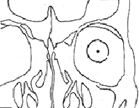
Figure 7 Schematic diagram of X-ray localization (anteroposterior view)
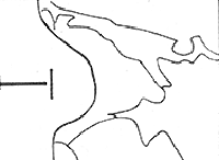
Figure 8 Schematic diagram of X-ray localization (lateral view)
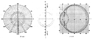
Figure 9 Intraocular foreign body localization measuring device
Recording method: After measuring the three aforementioned parameters, they can be plotted on an intraocular foreign body localization chart. This clearly visualizes the spatial position of the foreign body within the eyeball and its relationship to the eyeball wall.
Correction method: During radiography, eyeball deviation may occur, particularly during anteroposterior imaging, potentially causing errors in measuring the foreign body's meridian and distance from the sagittal axis. To correct these errors, two simple methods are available. When conditions permit, computer calculations may also be used.
① Vertical correction method: In addition to the anteroposterior radiograph, take a vertical radiograph using a similar technique. The patient lies prone with the head moderately extended, forming a 30° angle between the canthus line and the table. The eye gazes straight ahead, and the X-ray tube projects vertically from above, aligning the central beam with the corneal plane. Maintain a target-film to eye-film distance ratio of 10:1. If this proves difficult, ratios of 5:1 or 4:1 may be used, in which case the vertical scale on the intraocular foreign body locator can be employed for measurement. Measure the foreign body's position in millimeters nasal or temporal to the sagittal plane, then determine its position above or below the horizontal section of the eyeball from the original lateral radiograph. Correction can then be performed using calculation, drawing methods, or the intraocular foreign body localization chart.
②Indicator Rod Correction Method: If the eyeball is deviated when taking the anteroposterior film, the projection of the indicator rod will not appear as a circular dot but as an ellipse or elongated shape. Based on the extended length (L) of the indicator rod's projection and the distance (P) between the foreign body and the sagittal axis on the lateral film, the correction distance (d) is calculated. The formula is as follows: d = L · P / 20. After calculating d, move the distance d from the center of the positioning ring to the opposite side of the extended projection of the indicator rod to determine the new center. Using this new center as the origin of the coordinates for measurement, the measured meridian of the foreign body and its distance from the sagittal axis will be the corrected accurate values.
③Computerized Localization and Correction Calculation Method: By utilizing computer programming, only the required data needs to be measured from the skewed photograph. After inputting the data into the computer, it prints out five pieces of information: the meridian position of the foreign body, the perpendicular distance from the foreign body to the corneoscleral limbus plane, the distance from the foreign body to the sagittal axis, the distance from the foreign body to the outer surface of the eyeball, and the optimal incision location. If the axial length of the eye is measured using B/A ultrasound, a dynamic model of the eyeball can be generated, and the foreign body can be plotted on the eyeball diagram.
⑵ Geometric Localization Method
First Film: Similar to the lateral film in the direct localization method, a contact lens-type locator with an indicator rod is placed, but the affected eye should be positioned as close as possible to the film cassette (the eye-film distance is about 4 cm). The X-ray tube is raised as much as possible, with a target-eye distance of 100 cm. Second Film: Quickly change the film (or use a double-exposure method without changing the film), ensuring the patient's head and eye remain completely still. Move the X-ray tube toward the patient’s feet by half the target-eye distance (i.e., 50 cm). Tilt the tube to aim at the eyeball (the tube tilt angle should be 26°34′), and take the second lateral film.
Measurement and Calculation Method: On both lateral films, draw the extension lines of the indicator rod’s projection, representing the horizontal plane of the eyeball. Measure the perpendicular distances of the foreign body from the horizontal plane on both films. The measurement from the first film is denoted as ±a, and from the second film as ±a′, with "+" indicating the foreign body is above the horizontal plane and "−" indicating below. Substitute into the formula:
b = [(±a′) − (±a)] × 2 (Note)*
The value of b obtained from the formula represents the distance of the foreign body to the nasal or temporal side of the eyeball’s sagittal plane. If b is "+," the foreign body is on the nasal side; if "−," it is on the temporal side. The values of ±a and ±b can be plotted on the intraocular foreign body localization record chart. Additionally, by marking the distance of the foreign body behind the corneoscleral limbus, the spatial position of the foreign body within the eyeball can be accurately represented.
⑶ Physiological Localization Method (Physiological Methods of Localization): Also known as the eyeball rotation localization method. This method is used when the reading of the film shows the foreign body is exactly on the eyeball wall, and it is uncertain whether it is intraocular or extraocular. The common approach is: Take two lateral films using the same method as the direct localization lateral film. For the first film, have the patient look upward; for the second film, have the patient look downward. Compare the distances of the foreign body from the eyeball’s horizontal plane on both films. If identical, the foreign body moves entirely with the eyeball and may be intraocular or on the eyeball wall. If different, the foreign body must be extraocular.
⑷ Thin-Bone Localization Method (Thin-Bone Localization): For very small foreign bodies or those with low density that produce faint shadows and are not clearly visible on standard anteroposterior and lateral X-ray films, the thin-bone localization method can be used.
Filming Method: The patient lies prone on the examination table, first positioning the head in a posteroanterior view, then rotating the face 45° toward the affected side. At this point, have the affected eye look downward (i.e., inward rotation of 45°) and align the X-ray center with the eyeball’s sagittal axis to take the anteroposterior film. Then, have the affected eye gaze horizontally toward the affected side (i.e., outward rotation of 45°) and align the X-ray center with the corneoscleral limbus plane to take the lateral film. The filming distance and measurement methods for reading the films are similar to those of the direct localization method’s anteroposterior and lateral films.
(5) Bone-free method: For extremely small foreign bodies that cannot be visualized even by the thin-bone localization method, the bone-free method can be employed for localization. A small film cut into a tongue shape, 15–20 mm wide, is wrapped in black paper and placed inside a rubber finger cot or a specially made latex sleeve. It is then inserted into the upper or lower fornix or the medial/lateral canthus. This ensures that the X-rays pass only through the eyeball without traversing any bone, resulting in a clear image of the foreign body while simultaneously displaying the contour of the anterior part of the eyeball. Multiple films can be successively inserted at different locations, and multiple exposures can be taken.
3. Computed Tomography (CT): This method can display both metallic and non-metallic foreign bodies while clearly delineating the contours of the eyeball wall, making it a novel and specialized localization technique.
CT is more sensitive than plain X-ray films for detecting radiopaque and semi-radiopaque intraocular foreign bodies. CT can identify heavy metal radiopaque foreign bodies such as copper and iron as small as 0.06mm3. CT cannot directly visualize wood or mineral fragments but can reveal granulomatous reactions or localized air density around the foreign body.
CT axial and coronal imaging can clearly define the relationship between the foreign body and the eyeball, determining whether the foreign body is intraocular or orbital. It also identifies the foreign body's relationship to the optic nerve, which is clinically significant.
CT has the following limitations in foreign body diagnosis:
(1) Cannot detect radiolucent foreign bodies.
(2) Foreign bodies may be obscured by surrounding hemorrhage, inflammatory exudate, abscess, or granuloma, leading to misdiagnosis.
(3) Large metallic foreign bodies can produce artifacts, degrading image quality.
4. Magnetic Resonance Imaging (MRI): This method provides clearer images than CT but is generally unsuitable for magnetic foreign bodies. Ferromagnetic foreign bodies are contraindicated for MRI as they may migrate, causing further intraocular tissue damage.
Non-magnetic foreign bodies, depending on their hydrogen atom content and T1 and T2 relaxation times, may exhibit various signals on MRI. MRI can detect radiolucent foreign bodies like wood or plant matter, which are invisible on CT. It is also more sensitive than CT in displaying granulomatous reactions, hemorrhage, or orbital emphysema around foreign bodies.
5. Ultrasonic Localization: The advantages and clinical significance of ultrasound have been previously discussed. A-mode scanning can accurately measure the distance between the foreign body and the eyeball wall, which is clinically essential. Additionally, when the same foreign body is detected from different angles, the intersection of the transducer's extended axes indicates its location, helping estimate its meridional and anteroposterior position. B-mode scanning not only locates the foreign body and its relation to the eyeball wall but also reveals its size and approximate shape.
6. Electromagnetic Localization: Also known as the induction test, this method involves pressing an electromagnetic locator against the closed eyelid and rotating it in various directions to detect intraocular foreign bodies and approximate their location. Ferrous foreign bodies can be detected within 10 times their diameter, while non-magnetic metals like copper, aluminum, or lead are detectable only within 1–2 times their diameter. Another function is distinguishing magnetic from non-magnetic foreign bodies based on sound variations when the probe approaches. A sterilized rubber sheath over the probe enables intraoperative localization, serving as an important surgical aid.
7. Intraoperative Auxiliary Localization: Various auxiliary methods are used during intraocular foreign body removal to enhance accuracy, minimize surgical injury, and improve visual recovery. Common techniques include: (1) Scleral surface indentation localization, (2) Diathermy localization, (3) Transillumination localization, (4) Reverse transillumination localization, (5) Localization using inherent scleral landmarks, (6) Electromagnetic localization, (7) Direct scleral ultrasound localization, (8) Television X-ray examination, (9) Magnetic tests (black dot test and jump test), and (10) Grid localization.
1. Diagnosis of Intraocular Foreign Bodies
The diagnosis of intraocular foreign bodies is based on the following:
1. Medical history: A history of trauma, particularly injuries caused by hammering or explosions, carries the highest likelihood of intraocular foreign bodies. Additionally, flying debris from machinery and various projectiles from gunshots are common causes. Puncture wounds from branches, bamboo splinters, thin wooden sticks, or fine metal wires may also result in broken tips remaining inside the eyeball.
2. Penetrating eye injury: The entry of a foreign body into the eye inevitably causes a penetrating injury. Therefore, a penetrating eye injury is a crucial and necessary indicator for diagnosing an intraocular foreign body.
3. Manifestations of the foreign body or its pathway
(1) Anterior chamber foreign body: Most anterior chamber foreign bodies are located on the surface of the iris or the posterior layer of the cornea. A few may reside within the layers of the iris and are difficult to detect.
(2) Lens foreign body: Foreign bodies on the lens or its capsule are easily detected. If the lens has grade I opacity or the foreign body is transparent, making it hard to identify, an ophthalmoscopic examination (transillumination) can be used. The foreign body will cast a shadow due to light obstruction. For severe lens opacities, special diagnostic methods such as X-ray, ultrasound, CT, or MRI are required.
(3) Ciliary body foreign body: Except for foreign bodies located in the posterior part of the ciliary body's flat portion, which may be visible with indirect ophthalmoscopy combined with scleral indentation, other cases require special diagnostic methods.
(4) Anterior vitreous foreign body: Good focal illumination or slit-lamp microscopy can easily detect these. Those near the eyeball wall require indirect ophthalmoscopy or a three-mirror examination. However, if vitreous hemorrhage, opacity, traumatic internal visual obstruction, corneal opacity, iris adhesion, or non-dilating pupils prevent direct visualization, the aforementioned special diagnostic methods are necessary.
(5) Posterior eyeball foreign body: If the refractive media remain clear, foreign bodies or organized masses containing them may often be found in the posterior vitreous, retina, or optic disc. Only about 20–25% of posterior segment foreign bodies can be directly visualized with these methods; the majority require special diagnostic techniques for confirmation. Even when a foreign body (or an organized mass containing it) is directly observed, further confirmation via X-ray, ultrasound, electromagnetic locators, or magnetic testing is often needed.
(6) Detection of the foreign body pathway: If the foreign body cannot be directly seen, locating its entry pathway into the eye, combined with medical history, can aid diagnosis. For example:
① A fresh wound or healed penetrating wound (full-thickness scar) on the cornea, with no trauma to the iris or lens and a history of debris injury, suggests the foreign body may be in the anterior chamber or anterior chamber angle.
② If the cornea has such a wound or healed wound, with a corresponding perforation on the iris but no lens injury, the foreign body may be in the posterior chamber.
③ Corneal injury with localized or extensive opacity in the corresponding area of the lens suggests the foreign body is likely within the lens or has passed through it to a posterior location. Foreign body injuries in the pupillary area may bypass the iris, passing only through the cornea and lens.
④ Vitreous pathway: If the cornea, iris, and lens show corresponding injuries but most of the lens remains clear, or if the entry point is on the sclera, the vitreous pathway should be carefully examined. This appears as an oblique band with scattered pigment.
(7) Detection of retinal injury: The presence of typical retinal injury strongly suggests a foreign body.
After identifying the foreign body pathway or retinal injury, the trauma history must be considered. If the injury was caused by debris, the likelihood of a retained foreign body is higher, but special diagnostic methods are still required for confirmation.
2. Special Diagnostic Methods for Intraocular Foreign Bodies:
There are seven special diagnostic methods for intraocular foreign bodies:
1. Magnetic test method: Use an electromagnet to test suction in front of and around the eyeball. Determine whether there is a magnetic foreign body in the eyeball based on whether the affected eye feels pain and whether there is pulsation at the tested suction site of the eyeball.
2. Inductive Testing Method: Also known as electromagnetic positioning, the instrument used operates on the same principle as a mine detector, functioning as a miniature metal detector. If a metal object is present within the magnetic field formed at the front of its probe, an induction occurs. This is then amplified and indicated by needle deflection and sound signals. This method is particularly suitable for metallic foreign bodies, especially magnetic ones.
3. X-ray Examination Method: Typically involves X-ray imaging to diagnose intraocular foreign bodies. Standard imaging includes lateral and posteroanterior views of the skull and eyeball. The presence, size, shape, and approximate location of the foreign body can be determined from the radiographs. If localization imaging is performed, the exact position of the foreign body can be identified. This method is suitable for metallic foreign bodies, larger stone fragments, and glass.
4. Computed Tomography (CT): Can provide clearer visualization of the foreign body and its location. Suitable for both metallic and most non-metallic foreign bodies.
5. Magnetic Resonance Imaging (MRI): Offers even greater clarity than CT but is unsuitable for magnetic foreign bodies unless they are very small.
6. Ultrasonic Examination Method: Both A-scan and B-scan ultrasonography can diagnose foreign bodies. Due to its convenience and broad applicability, its use has become increasingly widespread. Ultrasonography can not only detect various metallic foreign bodies but also clearly visualize many non-metallic foreign bodies that are invisible on X-rays.
7. Chemical Analysis Method: Determines the nature of the foreign body by analyzing the increased copper content in ocular tissues and aqueous humor caused by copper foreign bodies.
bubble_chart Treatment Measures
The extraction of intraocular foreign bodies varies depending on whether the foreign body is magnetic or not. The removal of the foreign body aims to restore and preserve vision, with the extraction surgery being a means rather than an end. All procedures should be performed with extreme caution to minimize tissue injury and create conditions for the restoration and maintenance of vision.
1. Extraction of Anterior Chamber Foreign Bodies
(1) Extraction of anterior chamber foreign bodies: Generally, anterior chamber foreign bodies can be extracted by making an incision along the corneal limbus at the meridian where the foreign body is located and using an electromagnet to suction it out.
For foreign bodies behind the cornea, the pupil can first be constricted. Then, a careful incision is made on the cornea at the location of the foreign body, and it is suctioned out.
(2) Extraction of posterior chamber foreign bodies: If there is already traumatic cataract, a loop incision can be made at the corneal limbus opposite the foreign body, and the electromagnet can be used to suction the foreign body at the incision or slightly beyond. If the lens is completely clear, an incision can be made at the corneal limbus where the foreign body is located, and the root of the iris can be incised to suction out the foreign body.
(3) Extraction of lens foreign bodies: If there is already a cataract, the foreign body can be removed during cataract extraction surgery. During extracapsular extraction, the foreign body can be removed first, followed by the cataract, to prevent loss of the foreign body or its entrapment in the anterior chamber angle during surgery. After removing the foreign body and the cataract, an intraocular lens can be implanted.
If the lens is still clear, an electromagnet (sometimes a giant electromagnet is needed) can be used during surgery to suction the foreign body to a position 2–3 mm below the anterior capsule near the anterior pole. Then, it is suddenly made to jump, piercing the anterior capsule and entering the anterior chamber. The pupil is immediately constricted, and the foreign body is extracted using the method for anterior chamber foreign bodies. Postoperatively, the pupil is kept constricted for several days to form posterior synechiae, sealing the wound in the anterior capsule and preventing traumatic cataract. Alternatively, cyanoacrylate or plasma can be used to seal the wound in the anterior capsule.
(4) Extraction of ciliary body foreign bodies: Foreign bodies on the anterior surface of the ciliary body can be extracted by making an incision at the corneal limbus, similar to the method for posterior chamber foreign bodies. For foreign bodies in other parts of the ciliary body, an incision can be made on the sclera closest to the foreign body for extraction.
(5) Extraction of floating vitreous foreign bodies: An appropriate position is selected, and an incision is made at the pars plana of the ciliary body along the corresponding meridian. The foreign body is then suctioned out using an electromagnet (Figures 1, 2). If necessary, the magnetic rod relay method can be used. If the vitreous is opaque and the foreign body is not visible, a vitrectomy is performed first, and the magnetic rod is introduced once the foreign body becomes visible.
Method for the magnetic rod relay: A specially designed magnetic rod (a soft iron rod with a diameter of 0.9 mm or 1.5 mm and a length of 50 mm) is inserted through the pars plana of the ciliary body. Under the guidance of a surgical microscope or direct/indirect ophthalmoscope, the rod is brought close to the foreign body. The electromagnet is then applied to the external part of the rod, causing the foreign body to be attracted to the tip of the rod. The rod is slowly withdrawn, and the electromagnet is moved to the incision to suction out the foreign body (Figure 3).
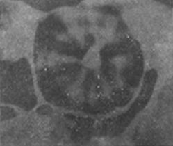
Figure 1: Method for extracting magnetic foreign bodies
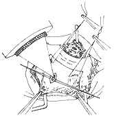
Figure 2: Extraction of floating vitreous foreign bodies.
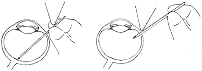
Figure 3: Magnetic rod relay method
(6) Extraction of foreign bodies adherent to the eyeball wall
1) Conventional posterior approach extraction method
Positioning suture: A marker is placed at the corneal limbus corresponding to the meridian where the foreign body is located (e.g., 210°, or 8 o'clock position). Then, a positioning suture is placed at the opposite corneal limbus (30°, or 2 o'clock position).
Conjunctival incision: Incise the bulbar conjunctiva along the meridian where the foreign body is located, make an incision along the limbus with a length of 1/2 the circumference or slightly more, and perform 1-2 radial incisions. Then, separate the bulbar conjunctiva and the Tenon's capsule posteriorly.
Temporarily detach the extraocular muscles.
Place traction sutures: ① Straighten the positioning suture, pass it across the cornea, follow the marked line, and run it straight along the surface of the sclera posteriorly. ② Measure the perpendicular distance from the corneal limbus using a perpendicular distance gauge based on the distance between the foreign body and the corneal limbus. It is best to use the calculated optimal incision site as the reference.
Auxiliary localization: Use the aforementioned auxiliary localization methods to confirm the position of the foreign body. Typically, the magnetic test is performed first. If the result is positive, other methods may not be necessary. If the result is negative, reverse transillumination localization or electromagnetic localization can be used. For ophthalmoscopically visible foreign bodies, scleral indentation, transillumination localization, or diathermy localization can be employed. If all these methods yield negative results, grid localization can be used as a last resort. For foreign bodies in or near the posterior pole, scleral anatomical landmarks can be used for localization, but other methods must also be applied for confirmation.
Partial scleral incision: Make a meridional incision, the length of which depends on the size of the foreign body. For large and thick foreign bodies, a "T"-shaped incision may be made (Figure 4).
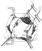
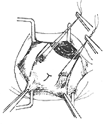
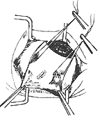
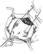
Figure 4: Posterior approach extraction method
Diathermy or cryotherapy: Perform diathermy or cryotherapy on both sides of the incision to prevent retinal detachment and reduce intraoperative bleeding.
Preplaced sutures: Depending on the size of the incision, place a single-knot suture or mattress suture in advance.
Incise the eyeball wall: Use an extremely sharp blade to incise the remaining sclera, choroid, and retina.
Extract the foreign body: Use an electromagnet (or permanent magnet) to extract the foreign body through the incision.
Complete the surgery: Tie the preplaced sutures, reattach the detached extraocular muscles, and suture the conjunctival incision.
2) Transvitreal extraction method: For foreign bodies located in the posterior eyeball wall, especially those at the posterior pole where the posterior approach is difficult, transvitreal extraction can be performed. The method is as follows: Make three incisions in the pars plana as in vitrectomy, insert the infusion cannula, and introduce the endoilluminator. Under the surgical microscope, use a contact lens to visualize the foreign body, dissect it to detach it from the retina, and move it into the vitreous. To prevent bleeding, underwater diathermy may be applied around the foreign body if necessary. Then, introduce a relay magnetic rod through the third incision and extract the foreign body as described earlier. If the foreign body is obscured due to preexisting vitreous opacity or traumatic white internal visual obstruction, perform vitrectomy or lensectomy first to clearly visualize the foreign body. After extraction, perform vitrectomy at the instrument entry and exit sites to prevent future proliferative vitreoretinopathy. Since this method does not involve penetrating the retina during extraction, it reduces retinal injury and the risk of retinal detachment.
(7) Extraction of optic disc foreign bodies
1) Magnetic rod relay extraction method.
2) Two-step extraction method: Step 1: Use a handheld electromagnet or giant magnet to repeatedly attempt extraction, causing the foreign body to detach from the optic disc and enter the vitreous. Step 2: Extract the foreign body using the floating foreign body extraction method in the vitreous. This method can also be used for extracting foreign bodies on the surface of the posterior pole retina, especially if the injury is recent and the foreign body is not firmly fixed to the retina. First, detach the foreign body from the retina, then extract it.
2. Extraction of non-magnetic foreign bodies
⑴ Extraction of non-magnetic foreign bodies in the anterior and posterior chambers: Foreign bodies in the iris can be removed through a corneal limbal incision using small forceps. For foreign bodies in the posterior chamber, an iridectomy is required to expose and extract the foreign body. For foreign bodies located in the posterior layer of the cornea, if deeply embedded, a corneal flap can be created in the anterior layer to lift and expose the foreign body. If most of the foreign body is in the anterior chamber with minimal corneal embedding, a needle aspiration method can be used. This involves inserting a needle through a partial incision at the corneal limbus, approaching the foreign body with the bevel, and aspirating it out.
(2) Extraction of non-magnetic foreign bodies in the lens: If there is already traumatic cataract, the foreign body can be removed simultaneously during cataract surgery. During the operation, the foreign body can be extracted first after capsulotomy, followed by lens delivery. If absolutely necessary, intracapsular extraction can also be performed, removing the foreign body along with the lens.
If the lens remains transparent, surgery is not urgent. Close observation is sufficient, and if no metallosis occurs, surgery is unnecessary.
(3) Extraction of non-magnetic foreign bodies in the ciliary body: An incision is made on the sclera corresponding to the foreign body, and the foreign body is carefully located and removed. For smaller foreign bodies, auxiliary localization methods such as electromagnetic locators or grid localization may be required.
(4) Extraction of floating non-magnetic foreign bodies in the vitreous: If visible under an ophthalmoscope, the preoperative positioning should be carefully selected to find an appropriate posture where the foreign body is relatively far from the retina, closer to the anterior part, easy to observe, and accessible. Ideally, it should be visible under oblique illumination. During the operation, a claw-shaped foreign body forceps is inserted through an incision in the pars plana of the ciliary body in the selected posture to carefully capture the foreign body. If not visible under oblique illumination, the surgery can be performed under observation with a surgical microscope (equipped with various contact lenses).
If the vitreous is too opaque to directly visualize the foreign body, a vitrectomy should be performed first, followed by foreign body extraction. Alternatively, only a vitrectomy may be performed initially, with the foreign body removed in a second surgery once it becomes visible.
(5) Extraction of non-magnetic foreign bodies adjacent to the retina
1) Grid localization extraction: This is the standard procedure for removing such foreign bodies. The method involves suturing a grid locator (Figure 5) onto the sclera at the site of the foreign body based on preoperative localization results. X-ray imaging (typically an anteroposterior and lateral view of the locator) is performed. The precise location of the foreign body can be determined from the anteroposterior view, while the lateral view shows the distance between the foreign body and the eyeball wall. The foreign body is then extracted by incising the eyeball wall at this location. After incising the eyeball wall, possible scenarios and corresponding approaches are briefly described as follows:

Figure 5: Grid localization method for foreign body extraction
1. For small foreign bodies, the incision is perpendicular. 2. For thin and elongated foreign bodies, the incision is offset to align with the foreign body. 3. For larger foreign bodies, the incision matches the foreign body.
If the blade touches the foreign body, the incision is halted. By retracting the incision with pre-placed sutures, the foreign body becomes visible and can be carefully grasped.
After incising the transparent eyeball wall, the foreign body may spontaneously exit through the incision.
The foreign body, along with the organized tissue encapsulating it, may spontaneously exit through the incision.
If a large mass of white organized tissue or part of it protrudes from the wound, this mass should be grasped and dissected to locate the foreign body within.
If a bead of transparent vitreous prolapses from the incision, flattening this bead may reveal the foreign body inside the incision. The vitreous bead can then be excised, and the foreign body quickly grasped afterward.
If the incision is incomplete and only a small opening is made, a large amount of liquefied vitreous may escape, causing the eyeball to soften and collapse. In this case, the pre-placed sutures should be lifted, and the incision quickly completed with scissors. The foreign body should then be carefully searched for within the incision or along its edges. Often, the foreign body or its encapsulating tissue can be seen on the inner surface of one of the wound edges.
If, after incising the eyeball wall, the incision is entirely filled with gray-white organized tissue and the foreign body or its encapsulating mass is not visible, the exposed organized tissue should be carefully dissected layer by layer with forceps. The foreign body is often found during this process. Occasionally, a pus-filled cavity may appear during dissection, and the foreign body is usually located within this cavity.
The search for the foreign body primarily involves repeated careful observation under focal illumination. At no point should forceps be blindly inserted into the incision for trial grasping.
2) Extraction method via the vitreous: Similar to the method of extracting magnetic foreign bodies through the vitreous, following the vitrectomy approach, the fiber optic and separator are inserted through the pars plana to peel the foreign body off the retina, and then the foreign body forceps are used to grasp and remove it.
3) Electromagnetic locator positioning extraction method: For relatively large non-magnetic metal foreign bodies, an electromagnetic locator can also be used for silent positioning. The possible situations encountered after determining the location and incising the ocular wall, as well as the methods adopted, are the same as those in the grid positioning extraction method.
3. Postoperative therapy after intraocular foreign body removal
(1) Prevent infection, control inflammation, stop bleeding, and relieve pain.
(2) For incisions made at the corneal limbus or the pars plana of the ciliary body, the patient should rest in bed for 1–2 days postoperatively. Examine the anterior segment and fundus; if the anterior chamber has formed or the fundus shows no abnormalities, the patient may get out of bed and move around.
(3) For surgeries involving retinal incisions, the patient should rest in bed for 4–10 days. Only after confirming the absence of retinal detachment may the patient get out of bed and move around, and the healthy eye can be uncovered.
(4) For patients who simultaneously undergo cataract surgery or vitrectomy, postoperative management should follow the routine protocols for these surgeries.
(5) Postoperatively, closely monitor for common complications such as vitreous hemorrhage, hyphema, vitreous organization formation, and retinal detachment. If any occur, manage them according to the standard protocols for each condition.
(6) Patients who undergo retinal incision for foreign body removal should avoid heavy physical labor, vibrations, and strenuous activities for six months postoperatively.
Discovery of complications from intraocular foreign bodies: Intraocular foreign bodies that remain for a long time often lead to certain complications. In such cases, diagnosis can be based on these complications and then confirmed by other methods. Common complications include the following:
1. **Ocular siderosis (siderosis bulbi)**: Iron foreign bodies retained for days to months can cause iron deposition, initially appearing around the foreign body and later spreading to various tissues within the eye, presenting as fine brownish-yellow granular deposits. In the cornea, deposits are mostly found in the stromal layer, particularly in the peripheral regions. The iris turns brown, and over time, iris atrophy, posterior synechiae, moderate pupillary dilation, and weakened or absent light reflexes may occur. In the lens, brown granules first appear beneath the anterior capsule or form round or oval spots, followed by diffuse cortical opacification with a brownish-yellow hue. The vitreous becomes liquefied and turbid, appearing brownish. The retina is susceptible to damage, leading to degeneration and atrophy, manifested as reduced vision and visual field constriction.
2. **Ocular chalcosis**: Copper entering the eye can lead to increased copper levels in the aqueous humor within hours. However, clinical manifestations of chalcosis typically appear months or longer after the injury. The severity of chalcosis correlates with the copper content of the foreign body, with copper exceeding 85% causing severe damage. Pure copper can induce acute sterile suppuration. When the foreign body is encapsulated by fibrous tissue, chalcosis is relatively milder. Small copper particles in the iris often do not cause chalcosis. In the cornea, chalcosis is most prominent in the peripheral Descemet’s membrane, clinically presenting as the typical **Kayser-Fleischer ring**. The iris also appears yellowish-green, with moderate pupillary dilation and sluggish reaction. The lens may develop yellowish-green granular deposits beneath the capsule or on the posterior capsule surface. A classic lens change is **sunflower cataract**, where a gray-yellow disc-shaped opacity forms in the central subcapsular cortex, surrounded by radiating petal-like opacities. Over time, this may progress to total cataract. The vitreous may contain a bright golden reflective mass resembling the foreign body itself, but it moves rapidly with changes in light direction. Slit-lamp microscopy reveals numerous fine deep yellow-green particles floating with eye movement. Along retinal vessels, golden reflexes may appear, while yellow-green particles elsewhere may gradually disappear. The macula may become a gray-white atrophic area, and the lens may turn milky white. Some copper foreign bodies (with very high copper content) may gradually migrate forward, causing sterile suppuration and eventually perforating the sclera to exit the eye, or they may move to the anterior chamber angle.
3. **Iridocyclitis**: Unexplained unilateral iridocyclitis or panuveitis with chronic inflammation warrants a detailed history of trauma and further examinations to confirm or rule out the presence of an intraocular foreign body.
4. **Cataract**: Unexplained cataract in young or middle-aged adults may sometimes be caused by an intra-lenticular foreign body or a foreign body that has traversed the lens.
5. **Other complications**: Unexplained vitreous opacities with fibrous membranes or strands, unilateral secondary retinal detachment, or unexplained unilateral secondary glaucoma should also raise suspicion of retained intraocular foreign bodies, prompting appropriate investigations.


