| disease | Congenital Diseases of the Trachea |
Congenital tracheal diseases are relatively rare and often associated with congenital malformations of other organs and tissues. Severe tracheal lesions can lead to death at birth, making them uncommon in clinical practice.
bubble_chart Clinical Manifestations
Tracheal atresia or absence: In such cases, the infant dies at birth, but the larynx and lungs may develop normally, and sometimes the bronchus communicates with the esophagus.
Tracheoesophageal fistula: This malformation is relatively common, with an incidence of approximately 1 in 3,000 newborns. Durston reported the first case of congenital esophageal atresia in 1670. Gibson described congenital esophageal atresia with tracheoesophageal fistula in 1697. In 1939, Ladd and Levin successfully performed staged corrective surgery on two patients. In 1941, Haight and Towsley reported the first successful initial stage [first stage] corrective surgery.
Embryology: Both the digestive and respiratory tracts originate from the foregut of the embryonic primitive gut. The primitive esophagus is located posterior to the respiratory organs. The primitive gut is divided into three parts: the foregut, midgut, and hindgut. In the early stages, both the cephalic and caudal ends of the primitive gut are closed. By the end of the third week of embryonic development, the pharyngeal membrane at the cephalic end of the primitive gut ruptures, connecting the foregut to the oral cavity. As the heart shifts downward, the length of the esophagus rapidly increases. Between the 21st and 26th days of embryonic development, the laryngotracheal groove appears on both sides of the foregut, followed by epithelial growth forming the esophagotracheal septum, which separates the esophagus from the trachea. If the esophagus and trachea do not fully separate, a communication between their lumens forms a tracheoesophageal fistula. If the esophagotracheal septum deviates posteriorly or the foregut epithelium overgrows into the esophageal lumen, esophageal atresia occurs. Additionally, during the early development of the esophagus, some foregut cells may separate from the esophagus and continue to grow, leading to esophageal duplication anomalies, most commonly presenting as cysts near the esophageal wall, some of which may communicate with the esophageal lumen.
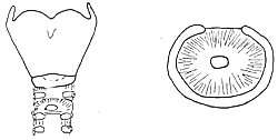
Tracheal web: A thin connective tissue membrane is present within the tracheal lumen, with a small central opening for air passage. Tracheal webs are often located below the cricoid cartilage. After diagnosis by tracheal tomography and endoscopy, the membrane can be excised via bronchoscopy. For long and thick membranes, a tracheostomy is first performed below the web, and a tube is inserted; the web is excised later when the patient grows older (Figure 1).
Figure 1: Tracheal web
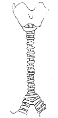
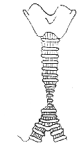
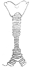
Congenital tracheal stenosis: The tracheal wall develops normally, but the lumen is narrowed. The extent and morphology of the stenosis can be classified into three types (Figure 2): ① Generalized tracheal stenosis: The internal diameter of the cricoid cartilage is normal, but the tracheal lumen below it is narrowed along its entire length, with the most severe narrowing above the carina, sometimes measuring only a few millimeters in diameter. The main bronchi are normal. ② Funnel-shaped stenosis: This can occur in the upper, middle, or lower trachea. The length of the stenotic segment varies. The tracheal diameter above the stenosis is normal, while the lumen gradually narrows into a funnel shape. ③ Short-segment stenosis: This often occurs in the lower trachea, with varying lengths of the narrow segment, and may be associated with bronchial anomalies. This type is the most common.
 |  |  |
| (1) Generalized tracheal stenosis | (2) Funnel-shaped stenosis | (3) Short-segment stenosis |
Figure 2: Congenital tracheal stenosis
Obstructive breathing difficulties of varying severity are present from birth. Stridor may occur during inhalation, accompanied by feeding difficulties and delayed growth and development. In cases with severe stenosis, suprasternal, intercostal, and subxiphoid soft tissue retractions are observed during inhalation. These symptoms worsen with concurrent respiratory infections. Diagnosis can be confirmed through X-ray tracheal tomography and endoscopy, although tracheography carries the risk of exacerbating the obstruction.
bubble_chart Treatment MeasuresFor mild cases of stenosis, a moving qi tube dilation procedure can temporarily improve symptoms, or a catheter can be inserted via tracheotomy. Short-segment tracheal stenosis or short-segment fistula disease with funnel-shaped stenosis may be treated with partial resection of the moving qi tube and end-to-end anastomosis. In infants and young children, the tracheal lumen is small, and postoperative mucosal edema can lead to tracheal obstruction, with an extremely high surgical mortality rate. Additionally, the anastomotic site may remain narrower than normal even after growth. Therefore, surgery should be postponed as much as possible until the child is older.


