| disease | Achondroplasia |
| alias | Chondrodystrophic Dwarfism, Fetal Chondrodystrophy |
Achondroplasia, also known as fetal chondrodystrophy (chondrodystrophia fetalis) or chondrodystrophic dwarfism, is a congenital developmental disorder caused by defective endochondral ossification, primarily affecting the long bones. Clinically, it manifests as a specific type of dwarfism—short-limbed dwarfism. Patients typically exhibit normal intellectual and physical development and are often employed as acrobatic clowns in theaters or circuses.
bubble_chart Etiology
It is a congenital developmental anomaly with evident hereditary and familial traits, inherited as an autosomal dominant disorder. If one parent is affected, 1/2 of the offspring may develop the condition; if both parents are affected, nearly all children will be involved. Due to many patients remaining unmarried or experiencing difficult deliveries, they may not have offspring, thus influencing the inheritance pattern. Therefore, sporadic cases account for 90% of sexually transmitted diseases. Of course, some cases may also result from gene mutations. In twins, one may be affected, or both may be affected, with females slightly outnumbering males.
bubble_chart Pathological Changes
The fundamental pathological changes occur in the process of endochondral ossification, where the longitudinal growth of long bones is impaired, while intramembranous ossification remains unaffected. As a result, the thickness of the bones remains normal, but their reduced length makes them appear relatively thicker. Epiphyseal cartilage cells can proliferate but fail to undergo normal calcification and ossification, leading to enlarged bone ends. Microscopically, the cartilage cells do not arrange in the regular columnar pattern as they normally would; instead, they are scattered and irregularly clustered. The distinct zones of the ossification process—such as the resting zone, proliferative zone, hypertrophic zone, and zone of provisional calcification—become disorganized. The metaphyseal capillaries fail to invade the epiphysis in an orderly manner, disrupting normal absorption. Mature cartilage cells cannot calcify, impairing bone growth. Additionally, widespread chondromyxoid degeneration can be observed, with swollen cells, enlarged nuclei, and a semifluid matrix structure. Ossification in the affected cartilage is delayed, appearing as patchy distribution, while calcification between these patches proceeds relatively normally.
bubble_chart Clinical Manifestations(1) Dwarfism This condition is the most common cause of dwarfism. At birth, the fetus exhibits normal body length but shorter limbs, a disparity that becomes more pronounced over time. The proximal limbs, such as the humerus and femur, are shorter than the distal bones. The child appears overweight. By maturity, the average height is 131±5.6 cm for males and 124±5.9 cm for females. Reports in the literature include heights of 97 cm and 104 cm. The midpoint of the body is above the navel, sometimes even at the lower end of the sternum. The hands can only reach below the lower tuberosity of the femur, unlike in normal individuals where they can reach the lower third of the thigh. Due to the short limbs, when the legs are extended, the face can touch the toes.
(2) Enlarged Head Some patients exhibit grade I hydrocephalus, with prominent vault and forehead, a saddle-shaped nasal bridge, flat nose, thick lips, and protruding tongue (in infants).
(3) Thoracic Kyphosis and Lumbar Lordosis The latter is more pronounced. The sacrum is more horizontal, causing the characteristic protrusion of the buttocks.
(4) Flat and Small Thorax The ribs are abnormally short.
(5) Thick, Short, and Splayed Fingers Often, the fourth and fifth fingers form one group, the second and third another, and the thumb stands alone, resembling a "trident" (Figure 1). Some patients exhibit grade I limitation in elbow extension.
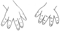
Figure 1 Trident-shaped finger deformity in achondroplasia
(6) Bowlegs and Rolling Gait The lower limbs are bowed, resulting in a rolling gait.
(7) Normal Intelligence, Good Teeth, Strong Muscles, and Normal Sexual Function Intellectual development is normal, teeth are healthy, muscle strength is strong, and sexual function is unaffected.
X-ray Findings: ① The cranial vault is large, with a prominent forehead, parietal, and occipital bones, but the cranial base is short. The foramen magnum is smaller and funnel-shaped, possibly only half the diameter of a normal person. If hydrocephalus is present, the lateral ventricles are dilated. ② Long bones are shortened, with thickened shafts and narrowed medullary cavities. The epiphyses may appear fragmented or irregular. At the knee joint, the bone ends often separate in a "V" shape, with the ossification center of the epiphysis fitting into this V-shaped notch. Due to the ossification center being close to the shaft, the joint space appears widened. The lower limbs are bowed, with the fibula longer than the tibia, and the ulna longer than the radius in the upper limbs. ③ Vertebral body thickness is reduced, but the overall shortening of the spine is much less pronounced compared to the limbs. The distance between the pedicles gradually decreases from the first to the fifth lumbar vertebra. Myelography reveals a narrow spinal canal with multiple posterior disc protrusions. ④ The pelvis is narrow, with flat and rounded iliac bones, all diameters reduced. The acetabulum is displaced posteriorly, close to the sciatic notch, with coxa vara and asymmetry between the acetabulum and femoral head. The ribs are short, and the sternum is wide and thick. The scapular angle is not sharp, and the glenoid cavity is shallow and small.bubble_chart Treatment Measures
Generally, it is not difficult to differentiate typical cases from dwarfism caused by other reasons. 1. **Hypochondroplasia**: The manifestations of dwarfism are less pronounced, and the skull is normal. 2. **Chondro-ectodermal dysplasia (Ellis-Van Creveld syndrome)**: This is a short-limbed dwarfism accompanied by chest deformities, cardiac lesions, syndactyly, and poor development of nails and teeth. The shortening of limbs usually occurs in the distal bones. 3. **Spondylo-epiphyseal dysplasia**: Also a short-limbed dwarfism, often involving damage to large proximal joints. The skull is normal, but the vertebral bodies are flattened with fusion of ossification centers. The thorax is poorly developed, resembling a bell shape. 4. **Rickets and cretinism**: Rickets has typical clinical and radiographic features, making it easy to distinguish. Cretinism is often associated with intellectual developmental disorders.
bubble_chart PrognosisIf the infant does not die prematurely, they can perform various jobs as adults with a good prognosis. A small number of patients may develop hydrocephalus due to a smaller foramen magnum. The incidence of spinal canal stenosis can reach 40%, mostly in the lumbar region. Occasionally, it occurs in the cervical or thoracic spine, causing compression on nerve roots or the spinal cord, requiring laminectomy for decompression or foraminotomy. Rarely, osteotomy may be performed due to lower limb deformities. There is no specific treatment for the disease cause.


