| disease | Chronic Frontal Sinusitis |
When inflammation persists for more than 30 days after the onset of acute frontal sinusitis, it is called chronic frontal sinusitis. It often flares up acutely under certain conditions and is frequently accompanied by chronic ethmoid sinusitis.
bubble_chart Etiology
1. Acute frontal sinusitis that is not promptly managed or improperly treated can severely damage the mucous membrane, impairing its normal function and leading to chronic inflammation.
2. Allergic frontal sinusitis, edema of the nasofrontal duct mucous membrane, and reduced ciliary transport function obstruct drainage during acute inflammation, resulting in chronic inflammation.
3. High deviation of the nasal septum, hypertrophy of the middle turbinate, nasal polyps, or obstruction of the ostiomeatal complex drainage.
4. Barotrauma, such as rapid descent during flight, diving, or underwater work, can cause chronic infection of the frontal sinus.
5. Systemic factors, such as reduced immune function, diabetes, malnutrition, or vitamin deficiencies.
bubble_chart Pathological Changes
The pathological changes are generally similar to those of chronic maxillary sinusitis, including thickening of the mucous membrane, loss of cilia, and accumulation of pus in the sinus cavity. In cases of allergic inflammation, there is mucous membrane edema and polypoid changes. The difference lies in chronic frontal sinusitis, where poor drainage can easily lead to osteitis and osteomyelitis, potentially causing fistulas on the anterior wall and floor that continuously discharge pus. The fistulas are often located on the inner upper wall of the orbit, with visible scar formation on the upper eyelid.
bubble_chart Clinical ManifestationsDullness and distension in the frontal region, more pronounced on the affected side. If the frontal sinus drainage is obstructed, headache may occur, with possible referred headache in the trigeminal nerve distribution area. Nasal congestion is significant, often worse in the morning, and persistent nasal congestion on the affected side may also be present. Nasal discharge is mucopurulent or purulent, more abundant in the morning, often related to postural drainage. Hyposmia may occur. If frontal osteomyelitis develops, a purulent fistula may form on the forehead, usually located on the anterior wall or floor of the frontal sinus, where the bone contains marrow.
(1) Anterior rhinoscopy: Congestion of the mucous membrane can be observed, with purulent discharge present in the anterosuperior part of the middle meatus. In maxillary sinusitis, pus is often located in the {|to be decocted later|} area of the middle meatus, while in ethmoid sinusitis, purulent secretions may be seen in both the middle meatus and the olfactory cleft, which can aid in differentiation.
(2) Head-position test: If no purulent discharge is observed during anterior rhinoscopy, 1% Ephedrine can be used to shrink the middle turbinate and the mucous membrane of the middle meatus. The patient should then maintain an upright head position for 5 minutes, after which the nasal cavity is re-examined to check for the presence of pus in the middle meatus. For cases suspected of maxillary sinusitis, maxillary sinus puncture and irrigation may be performed first to remove the pus, followed by positional drainage to assess the presence of frontal sinusitis.
(3) Frontal sinus X-ray: Nasofrontal and lateral views are taken to compare the radiolucency of both frontal sinuses and evaluate any pathological changes. Asymmetry in the size of the bilateral frontal sinuses is normal and unrelated to the diagnosis of frontal sinusitis. A well-developed frontal sinus may also have bony septa, which is also a normal finding.(4) CT scan: Coronal and axial scans are performed to assess the size and extent of the frontal sinus, the condition of the anterior and posterior bony walls, and whether there is any thickening of the mucous membrane within the sinus cavity.
bubble_chart Treatment Measures
(1) Non-surgical therapy includes the use of nasal mucosal vasoconstrictors and antibiotic nasal drops, replacement therapy, physiotherapy, etc., which may only be effective for early mild cases.
(2) Intranasal surgery includes correction of high septal deviation, nasal polyp removal, partial middle turbinectomy, etc. This type of surgery is suitable for chronic suppurative frontal sinusitis that has not responded to non-surgical treatment, but it is not recommended for patients with a history of frontal sinus trauma or complications of frontal sinusitis. This surgery is also referred to as adjunctive surgery.
(3) Intranasal frontal sinus surgery: The patient is placed in a supine position, and either topical or general anesthesia is administered. A "V"-shaped incision is made on the lateral nasal wall near the nasal root, followed by mucosal dissection and removal of the uncinate process to open the anterior ethmoid sinuses. If the middle turbinate is hypertrophic, it should first be fractured and repositioned or partially resected. The posterior edge of the maxillary process is then chiseled away to widen the nasofrontal duct. Care must be taken to avoid the cribriform plate located posteromedially in the nasofrontal duct. Postoperatively, the mucosal flap is repositioned, and a 6mm silicone tube can be used for frontal sinus drainage, with irrigation performed after 6 days. This procedure is relatively simple, causes minimal mucosal injury, and is safer, with a lower risk of nasofrontal duct stenosis. Additionally, it leaves no forehead scars and avoids the need for more complex intranasal frontal ethmoid sinus surgery. If the results are unsatisfactory, an external frontal sinus surgery may be performed.
1. Lynch procedure
(1) Indications
① Cases where intranasal surgery or intranasal frontal sinus surgery has failed;
② Chronic frontal sinusitis complicated by osteomyelitis or fistula;
③ Chronic frontal sinusitis with intraorbital or intracranial complications;
④ Fungal frontal sinusitis;
⑤ Foreign bodies in the frontal sinus or frontal sinus fracture.
(2) Surgical procedure: The patient is placed in a supine position, and the face is disinfected with alcohol. Topical anesthesia is applied to the nasal mucosa, while the inner canthus and eyebrow arch are infiltrated with 1% procaine or lidocaine mixed with a few drops of 0.1% adrenaline. Eyebrows are not shaved, and the affected eye is not covered with a surgical drape to allow continuous monitoring of vision and ocular conditions during the procedure.
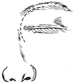
An incision is made along the eyebrow, with its medial end turning downward slightly below the plane of the inner canthus on the maxillary frontal process. Care is taken to avoid damaging the orbital wall while dissecting the periosteum. The lacrimal sac and the trochlea of the superior oblique muscle, located about 0.5cm deep at the superomedial angle of the orbit, are carefully dissected and displaced medially, then protected with a small gauze strip. After proper handling, the lacrimal bone and the lamina papyracea of the ethmoid bone are exposed medially, and the anterior ethmoid artery is ligated. The floor of the frontal sinus is chiseled away to access the sinus, and the diseased mucosa is dissected and removed. The maxillary frontal process, lacrimal bone, and lamina papyracea are further chiseled to complete ethmoid sinus opening. If necessary, the anterior wall of the sphenoid sinus may also be opened to facilitate drainage and treat inflammation. Finally, a 0.6cm-thick silicone drainage tube is inserted. The skin and subcutaneous tissue are sutured in two layers with silk thread. Before closing the incision, the trochlea of the superior oblique muscle must be repositioned to avoid postoperative diplopia (Figure 1).

Figure 1 Incision for radical frontal sinus surgery.
2. Frontal Sinus Anterior Wall Osteoplastic Flap Approach with Obliteration This procedure was first reported by Schonborn (1894) and Breiger (1895), who lifted the anterior wall bone flap of the frontal sinus and obliterated the sinus cavity with transplanted fat, calling it frontal sinus osteoplasty. In 1904, Beck, Winker, and Hoffmann modified the technique, but it failed to gain widespread adoption due to the difficulty in detecting infections at the time. In 1981, Gibso, Kergera, and Itoiz reported successful experiences with this method. In 1954, Macbeth further reported using the osteoplastic flap approach to treat frontal sinus inflammation, cysts, and bone tumors. In 1972, Bosley and Session reported over 100 cases of frontal sinus osteoplasty and fat obliteration, achieving satisfactory results. Domestically, Luo Zhaoping (1956), Wang Tianfeng (1964), and Gu Zhiping (1980) also reported cases, though the numbers were limited, possibly due to the relatively low incidence of the condition.
(1) Indications
① Chronic frontal sinusitis with recurrent episodes that is refractory to treatment;
② Chronic frontal sinusitis with fistula formation in the anterior wall;
③ Failed intranasal frontal sinus surgery or Lynch procedure;
④ Frontal sinus cysts, bone tumors, or anterior wall fractures due to trauma.
(2) Contraindications
① Multisinusitis—other sinus conditions should be treated first;
② If intraoperative findings reveal disease invasion of the posterior wall of the frontal sinus or adhesion between the diseased mucosa and the dura mater, fat grafting should not be performed.
(3) Preoperative Preparation
① Obtain frontal sinus X-rays in the nasofrontal and lateral views to determine the extent of the frontal sinus cavity. Cut out the bilateral frontal sinus shapes from the nasofrontal X-ray films, aligning the lower edge with the supraorbital margin, and sterilize for later use.
② Shave the eyebrows and prepare the abdominal skin.
③ Routine preoperative tests, including hematuria tests, liver and kidney function tests, and penicillin allergy testing.
(4) Anesthesia and Positioning Due to the lengthy procedure, general anesthesia with endotracheal intubation is typically used. To minimize intraoperative bleeding, infiltrate the incision site with 1% procaine or xylocaine mixed with a few drops of 0.1% epinephrine. The patient is placed in a supine position with the head slightly elevated to keep the forehead horizontal.
(5) Surgical Procedure
① Incision Place the pre-cut nasofrontal X-ray film of the frontal sinus over the forehead and outline the sinus boundaries on the skin using gentian violet. Make a curved incision starting 1 cm above the inner canthus and extending outward to the lateral edge of the frontal sinus. For bilateral frontal sinus surgery, extend the incision to the contralateral side with a transverse incision at the nasal root. If the frontal sinus is large and bilateral surgery is required, a hairline incision may be used, with the skin flap folded downward to minimize scarring and ensure full exposure, taking care not to penetrate the periosteum.
② Skin Flap Dissection Incise the skin, subcutaneous tissue, and muscle layer, then dissect the skin flap to fully expose the entire frontal sinus with slight lateral separation.
③ Periosteal Incision Place the sterilized frontal sinus template on the corresponding periosteal area to mark the sinus position and shape. Make a periosteal incision along the frontal sinus outline, preserving the supraorbital margin periosteum. Use a dissector to slightly separate the periosteum at the incision site by about 0.5 cm.
④ Bone Flap Elevation At the periosteal incision site, drill a row of small holes using a small round burr, spaced about 0.5 cm apart. After each hole is drilled, probe the sinus cavity extent before continuing to drill laterally until reaching the supraorbital margin. Ensure drilling does not exceed the frontal sinus boundaries to avoid intracranial entry. Use a small chisel or wire saw to divide the bone between the holes, angling the chisel toward the sinus cavity center to create a beveled edge for better bone flap repositioning and to prevent depression. The supraorbital margin bone is thicker and requires more force to chisel. Then, insert a periosteal elevator or flat chisel into the frontal sinus cavity to gently lift and invert the bone flap downward. The thin bone at the sinus floor may fracture cleanly in a linear fashion during inversion, fully exposing the frontal sinus cavity.
⑤ Mucosa Removal Use a dissector and gauze to remove all sinus mucosa, including that on the bone flap. For the mucosa around the nasofrontal duct, perform cylindrical dissection and invert it downward, pushing it toward the nasal cavity to promote adhesion and closure. Lightly polish the inner cortical bone surface with a burr to remove residual mucosa and create a rough surface, enhancing blood supply to the grafted fat. Use a surgical microscope to inspect for any remaining mucosa, which must be thoroughly removed to prevent postoperative mucocele formation.
⑥ Fat Grafting Harvest subcutaneous fat from the lower left abdomen, mix with 400,000 units of penicillin powder (if preoperative skin test is negative), and fill the sinus cavity.
⑦ Reposition the bone flap.
⑧ Suture the periosteum, subcutaneous tissue, and skin in layers with catgut and silk sutures, without drainage, and apply a pressure dressing to the forehead.
(6) Postoperative Management Administer broad-spectrum antibiotics for 10–14 days, remove sutures after 5–7 days, and discontinue the pressure bandage.
3. Endoscopic Sinus Approach This method is a new technique developed in the past 20 years. Its principle is to maintain adequate ventilation and drainage of the openings of each sinus, allowing the inflammation of the sinus mucosa to gradually subside. When treating chronic frontal sinusitis, it is necessary to remove the lesions of the anterior and middle ethmoid sinuses.
(1) Preoperative Preparation Both patient preparation and surgical instrument preparation are the same as those for functional endoscopic sinus surgery (FESS).
(2) Position and Anesthesia
① Position The patient is placed in a supine position.
② Anesthesia First, apply 15 ml of 2% tetracaine mixed with 2 ml of 0.1% adrenaline for topical anesthesia to the middle meatus, olfactory cleft, and the entire nasal cavity in two applications. This effectively prevents intraoperative bleeding. Then, perform submucosal infiltration anesthesia at the middle turbinate and agger nasi using 1% lidocaine with a small amount of adrenaline.
(3) Surgical Procedure
① Incision Make a vertical or "L"-shaped incision along the lateral nasal wall at the root of the anterior end of the middle turbinate. Dissect the mucosa to expose the ethmoid bulla bone.
② Removal of Anterior Ethmoid Cells Gently press the ethmoid bulla with a nasal septum elevator. Under the guidance of a 0-degree endoscope, open the ethmoid bulla with ethmoid forceps. Switch to a 70-degree endoscope and 70-degree ethmoid forceps to clear the anterior and superior ethmoid cells. Proceed upward to locate the frontal sinus opening. If the frontal sinus opening is obscured by polyps or swollen tissue, use a probe to locate it.
③ Frontal Sinusotomy After identifying the sinus opening, use a curette to open the floor of the sinus. The floor of the frontal sinus is located at the top of the anterior and superior ethmoid cells and is the thinnest part of the frontal sinus walls, making it relatively easy to open. However, care must be taken to avoid excessive posterior opening to prevent injury to the anterior skull base. During the procedure, aspirate secretions from the frontal sinus and insert a 70-degree endoscope for observation. Postoperatively, the frontal sinus cavity is not packed to facilitate drainage.
4. Frontal Sinus Cranialization (Craniumlization) This is a novel technique first introduced by Donald in 1982. It is suitable for fractures of the posterior wall of the frontal sinus and offers the advantages of preventing intracranial infection and maintaining the aesthetic contour of the forehead.
(1) The position and anesthesia methods are the same as above.
(2) Make a coronal incision on the forehead and reflect the skin flap downward.
(3) Completely drill open and remove the anterior wall bone plate of the frontal sinus. After cleaning, immerse it in Betadine iodine solution for preservation.
(4) Use bone forceps to remove the posterior wall of the frontal sinus. Strip the mucosa from both the anterior and posterior walls, and use an electric drill to thoroughly remove any residual mucosa.
(5) After fully dissecting the mucosa of the nasofrontal duct, invert it into the nasal cavity. Then pack the duct with muscle to completely isolate the nasofrontal duct from the nasal cavity.
(6) Retrieve the anterior wall of the frontal sinus from the soaking solution, rinse it with saline, and fix it to the defect site with stainless steel wire. Finally, suture the skin of the coronal incision on the forehead. Postoperatively, the dura mater of the forehead bulges forward and contacts the anterior wall of the frontal sinus, effectively transforming the anterior wall of the frontal sinus into cranial bone.


