| disease | Ureteral Injury |
The ureter is a slender, tubular organ composed of muscular and mucous membranes, located in the retroperitoneal space, well-protected and with considerable mobility. Therefore, injuries to the ureter caused by external violence (except for penetrating injuries) are quite rare; however, ureteral injuries often occur during internal examinations and extensive pelvic surgeries. The incidence of ureteral injuries is difficult to determine and is likely higher than generally reported. When the ureter is injured by external violence, the symptoms are almost always masked by concurrent injuries to other internal organs, often only discovered during surgical exploration. Ureteral injuries caused by pelvic surgeries and the use of transureteral instruments may go undiagnosed in some cases due to the lack of obvious symptoms. With the advancement of endourology, the incidence of ureteral injuries caused by instrumental procedures has increased.
bubble_chart Etiology
(1) Traumatic injury is commonly seen during wartime. Ureteral injury is often accompanied by injury to other internal organs or penetrating wounds. Some casualties experience excessive bleeding and severe shock, leading to death due to delayed rescue. Among cases brought to the hospital for emergency treatment, ureteral injury is often discovered during surgical exploration or when urinary extravasation or fistula occurs. According to records of 25 cases of ureteral war injuries, only 7 cases were diagnosed at the initial stage [first stage].
(2) Instrumental injury occurs during retrograde ureteral catheterization, ureteropyeloscopy, or endourological procedures that perforate the ureteral wall, or when a stone basket becomes lodged or the ureter is avulsed during ureteral catheterization for stone extraction. Ureters with a history of stones, trauma, or infectious inflammation are more susceptible to injury due to ulceration or tissue fragility. Grade I injury to a normal ureter usually does not cause permanent damage, but severe injury can lead to ureteral stricture.
(3) Surgical injury is commonly seen during extensive abdominal or pelvic surgeries, such as hysterectomy or radical resection of rectal cancer. Injury can result from ligation, clamping, incision, transection, partial resection, or damage to the ureteral blood supply leading to wall necrosis. It may not be detected during surgery and is often discovered postoperatively when fistula disease, urinary leakage, or anuria (in cases of bilateral injury) occurs. Surgical injury is more common in the lower ureter due to its complex anatomy and deep surgical field, making it difficult to identify the ureter's position.(4) Radiation injury, such as that following radiotherapy for cervical carcinoma, can affect the ureter, causing wall edema, hemorrhage, necrosis, fistula formation, or fibrous scar tissue, leading to ureteral obstruction.
bubble_chart Clinical Manifestations
The symptoms of ureteral injury are highly inconsistent. If other vital organs are injured simultaneously, patients often experience symptoms such as shock and peritonitis, making the symptoms of ureteral injury difficult to detect early. Common symptoms after ureteral injury include:
(1) Laceration of the ureteral mucosa only presents with hematuria and localized pain, which generally resolves and disappears quickly.
(2) Urinary extravasation can occur immediately after the injury or 4-5 days later due to blood supply impairment (such as clamping, ligation, or ischemia after external membrane stripping), leading to delayed urinary extravasation as the ureteral wall necroses. Urine leaks from the ureteral injury site into the retroperitoneal space, causing localized swelling and pain, abdominal distension and fullness, muscular rigidity on the affected side, and significant tenderness. If the peritoneum ruptures, urine can enter the abdominal cavity through fistula disease, causing peritoneal irritation symptoms. Once secondary infection occurs, sepsis such as shivering and high fever may develop.
(4) Ligation of the ureter can cause distending pain and percussion tenderness in the lumbar region on the affected side. During physical examination, an enlarged kidney may be palpable. If there is no secondary infection, ligation of one ureter may not cause severe symptoms and may be overlooked. However, the patient often loses a kidney as a result. Anuria may occur in cases of a solitary kidney or bilateral ureteral ligation. Therefore, if no urine is produced 12 hours after pelvic or abdominal surgery, the possibility of ureteral injury should be considered.
(5) Radiation-induced ureteral scar stenosis is difficult to treat surgically. If necessary, urinary diversion should be performed as early as possible.
During abdominal surgery, especially in retroperitoneal and pelvic surgeries, one should always be vigilant for the possibility of ureteral injury. When ligating or cutting tubular tissues during surgery, the possibility of the ureter should be considered. If a continuous flow of bloody fluid is observed in the wound during surgery, indigo carmine can be injected intravenously to check for the presence of blue fluid in the wound, which can help in the early detection of ureteral injury. This diagnosis is often considered only after trauma or post-surgery due to urinary extravasation or anuria. However, it needs to be differentiated from renal and bladder injuries. A renal scan often shows obstruction of the upper urinary tract on the ligated side, while excretory urography or retrograde ureterography can usually confirm the diagnosis.
bubble_chart Treatment Measures

When the ureter is injured, it should be repaired as early as possible to ensure patency and protect renal function. Urinary extravasation should be thoroughly drained to avoid secondary infection. For grade I ureteral mucosal injury, hemostatic and antibacterial medications can be used, and symptom changes should be closely monitored. Small perforations may heal on their own if a suitable ureteral stent can be inserted and retained.
Upper ureteral injuries can be explored through a lumbar incision, while mid-to-lower ureteral injuries can be explored through a curved lower abdominal incision or a rectus abdominis incision on the injured side. During exploration, it should be noted that the mid-to-lower ureter is often pushed forward along with the peritoneum, making it difficult to locate.
(1) If ureteral trauma is accompanied by severe injuries to other organs and the patient is in critical condition, priority should be given to saving the patient's life. Extravasated urine should be thoroughly drained, and a nephrostomy on the injured side can be performed to allow for intermediate-stage [second-stage] repair of the ureteral injury.
(2) Ureteral injuries caused by retrograde catheterization are generally not severe and can be treated conservatively. However, if urinary extravasation, infection, or a large laceration occurs, surgery should be performed as soon as possible. During stone extraction, excessive force should not be used. If a stone basket becomes lodged and cannot be removed, immediate surgical intervention is necessary to extract the stone. Excessive traction can lead to ureteral rupture and avulsion, complicating repair efforts.
(3) If a ureteral injury occurs during surgery, it should be repaired promptly. If clamping or accidental ligation occurs, the sutures should be removed, and a ureteral stent should be placed to drain urine. However, if the blood supply to the ureter is compromised and stenosis is likely, the injured segment of the ureter should be resected and re-anastomosed. To ensure surgical success, non-viable injured ureteral tissue should be completely removed. The anastomosis must be tension-free, well-aligned, and sutured with absorbable interrupted sutures. For injuries near the bladder in the lower ureter, anti-reflux methods such as the submucosal tunnel or nipple technique can be used for re-anastomosis with the bladder. If the ureteral defect is long and anastomosis is difficult, the bladder on the injured side can be mobilized, and a psoas hitch or tubular bladder flap ureteroplasty can be used to replace the lower ureter up to the pelvic brim (Figure 1). Mobilizing the injured kidney and pulling it downward can also help reduce tension. For long-segment ureteral defects, autotransplantation of the kidney to the iliac fossa can be considered (Figure 2). If there is no significant infection, stenting is generally unnecessary. If stenting is required, a double-J stent is preferred (Figure 3). A suture can be tied to the bladder end of the catheter, allowing it to be expelled with urine flow during urination. After two weeks, the suture can be pulled to remove the catheter. If the suture does not exit the urethral meatus, it can be retrieved using a cystoscope and foreign body forceps. If the ureter is anastomosed to the bladder, a urinary catheter should be retained for at least one week. The surgical field must be thoroughly drained, preferably with a silicone suction drain. If a ureteral injury or ligation is discovered postoperatively, early surgical intervention should be pursued. Postoperative patients often lack the conditions for reoperation, and urinary fistulas typically occur around 10 days post-surgery when the wound is edematous, hyperemic, and fragile, increasing the risk of repair failure. Therefore, if surgical repair is not feasible, a nephrostomy can be performed first, followed by intermediate-stage [second-stage] repair. There are reports of cases where ureteral obstruction was relieved 40–50 days after ligation, and renal function was restored, so even if discovered late, efforts should be made to salvage renal function. To prevent intraoperative ureteral injury, a ureteral catheter can be placed preoperatively via the bladder as a surgical landmark. Intestinal substitution for the ureter has a high complication rate and should be used cautiously.
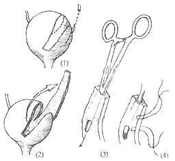
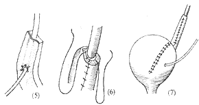
Figure 1: Repair of lower ureteral injury defect using bladder flap ureteroplasty, showing steps (1) to (7).
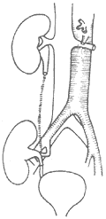
Figure 2 Repair of long-segment ureteral defect with autotransplantation during a seasonal epidemic
Figure 3 Application of double-J ureteral stent catheter


