| disease | Patulous Eustachian Tube Dysfunction |
The pharyngeal opening of the Eustachian tube is normally closed and only opens momentarily during actions such as swallowing, yawning, opening the mouth wide, and blowing the nose to allow for gas exchange in the tympanic cavity. If the muscles of the pharyngeal opening become paralyzed or atrophied, the opening may remain in a constant open state, leading to symptoms known as abnormal patency of the Eustachian tube.
bubble_chart Etiology
1. Defects in the soft tissue around the pharyngeal opening of the Eustachian tube, scar adhesions, atrophy, and muscle paralysis, commonly seen in conditions such as atrophic rhinitis, pharyngitis, post-radiation nasopharyngeal membrane atrophy, and myasthenia gravis.
2. Psychological factors: Excessive mental stress can cause muscles to remain in a state of tonic contraction.
bubble_chart Clinical Manifestations
1. Low-pitched tinnitus, synchronized with the breathing rate, worsens when speaking, opening the mouth, or swallowing, and alleviates when lying flat, bending over, or closing the mouth and inhaling through the nose. Therefore, patients frequently perform nasal inhalation and sometimes remain silent, unwilling to speak.
2. Hyperacusis - Difficulty hearing others clearly while perceiving one's own voice as excessively loud, caused by the direct transmission of speech sounds through the Eustachian tube into the middle ear.
3. Examination reveals a cloudy tympanic membrane, characterized by inward invasion and outward fluttering movements synchronized with breathing.
Based on medical history and signs, the diagnosis is not difficult. For cases with incomplete closure and intermittent symptoms, the diagnosis may be more challenging, and acoustic immittance testing can be performed. A wavy immittance graph may be detected during forced breathing.
bubble_chart Treatment Measures1. In the past, methods such as corroding or burning the pharyngeal orifice mucosa to cause scar contraction were used, but the efficacy was uncertain and is now rarely adopted. A 4:1 mixture of boric acid and salicylic acid powder can be sprayed, or 30% trichloroacetic acid or 5% phenol can be applied locally.
2. Liquid silicone is injected under the mucosa of the anterior lip of the pharyngeal orifice, performed under nasopharyngoscopy, until the pharyngeal orifice is narrowed to half its original size.
3. Laser cauterization is used to burn the anterior lip tissue, causing scar contraction and narrowing.
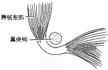
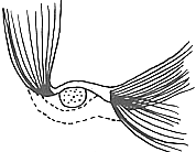
4. Tensor veli palatini tendon release: The patient lies supine with the head tilted back, and a mouth gag is used to open the mouth. Local submucosal infiltration and greater palatine nerve block anesthesia are performed using 1% lidocaine. A small curved incision is made posterior to the eighth molar at the junction of the soft and hard palate. The pterygoid process is separated, and a chisel is used to cut it from its root. The posterolateral tendon is moved to the anteromedial side of the pterygoid process to relax the tensor veli palatini. The tendon is then fixed with silk sutures behind the pterygoid process, allowing the pharyngeal orifice to relax and close.
5. For patients with functional disorders, local block and electroacupuncture stimulation therapy can be used (Figure 1).


Figure 1: Tensor veli palatini tendon release
(1) Normal anatomical position of the tensor veli palatini tendon (2) Position of the tendon after redirection


