| disease | Chronic Pancreatitis (Surgery) |
The ratio of chronic pancreatitis to acute pancreatitis is approximately 1:10, while in adolescents, the ratio is about 3:1. The exact cause of this disease remains unclear, and the reasons for its occurrence in young people and adolescents are not fully understood.
bubble_chart Pathogen
From a modern perspective, chronic relapsing pancreatitis is classified into alcoholic and obstructive types.
1. **Alcoholic Chronic Pancreatitis**: This is the common type of chronic pancreatitis in Western countries. The pancreas appears fibrotic and hardened, often involving the entire organ with a grayish-white appearance. In severely affected areas, the pancreatic lobules are indistinct, and significant fibrosis leads to pancreatic atrophy, pale sections, and minimal hemorrhage. Ultrastructural examination of pancreatic tissue reveals hyperactive acinar secretion, reduced mature zymogen granules, increased numbers of prozymogen granules, enlarged nuclei and nucleoli, Golgi complexes, rough endoplasmic reticulum, and mitochondria. The number of ductal and centroacinar cells also increases.
Alcohol consumption increases the protein content in pancreatic juice. Microscopically, proteinaceous deposits can be seen in small pancreatic ducts, composed of pancreatic juice proteins and glycoproteins. If calcium carbonate is incorporated, calcifications or pancreatic duct stones may form. When protein plugs and necrotic debris obstruct small ducts, the distal pancreatic ducts dilate. Larger pancreatic ducts may exhibit segmental or diffuse dilation. The parenchyma around the ducts shows inflammatory infiltration. As the disease progresses, acinar tissue disappears and is replaced by fibrous tissue, which may pull on the ducts, causing dilation. However, beaded ductal appearance is rare. Fibrotic tissue compresses the small veins around the islets, preventing insulin from entering the bloodstream, which can lead to diabetes.
2. **Obstructive Chronic Pancreatitis**: Pancreatic duct obstruction can result from various causes, such as fibrosis of the Vater’s papilla, papillary inflammation, congenital or acquired stenosis of the main pancreatic duct, or tumor compression. In China, chronic pancreatitis observed via ERCP, surgery, or autopsy is often secondary to gallstones or other biliary diseases, accounting for about 40–50% of cases. Acute necrotizing pancreatitis, pancreatic abscesses, etc., can gradually lead to chronic inflammation, often presenting as chronic relapsing pancreatitis.Obstructive chronic pancreatitis differs from alcoholic pancreatitis in the absence of proteinaceous plugs in the ducts, though the gross appearance is similar. Microscopically, the findings are distinct: the lesions are diffuse, with loss of lobular architecture and widespread involvement of exocrine tissue. Large ducts show moderate to severe dilation, while small ducts often remain normal, with intact epithelium and empty lumina, rarely obstructed by protein plugs or calcifications. Some suggest that the fibrotic tissue in obstructive chronic pancreatitis consists of collagen with a short half-life, making the obstructive changes sometimes irreversible.
bubble_chart Clinical Manifestations
The clinical symptoms of chronic pancreatitis are recurrent and intractable abdominal pain, primarily located in the left upper abdomen or upper abdomen, radiating to the left shoulder and lumbar region, sometimes accompanied by fever, jaundice, and steatorrhea. Localized portal hypertension (left-sided) may occasionally occur. If caused by biliary tract diseases, symptoms or signs of cholangitis often appear during episodes or intervals of chronic pancreatitis.
1. Intractable pain: This is a prominent symptom of chronic pancreatitis, which may occur without obvious triggers or after consuming high-fat meals. The exact cause remains unclear. Previously, increased pancreatic duct pressure was considered one of the contributing factors. Recent theories suggest that pancreatitis is associated with neuritis. Studies have revealed an increased number of nerves (>100%) in inflamed pancreatic tissue, along with enlarged diameters, infiltration of oval cells, and significant degeneration of the nerve sheath membrane. Additionally, elevated levels of neuropeptide substance P and calcitonin gene-related peptide have been found in sensory nerve fibers of chronic pancreatitis patients. Furthermore, sensory nerve endings around the pancreas may respond to chemical and physical disturbances, meaning that ongoing pathological processes within the pancreas can trigger pain. Neither gross pancreatic changes, histomorphology, nor imaging studies can definitively correlate intractable pain with morphological abnormalities. Therefore, the pain may be related to the dynamic progression of the disease rather than static structural changes.2. Jaundice: This is often transient, occurring in about 30% of chronic pancreatitis cases. Jaundice usually results from edema of the pancreatic head compressing the distal common bile duct, while in some patients, it is caused by fibrotic strictures of the distal bile duct. Such obstructions are typically partial.
3. Others: Prolonged obstruction of the pancreatic duct may lead to inflammatory cysts.
If adjacent blood vessels are affected by compression or thrombosis, left-sided portal hypertension may develop. Meanwhile, pancreatic nerves thicken (approximately 50%), and the destruction of the peripheral protective sheath eliminates the barrier against potentially toxic pancreatic enzymes and kinins.
bubble_chart Auxiliary Examination1. Pancreatic Function Tests
Pancreatic Secretion Test (PST): The bicarbonate concentration in duodenal fluid stimulated by cholecystokinin-pancreozymin (CCK-PZ) and secretin decreases, while pancreatic enzymes and fluid volume may or may not show typical reductions; in PST, only bicarbonate concentration decreases or pancreatic fluid and enzymes are reduced; in Pancreatic Function Diagnostic (PFD) tests, urinary para-aminobenzoic acid (PABA) excretion may be less than 70%.
2. Ultrasound Examination
A diagnosis can be confirmed if pancreatic stones are present, the pancreatic duct is dilated (>3mm), and any of the following imaging findings are observed:
(1) Irregular pancreatic duct walls with intermittent strong echoes.
(2) Continuous images of pancreatic duct cysts.
(3) Pancreatic atrophy and regional enlargement.
For pancreatic atrophy or regional swelling, assessment should be made along both the long and short axes of the pancreas. An anteroposterior diameter of <10mm indicates atrophy, while >30mm indicates swelling.
3. CT Examination
Varying degrees of dilation in the main pancreatic duct may be observed, sometimes accompanied by pancreatic cysts; pancreatic atrophy or regional swelling may also be seen alongside main pancreatic duct dilation; pancreatic calcification or stones may be present.
4. ERCP Examination
The main pancreatic duct and its branches show varying degrees of irregular dilation. Sometimes, the main pancreatic duct may be obstructed, preventing contrast medium from reaching the distal duct. In some cases, the main pancreatic duct dilates with localized cystic dilation, resembling pancreatic cysts; most patients exhibit pancreatic calcification or stones.
A history of acute pancreatitis, frequent episodes of cholangitis due to gallstones, long-term alcohol abuse, and typical symptoms such as abdominal pain, fever, and jaundice, often triggered by alcohol consumption or high-fat diets. In emaciated individuals, a transverse, elongated mass may be palpable in the pancreatic region with varying degrees of deep tenderness. Diagnosis can be readily established through pancreatic secretion tests (PST), ultrasound, CT scans, or ERCP.
The diagnostic criteria for chronic pancreatitis require the presence of one or more of the following:
(1) Recurrent episodes of upper abdominal pain, tenderness or a palpable mass in the pancreatic region, with positive findings on pancreatic function tests or imaging studies.
(2) Significant pancreatic calcifications or stones.
(3) Histopathological changes in pancreatic tissue.
bubble_chart Treatment Measures
I. Medical Treatment of Chronic Pancreatitis
Resting the pancreas: Administer H2 receptor antagonists to inhibit gastric acid secretion, along with anti-enzyme agents and dietary adjustments.
Treatment of underlying causes: Pay special attention to improving biliary tract diseases and hyperlipidemia, and strictly prohibit alcohol consumption.
Support for systemic condition: Due to recurrent episodes, the overall condition is generally poor, especially in long-term alcoholics with worse nutritional status. Active support should be provided. II. Surgical Treatment of Chronic Pancreatitis
1. The objectives of surgical treatment for chronic pancreatitis are:
(1) Relieving pancreatic duct obstruction.
(2) Relieving obstruction at the distal bile duct.
(3) Alleviating pain.
(4) Reducing complications caused by chronic pancreatitis.
2. Indications for surgery in chronic pancreatitis:
(1) Recurrent acute episodes or intractable pain despite appropriate lifestyle adjustments and medical treatment.
(2) Presence of biliary stones, especially at the distal bile duct or ampulla, which often cause pancreatitis due to impaction.
(3) Stenosis of the distal bile duct or the opening of the biliary-pancreatic duct.
(4) Local portal hypertension or gastrointestinal bleeding.
(5) Associated pancreatic cysts or pancreatic fistulas.
(6) Suspected pancreatic cancer.
3. Common surgical procedures for chronic pancreatitis:
(1) Pancreatic duct decompression: Papillary pancreatic ductotomy with成形术; pancreaticojejunostomy (side-to-side); distal pancreaticojejunostomy.
(2) Pancreatic resection: Total pancreatectomy; pancreaticoduodenectomy; duodenum-preserving pancreatic head resection; distal pancreatectomy.
(3) Management of biliary tract (for chronic pancreatitis caused by biliary diseases): Treat accordingly to relieve obstruction, ensure drainage, and remove stones or other lesions.
(4) Treatment of complications caused by pancreatitis, such as pancreatic cysts or abscesses.
(5) Neurolysis for intractable pain: Pancreatic head plexus neurectomy; splanchnic neurectomy; celiac plexus neurectomy. Changes in pancreatic duct pressure in chronic pancreatitis:
In chronic pancreatitis, pancreatic duct pressure (perfusion pressure) is higher than normal. Sato reported that the perfusion pressure in chronic pancreatitis patients was 3.73 ± 1.23 kPa, with a residual pressure of 2.18 ± 0.33 kPa, compared to 1.49 ± 0.451 kPa and 0.784 ± 0.274 kPa in the control group. Endoscopic measurements also showed higher pancreatic duct pressure in chronic pancreatitis, especially in cases of intractable pain. Many scholars advocate early surgery to prevent or slow disease progression, improve declining endocrine and exocrine functions, and alleviate pain. However, practice has shown otherwise. Warshaw found that exocrine insufficiency increased by 20–60% one year after pancreaticojejunostomy, 9–24% after five years, and 41–80% after ten years. Endocrine insufficiency also increased: 30–50% after one year, 24–59% after five years, and 29–57% after ten years. These data suggest that early decompression surgery is not justified from the perspective of preserving pancreatic function. III. Side-to-Side Pancreaticojejunostomy
Before performing this procedure, endoscopic retrograde pancreatography should be conducted, supplemented by B-ultrasound or CT, to assess the degree of pancreatic duct dilation, the location of stenosis, and whether multiple or single strictures are present. If the pancreatic duct is stenotic and the dilated duct diameter exceeds 7 mm, pancreaticojejunostomy is suitable and effective, with pain relief exceeding 80% and lasting over five years. If the pancreatic duct is not dilated, internal drainage is not advisable. Poor outcomes are expected in cases with pancreatic calcification.
1. Pancreaticojejunostomy is generally performed using Puestow's modified technique. However, Buval's method of resecting the pancreatic tail and performing Roux-en-Y pancreaticojejunostomy with end-to-side anastomosis between the resected pancreatic tail and jejunum, as well as Puestow and Gillesby's approach of resecting the pancreatic tail, longitudinally incising the pancreatic duct to the right of the mesenteric vessels, embedding the pancreas into the jejunal loop, performing Roux-en-Y pancreaticojejunostomy, and splenectomy, are less commonly used.
2. The advantages of the modified Puestow procedure (Parlington) are: ① a large anastomotic stoma; ② more importantly, since islet cells are concentrated in the pancreatic tail, preserving the tail without resection retains a large number of islet cells, which is highly beneficial for patients with diabetes; ③ preservation of the normal spleen.
In Parington's side-to-side pancreaticojejunostomy, the anastomotic stoma should be made as long as possible, with the pancreatic duct fully split open, reaching a length of up to 10 cm. During the anastomosis, pancreatic stones should be removed as thoroughly as possible (Figure 3).
3. Chronic pancreatitis with obstruction of the distal common bile duct: Approximately 3–40% of chronic pancreatitis cases are accompanied by varying degrees of common bile duct obstruction, though few require surgical intervention to relieve the obstruction. The causes include compression by an enlarged pancreatic head or cyst, as well as fibrotic inflammatory tissue contraction in the pancreatic segment of the bile duct. Indications for surgical decompression:
(1) Frequent episodes of cholangitis with persistent jaundice.
(2) Confirmation via B-ultrasound, CT, or ERCP that the bile duct obstruction is caused by the pancreas.
(3) Common bile duct stenosis accompanied by stones.
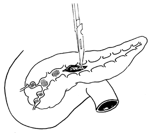
(1) Splitting the pancreas
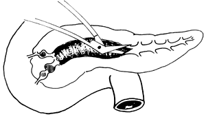
(2) Complete splitting of the pancreas
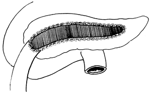
(3) Completion of anastomosis
Figure 3 Side-to-side pancreaticojejunostomy
For such cases, in addition to fully splitting the pancreatic duct and performing a side-to-side anastomosis with the jejunum, a jejunum-to-bile duct anastomosis is also added (Figure 4).
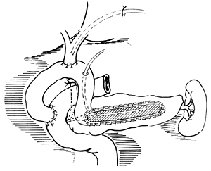
Figure 4 Roux-en-Y choledochojejunostomy and pancreaticojejunostomy
IV. Pancreaticoduodenectomy
Pancreaticoduodenectomy is a highly destructive procedure and should not be undertaken lightly. Strict adherence to its indications is essential. The indications are as follows:
1. Difficulty in performing longitudinal pancreatic incision and jejunal anastomosis (due to an overly narrow pancreatic duct), with chronic inflammation localized to the pancreatic head.
2. Inflammatory mass in the pancreatic head (including the uncinate process) that is difficult to distinguish from a tumor.
3. Inflammatory mass in the pancreatic head compressing the duodenum and causing duodenal stenosis.
4. Cyst in the pancreatic head compressing the duodenum and common bile duct, causing stenosis in both.
The mortality rate for pancreaticoduodenectomy in chronic pancreatitis is approximately below 2%. Reported efficacy varies, with pain relief rates around 70–80%.
V. Duodenum-preserving pancreatic head resection
This procedure does not require resection of the duodenum or the distal common bile duct, offers good pain relief, and has low surgical mortality and complication rates. It is an effective approach for treating chronic pancreatitis localized to the pancreatic head. Beger reported 141 cases of chronic pancreatitis with pancreatic head enlargement treated with this method, showing a hospital mortality rate of 0.7% and a late-stage [third-stage] mortality rate of 5%. Complete pain relief was achieved in 77% of cases. After a follow-up of 3.6 years, 81.7% showed no changes in glucose metabolism, 10.1% worsened, and 8.3% remained unchanged.
The surgical technique is illustrated in Figure 5. Key points of the procedure: During subtotal pancreatic head resection, a small portion of pancreatic tissue may be left between the common bile duct and the duodenal wall. When resecting the uncinate process, the duodenal mesenteric vessels should be protected. During Roux-en-Y pancreaticojejunostomy, the pancreatic duct opening should be enlarged as much as possible for anastomosis with the jejunal mucosa. Other aspects are the same as in the Whipple procedure.
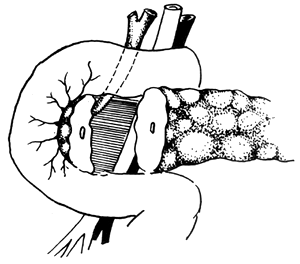
Figure 5 Pylorus-preserving pancreaticoduodenectomy
VI. Total Pancreatectomy
Total pancreatectomy removes the source of pancreatic pain but has a high mortality rate (5%) and makes postoperative endocrine and exocrine control difficult. Cooper reported 83 cases of this procedure, with 28 patients still experiencing pain 1.5 years after surgery, 38% of patients struggling with endocrine control, and 10 late-stage [third-stage] deaths. Therefore, total pancreatectomy for chronic pancreatitis must be approached with extreme caution.
VII. Sphincterotomy
In chronic pancreatitis, pancreatic duct stenosis is often multifocal and unevenly dilated, with rare cases of uniform dilation caused by ampullary stenosis. Thus, common bile duct sphincterotomy or pancreatic duct sphincterotomy is not feasible.
In summary, the disease causes of chronic pancreatitis are multifactorial and rarely singular. The resulting pathological changes are also inconsistent, with significant variations in age and regional differences. Consequently, there is no single treatment model. Treatment plans should be tailored based on specific conditions to improve cure rates and reduce complications and mortality.
Complications such as pancreatic cysts, pancreatic abscesses, obstruction of the lower common bile duct, pancreatic stones, and pancreatitis.


