| disease | Nasal Septum Perforation |
Perforation of the nasal septum is often the result of injury during nasal septum surgery. Although it does not cause serious consequences, its symptoms frequently trouble patients.
bubble_chart Etiology
1. During submucous resection of the nasal septum, injury to the corresponding soft tissues on both sides of the septum occurs. This is the most common cause of nasal septal perforation. Improper nasal septum electrocautery can also lead to perforation.
2. Traumatic injury of the nose results in improper management of nasal septum injury.
3. Improper handling of a nasal septal abscess.
4. Prolonged course of diseases causing nasal septal ulcer.
5. Industrial or chemical burns.
6. Special nasal pestilential diseases or systemic acute pestilential diseases (diphtheria, scarlet fever). {|105|}
bubble_chart Clinical Manifestations
Dryness in the nasal cavity often leads to the formation of crusts, causing a stuffy nose and headache. Blood-streaked nasal discharge or nosebleeds are common. If the perforation is in the anterior part of the nasal septum, a whistling sound may occur during breathing. Perforations in the posterior part of the nasal septum may be asymptomatic. Diagnosis is usually straightforward with nasal endoscopy.
bubble_chart Treatment Measures
For perforated edges with membrane adhesion showing ulcers or granulation tissue, the scabs should be removed, cauterized with 25% silver nitrate solution, and coated with ointment to promote healing. Some small perforations may cause no clinical symptoms and do not require surgical repair. For larger perforations with obvious symptoms, perforation repair should be performed.
Small nasal septum perforations are easier to repair successfully, while those larger than 1cm are considerably more difficult. The success of repair depends on many factors, primarily the following: ① Healthy nasal mucosa. Preoperative examination of the nasal cavity should be thorough, and the condition of the nasal mucosa should be improved. Active treatment is required for inflammation, and surgery should not be rushed. Smokers and drinkers should abstain from tobacco and alcohol for at least one month. Ulcers and erosions at the perforation edges require particularly aggressive treatment. ② Rational incision selection. The principle for choosing an incision is to facilitate operation, ensuring that the flap used for repair can be perfectly aligned and sutured with appropriate tension. If a labiogingival groove incision is selected, attention should also be paid to whether there are infectious diseases in the oral cavity or teeth. ③ The tissue flap covering the perforation should have a good blood supply, requiring the flap to be sufficiently wide.(1) Nasal Septal Mucosal Flap Repair Method
1. Tension-Relieving Suture Method: Suitable for small perforations located in the anteroinferior part of the nasal septum. After local surface and infiltration anesthesia, the edges of the perforation are slightly trimmed to create fresh wound surfaces. A curved incision is made 1–2 cm above and anterior to the perforation edge, with a length exceeding the diameter of the perforation. The mucoperichondrium and mucoperiosteum on both sides of the nasal septum are separated from the perforation edge to the curved incision to release tension. The mucoperichondrium is then pulled downward to cover the perforation, and the soft tissue flap is advanced 1–2 cm forward and upward to cover the perforation, with sutures placed at the anterosuperior edge of the perforation.
2. Nasal Septal Mucosal Flap Transposition Repair Method: On the left side of the nasal septum, a curved incision is made above the perforation, extending around the posterior edge to below the perforation. Another curved incision is made from the starting point of the first incision, extending around the anterior edge of the perforation, forming a spindle shape with the perforation in the middle and triangular mucosal flaps above and below. The two triangular mucosal flaps are then dissected from their tips toward the perforation edges, forming mucosal flaps pedicled at the perforation edges. The upper mucosal flap is flipped downward, and the lower flap is flipped upward, creating mucosal coverage on the right side of the perforation. The coverage on the left side of the perforation is similar to the unilateral tension-relieving suture method, but the width of the mucosal flap must be ensured.(2) Turbinate Mucosal Flap Repair Method
1. Middle Turbinate Mucosal Transposition Method: The perforation edges are slightly trimmed to create fresh wound surfaces. A "U"-shaped incision is made on the ipsilateral middle turbinate, and the mucosal flap is dissected from top to bottom to its pedicle. This flap is then flipped downward to cover the perforation and sutured to the wound edges around the perforation. The contralateral nasal cavity is packed, and the pedicle is severed after 2–3 weeks.
2. Inferior Turbinate Mucosal Transposition Method: The method is similar, except a standard "U"-shaped incision is made on the surface of the inferior turbinate, and the mucosal flap is flipped upward to cover a perforation located above the level of the inferior turbinate.
(3) Nasal Floor and Septal Mucosal Repair Method: A longitudinal incision is made on the lateral wall of the inferior meatus in one nasal cavity, extending downward to the nasal floor and then upward to separate the mucoperiosteum and mucoperichondrium. The dissection should be as extensive as possible, reaching above the superior edge of the perforation. The same procedure is performed on the contralateral side. A narrow strip of mucosal flap is then harvested above the perforation edge in an anteroposterior direction. The bilateral mucosal flaps are transposed upward to cover the perforation and sutured in place on both sides.(4) Extranasal Free Tissue Graft Method: Extranasal tissues used for nasal septum perforation repair include temporal fascia, fascia lata, and tibial periosteum. The free graft should be slightly larger than the perforation. A vertical incision is made anterior to the perforation on the left side of the nasal septum, and the surrounding mucosa is dissected with an elevator, extending about 0.5 cm from the perforation edge. The harvested fascia is inserted through the incision and placed between the two layers of dissected mucosa around the perforation, then fixed with sutures, and the nasal cavity is packed for compression.
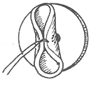
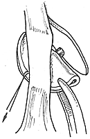
(5) Non-surgical closure method Depending on the size of the perforation, select an appropriately sized "H"-shaped silicone button and embed it at the perforation site. The two leaves of the button should be positioned on either side of the nasal septum, with the central axis of the button located precisely at the center of the perforation. Each leaf of the silicone button should be 1 mm thick, with a central axis diameter of 5 mm and a width of 3 mm. Facer reported a success rate of 72% with this method (Figure 1).
|
|
|
|
Figure 1 "H"-shaped silicone button closure method for septal perforation


