| disease | Tracheal and Bronchial Stenosis |
The application of mechanical ventilation therapy can improve respiratory function, with good clinical efficacy, and the number of treated cases is increasing. With the widespread use of mechanical ventilation therapy, complications such as tracheal stenosis following tracheotomy and intubation are becoming more common.
bubble_chart Etiology
A tracheostomy performed too high, injuring the first cartilage ring, can lead to cricoid cartilage erosion, inflammatory sexually transmitted disease changes, and difficult-to-correct grade III subcricoid stenosis. During tracheostomy, excessive removal of the anterior tracheal wall tissue can later result in the formation of abundant granulation tissue and fibrous scar tissue. Pressure from the tracheal tube on the anterior tracheal wall causes the tissue above the incision to collapse inward, while excessive weight from externally connected tubing compresses the tracheal wall, leading to tissue pressure erosion and subsequent fibrous scar formation. Additionally, overinflation of the external cuff used to seal the tracheal lumen can exert excessive circumferential pressure on the tracheal wall, causing tissue erosion and necrosis. In severe cases, this may lead to annular cicatricial stenosis or even tracheoesophageal fistula and tracheoinnominate stirred pulse fistula. The latter two conditions have high mortality rates. Therefore, during moving qi tracheostomy and intubation, attention should be paid to the tracheostomy site, avoiding excessive removal of anterior tracheal wall tissue. The selected tracheal tube should be of appropriate size and length, cuff inflation pressure should not be too high, and connected tubing should be lightweight and flexible to reduce the incidence of tracheal stenosis complications.
|
|
|
|
|
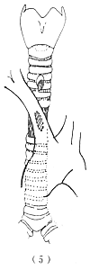
Figure 1 Tracheal stenosis after tracheostomy and intubation
(1) Stenosis caused by a high incision (2) Stenosis at the cuff site (3) Tracheal incision stenosis and cuff site (4) Tracheal stenosis and tracheoesophageal fistula (5) Tracheo-right innominate stirred pulse fistula with intermediate qi tracheal wall softening and inflammatory changes
bubble_chart Clinical Manifestations
Common symptoms include airway obstruction leading to shortness of breath and difficulty breathing, which worsen with physical activity and increased respiratory secretions, often accompanied by wheezing. In cases where tracheostomy and intubation have been performed, the presence of these symptoms should first raise suspicion of tracheal scar stenosis. Anteroposterior, lateral, and oblique tracheal tomograms can clearly reveal the location, severity, length, and morphological changes of the stenosis.
bubble_chart Treatment Measures
The endotracheal tube has been removed, and patients who no longer require mechanical ventilation but have severe tracheal stenosis generally need to undergo tracheal reconstruction surgery. For cases where ventilation function has not fully recovered, conservative treatments such as periodic tracheal dilation, tracheal reconstruction, tracheostomy tube insertion, or placing a ventilation tube in the stenotic segment to support the tracheal lumen can be adopted to maintain ventilation function and prolong life.


