| disease | Myopia |
| alias | Myopia, Myopia |
Myopia, also known as nearsightedness, is a condition where the eye can only see nearby objects clearly but not distant ones. When the eye is at rest, parallel light rays from infinity are refracted by the eye's optical system and converge to form a focal point in front of the retina, resulting in a blurred image on the retina. Far vision is significantly impaired, but near vision remains normal.
bubble_chart Etiology
The causes of myopia have been a subject of differing opinions in the past, but they can generally be attributed to two categories: internal and external factors, as outlined below:
1. Internal Factors
(1) Genetic predisposition: It is widely accepted that myopia has a certain hereditary tendency, especially for high myopia. However, this tendency is less pronounced for general myopia. Individuals with genetic factors tend to develop the condition at an earlier age, often with a refractive error exceeding 6.00D. Nevertheless, some cases of high myopia occur without any family history. High myopia is typically inherited as an autosomal recessive trait, while general myopia is considered a multifactorial genetic disorder.
(2) Developmental factors: Infants typically have smaller eyeballs, resulting in farsightedness. As they grow, the axial length of the eye gradually increases, reaching normal development by adolescence. Excessive growth can lead to myopia, known as simple myopia, which usually begins during school years and rarely exceeds 6.00D, stabilizing around the age of 20. However, if progression is rapid in early childhood and accelerates between ages 15–20 before slowing down, the myopia often exceeds 6.00D, sometimes reaching 20D–25D or even 30D. This type is referred to as high myopia, progressive myopia, or pathological myopia. In later years, degenerative changes may occur, leading to gradual vision deterioration that cannot be corrected with glasses. Congenital myopia is rare, though a few cases exist at birth.
2. External Factors Environmental influences play a significant role. Individuals engaged in prolonged near work, such as reading or writing, are more prone to myopia. Among adolescents, myopia is particularly prevalent, with a noticeable increase in incidence starting from grades five and six in primary school. This trend highlights the strong correlation between myopia development and near work. During adolescence, the eyeball is still growing, with strong accommodative ability and high elasticity of the sclera. Prolonged near tasks like reading or writing require constant focusing and convergence, exerting pressure on the extraocular muscles (especially the medial rectus) and raising intraocular pressure. As near work increases, so does the frequency and duration of accommodation and convergence, placing the ciliary and extraocular muscles under prolonged strain. Overuse of accommodation can lead to ciliary muscle spasms, causing temporary vision impairment. However, rest or the use of cycloplegic agents may restore vision completely. Hence, this type of myopia is sometimes called functional or pseudomyopia. Over time, the mechanical pressure from extraocular muscles can stretch the sclera, elongating the axial length and deepening myopia, which can no longer be reversed by drugs like atropine. Poor visual hygiene during adolescence is a direct cause of myopia, and neglecting overall health can exacerbate its progression.Recent prospective studies have examined the roles of environment and genetics in myopia development. These studies tracked students with initially normal vision over two years, analyzing factors contributing to myopia. The results showed: - **Genetic influence**: The ratio of new myopia cases among children with no parental myopia, one parent with myopia, and both parents with myopia was 1:2.6:3.8. - **Environmental influence**: The ratio of new myopia cases among students with 1–2 hours, 3 hours, and 4–5 hours of after-school reading was 1:2.1:3.2. Thus, both genetics and environment are critical factors in myopia development among students.
bubble_chart Clinical Manifestations
1. Classification of Myopia
1. By degree of myopia
⑴ Less than 3.00D is called Grade I myopia.
⑵ 3.00D to 6.00D is Grade II myopia.
⑶ Above 6.00D is high myopia, also known as pathological myopia.
2. By refractive components
⑴ Axial myopia is caused by excessive elongation of the eyeball's anteroposterior axis.
⑵ Curvature myopia is caused by excessive curvature of the cornea or lens surface.
⑶ Refractive index myopia is caused by an excessively high refractive index of the optical media.
3. Pseudomyopia, also known as accommodative myopia, is caused by the failure of relaxation of accommodation when viewing distant objects. It is fundamentally different from true myopia, which involves changes in refractive components.
2. Clinical Manifestations of Myopia
1. Visual acuity: The most prominent symptom of myopia is reduced distance vision, but near vision may remain normal. Although higher degrees of myopia generally result in poorer distance vision, there is no strict proportional relationship. Typically, myopia above 3.00D results in distance vision no better than 0.1; 2.00D myopia ranges between 0.2 and 0.3; and 1.00D myopia may achieve 0.5 or sometimes even better (see Figure 1).
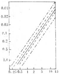
Figure 1: Myopia Degree and Distance Vision
— Average value; —— 50% confidence limit;
-·-·- 95% confidence limit.
2. Asthenopia: Particularly common in low myopia, though less pronounced than in hyperopia. It is caused by the incoordination between accommodation and convergence. In high myopia, because the viewing target is too close to the eye, convergence cannot match it, so monocular fixation is often adopted, which surprisingly does not cause asthenopia.

4. Eyeball: High myopia is mostly axial myopia, with elongation of the anteroposterior axis primarily affecting the posterior pole. This often manifests as prominent eyeballs, deeper anterior chambers, and larger pupils with sluggish reflexes. Due to the lack of accommodative stimulation, the ciliary muscle, especially the circular portion, becomes atrophic. In extreme high myopia, the lens may completely fail to support the iris, leading to Grade I iris tremor.
5. Fundus: Low myopia shows no significant fundus changes, but high myopia, due to excessive axial elongation, can lead to degenerative fundus changes.
⑴ Tessellated fundus: Retinal vessels become thin and straight after leaving the optic disc. Meanwhile, elongation of the choroidal capillaries may impair the nutrition of the retinal pigment epithelium, causing superficial pigment loss and exposing choroidal vessels, giving the fundus a leopard-spot appearance.
⑵ Myopic crescent: The choroid around the optic disc detaches from the temporal side of the disc under the stretching force of the sclera, exposing the underlying sclera and forming a white crescent. If the posterior pole continues to expand, the choroidal detachment may extend around the optic disc, eventually forming a circular patch. This patch may contain irregular pigment and sclerosed choroidal vessels.
⑶ Macula: Irregular, solitary, or confluent white atrophic patches may develop, sometimes accompanied by hemorrhage. Additionally, near the macula, degenerative lesions may occasionally appear as a black annular zone, slightly smaller than the optic disc, with clear borders and small round hemorrhages at the edges, known as Foster-Fuchs spots.
⑷ Posterior scleral staphyloma The stretching of the posterior part of the eyeball, when limited to a small area, can be seen as a sharp protrusion in the section, called posterior scleral staphyloma. If such atrophic sexually transmitted disease lesions occur in the macula, they may be associated with the operation of central vision.
5. Cystoid degeneration of the serrated edge.
6. Complications and sequelae
(1) Liquefaction, opacification, and posterior detachment of the vitreous: The most common subjective symptom is floaters, where patients perceive black spots drifting in their field of vision, resembling the movement of mosquitoes. It is often accompanied by sensations of flashes of light or sparks, particularly more pronounced in high myopia.
(2) Lens opacification.
(3) Retinal membrane tears, retinal detachment.
(4) Glaucoma—Some studies using applanation tonometry have shown that the prevalence of open-angle glaucoma in high myopia is 6–8 times higher than in normal individuals.
(5) Prolonged dark adaptation—This occurs because pathological changes in the pigment epithelial cells of highly myopic eyes inevitably affect the photochemical reaction process of visual cells.
bubble_chart Treatment Measures
1. True myopia eye
(1) Lens correction Before prescribing glasses, the first step is to determine the true degree of myopia through retinoscopy. For adolescents, optometry should be performed under cycloplegia to control accommodation and exclude pseudomyopia. The principle of prescribing glasses is to use the lowest degree of lens that can still correct the myopic eye to the best visual acuity. Generally, for myopia below 6.00D, full correction and regular wearing are recommended. For high myopia, complete correction is ideal for better vision, but patients often cannot tolerate it. Therefore, the lens degree is usually reduced (generally between 1.00D and 3.00D) to ensure comfort and maintain binocular visual function.
(2) Corneal contact lenses Wearing contact lenses can expand the visual field, provide better cosmetic effects, and significantly reduce anisometropia, thereby maintaining binocular visual function. For adolescents with myopia, contact lenses not only improve vision but also press on the cornea to prevent further progression of myopia. However, strict hygiene must be observed, including proper disinfection, maintenance, and regular replacement.
(3) Telescopic glasses Patients with extreme high myopia or macular lesions often use telescopic glasses for reading or close work. These glasses magnify by 1.8 times, enhancing farsightedness by 2%–3.5% and near vision by up to 5 times. Due to the extremely narrow field of view, they cannot be used while walking.
(4) Radial keratotomy This method was first attempted by the Soviet scholar Kranov (1970). There have been multiple reports in China. The procedure involves making 8–16 radial incisions between the central 3–5mm of the cornea and the corneoscleral limbus, with a depth of 0.36–0.50mm. After the corneal surface is incised, the curvature of the cornea flattens, reducing the degree of myopia. It is generally believed to correct up to 3.00D of myopia. However, the long-term effects remain uncertain, and strict selection of indications and contraindications is required, along with potential surgical complications. Thus, it has not been widely adopted.
(5) Scleral shortening This is the most commonly used surgical therapy, proven effective for high myopia and widely applied.
(6) Keratomileusis This method involves using a specially designed instrument to perform a lamellar resection of the central cornea. The removed corneal tissue is cryogenically treated and then ground on a precision lathe to achieve the desired refractive power before being sutured back in place. It is used to correct high myopia, but the surgery is extremely complex and carries certain risks, making it difficult to popularize.
2. Treatment of pseudomyopia Pseudomyopia occurs when the eye retains a certain degree of accommodation while viewing distant objects. In other words, it is a refractive state where the eye’s accommodation relaxes slowly when shifting from near to far vision. It increases with prolonged near work and higher accommodation levels and decreases or disappears with distance viewing and relaxation of accommodation. Therefore, pseudomyopia can disappear with treatment (including rest) but may recur if untreated. Various methods may show some effectiveness, but none provide lasting results. Thus, treatment should be selected based on the following principles: ① Harmless to the eye, with no impact on visual development even with long-term use. ② Since pseudomyopia can resolve on its own, the chosen method must have a scientific basis and objective proof of relaxing accommodation. Relying solely on improved visual acuity to assess efficacy is unreliable. ③ Simple and practical for widespread application.
Current methods include:
(1) Using various methods to increase visual excitability and lower visual thresholds: Such as qigong, cold water baths, or taking stimulant drugs. These therapies can improve farsightedness, and theoretically, visual acuity should also improve. However, they are not ideal treatments.
⑵ Local drug treatment: such as atropine-like drugs, which have a fast and obvious effect in relaxing accommodation, is a unified method used to distinguish between true and false myopia. However, these drugs inevitably cause side effects such as difficulty in near vision and photophobia. Some have attempted to use lower concentrations to achieve a certain degree of accommodation-relaxing effect without side effects, but research results show that as the side effects disappear, the therapeutic effect also disappears.
(3) Instruments that utilize optical principles to relax accommodation.
(4) Physiological therapy methods: such as distance viewing, fogging therapy, and crystalline lens exercises. All require distant targets as stimuli to relax accommodation, making them ineffective in near environments like evening self-study sessions. Xu Guangdi designed the binocular fusion method in 1983 to treat pseudomyopia, now updated as the Xu-Myopia Prevention and Treatment Device. It consists of a 10×9×3cm 3 dark box equipped with two sets of flashing lights. One set includes two bulbs positioned along the line of sight for distance viewing, simulating a distant target. When these lights flash, the eyes automatically fuse the images, directing the gaze toward infinity. Based on the synkinetic relationship between accommodation and convergence, as the eyes diverge, accommodation also relaxes. The other set consists of a single bulb placed between the two fusion lights, serving as a near target for binocular viewing. When the two sets of lights alternate flashing, the eyes shift between near and far focus, exercising the extraocular and intraocular muscles to treat pseudomyopia and prevent true myopia in near environments.
It is worth noting that various types of pinhole glasses for myopia treatment have recently appeared on the market. The vision-improving effect of pinholes is well-known. Recent literature reports that pinholes with a diameter of 0.5mm can maintain visual acuity at around 0.5 for refractive errors up to 5.0D. However, pinholes only increase depth of focus to improve vision and do not prevent or treat myopia. Moreover, although these lenses have multiple small holes, as students wear them in class, the interpupillary distance constantly changes with viewing distance, making it impossible to maintain binocular single vision regardless of the number of holes. The wearer can only rely on the dominant eye for monocular vision, while the other eye remains suppressed. The resulting visual disturbance is secondary; the primary concern is that prolonged suppression of one eye during childhood and adolescence can impair visual development. Therefore, pinhole glasses are not only ineffective but also harmful for myopia treatment and should not be used.
The causes of myopia are relatively complex, with two main factors being genetics and environment. Currently, since genetic engineering methods cannot yet be used to modify genetic genes, the focus of myopia prevention and treatment should be on improving the visual environment.
1. Strengthen publicity and education efforts: Establish specialized institutions for myopia prevention and treatment. Ensure that myopia prevention and treatment efforts are organized and carried out systematically. Conduct regular vision checks for students, establish vision records, and promptly examine and correct those with declining vision.
2. Improve the visual environment
(1) Strive to standardize classroom lighting and illumination. The ratio of window light-transmitting area to indoor floor area should not be less than 1:6. Blackboards should not reflect light and should remain jet black or dark green. The illumination on desktops should not be less than 100Lx. Light should come from the left or left front to prevent hand shadows from blocking the light while writing.
(2) Desks and chairs should meet ergonomic requirements, suiting the height and age characteristics of children, ensuring correct posture during class or homework. When reading or writing, the distance between the eyes and the desktop should be maintained at about 30cm and should not be less than 23cm.
(3) Develop good reading and writing habits and posture: Study sessions should not be too long. After 45 minutes of study, take a 10–15 minute break or gaze into the distance to allow the ciliary muscles to relax. Eye healthcare gymnastics can also be performed. Avoid reading in bed, while walking, or in moving vehicles. Do not read or write under intense sunlight or dim streetlights, and avoid prolonged close-up TV viewing to prevent asthenopia and eye strain.
3. Pay attention to physical exercise and nutrition, reduce academic workload, and enhance constitution.
4. Reduce the impact of genetic factors: Myopia is closely related to genetics. If both parents have high myopia, the probability of inheritance after marriage is very high, so eugenics should be considered.


