| disease | Cervical Tuberculosis |
Among cervical spine inflammatory diseases, cervical spine subcutaneous nodules are the most common. Spinal subcutaneous nodules rank first in the incidence of bone and joint subcutaneous nodules, accounting for approximately 40–50%. Subcutaneous nodules occurring in the cervical spine are relatively rare, making up only 2.2%–6.3%. Cervical spine subcutaneous nodules can cause spinal cord compression, leading to high-level paraplegia, resulting in severe disability. Therefore, early diagnosis, treatment, and prevention of this condition should be emphasized. The general trend in the age of onset for spinal subcutaneous nodules is that they are more common in children and adolescents, with incidence decreasing as age increases. It is generally believed that the occurrence of subcutaneous nodules is related to the body's immunity. In recent years, foreign reports indicate a rising trend in the incidence of subcutaneous nodule diseases, particularly extrapulmonary subcutaneous nodules, with an increasing proportion of adult cases. The main reasons for this include: (1) The prevalence of Acquired Immune Deficiency Syndrome (AIDS) in certain countries and regions; (2) The rising incidence of tumors, improved diagnostic capabilities for immune-related diseases, and advancements in organ transplantation, leading to an increasing number of patients undergoing chemotherapy, radiotherapy, and immunosuppressive therapy; (3) A relative relaxation in the prevention and control efforts for subcutaneous nodule diseases compared to the past.
bubble_chart Etiology
Cervical subcutaneous node, like other bone and joint subcutaneous nodes, is a secondary lesion, that is, a local manifestation of systemic subcutaneous node disease. The primary focus is mostly in the lungs, with a few in the lymph nodes, digestive system, and genitourinary system.
The subcutaneous node bacillus belongs to the Schizomycetes class and Actinomycetales order. The branched subcutaneous node bacillus is further divided into four types: bovine, human, avian, and murine. Among them, the human and bovine types of subcutaneous node bacteria are the main pathogenic bacteria causing human subcutaneous node disease. The subcutaneous node bacillus is slender, slightly curved, and blunt at both ends. In dry environments, the subcutaneous node bacillus can survive for a long time without dying, but it is relatively sensitive to dampness-heat.After the initial infection with subcutaneous node disease, the lesion quickly spreads to local lymph nodes. The subcutaneous node bacteria enter the bloodstream through the lymph nodes and then disseminate throughout the body. Three to nine weeks later, the body develops an allergic reaction or immunity to the invading subcutaneous node bacteria and their metabolic products. At this time, the subcutaneous node bacillus test turns from negative to positive. After infection, antibodies in the serum and intracellular antibodies appear.
Subcutaneous node lesions often undergo caseous necrosis. The production of caseous necrosis may be due to the accumulation of local inflammatory cells, which compress capillaries, causing local ischemia and tissue death; or it may be related to the allergic reaction caused by bacterial proteins. Caseous tissue rarely attracts white blood cells, so it often lacks the characteristics of general suppurative infections. Its necrotic debris is also not as quickly removed by phagocytes as in general necrotic tissue. The autolysis of caseous tissue is inhibited, making it resistant to absorption for a long time. The interior of caseous tissue is generally acidic, sometimes with a pH as low as 4.0. When caseous tissue softens, its pH gradually increases, shifting toward alkalinity. After the pH rises, caseous tissue is prone to calcification. Caseous lesions heal through softening, absorption, and fibrous tissue proliferation, or they may heal by calcification. In some lesions that have already fibrosed or calcified, subcutaneous node bacilli may still survive in a dormant state. After softening, caseous material often spreads with pus to other parts of the body, causing new lesions.

The onset of cervical subcutaneous node is related to chronic strain or cumulative injury of the cervical spine. A large amount of clinical evidence shows that traumatic fractures, dislocations, or sprains do not locally induce subcutaneous node disease. Among the bones of the trunk, spinal subcutaneous node cases are the most numerous, possibly due to the spine bearing the most weight. Within the spine itself, the lumbar vertebrae bear the most weight, so lumbar vertebral disease cases are the most numerous. The lower limbs bear more weight than the upper limbs, so cases in the lower limbs are also more numerous than in the upper limbs. From these facts, it can be seen that strain has a certain relationship with the occurrence of this disease.
bubble_chart Pathological Changes
Cervical subcutaneous nodes are most commonly found at the cervical 6

Subcutaneous node bacilli primarily enter the vertebral body through the stirred pulse system from the primary lesion, with a minority entering via the venous system and lymphatic reflux. When the body's resistance declines, the bacterial emboli in the vertebral body form lesions. In most cases (about 90%), there is only one vertebral lesion. A minority of cases have lesions in two or more locations, separated by relatively healthy vertebral bodies or intervertebral discs, hence termed skip lesions. Based on the location of the lesions, vertebral subcutaneous nodes are classified into three types: marginal, central, and subperiosteal.
Marginal Type: Clinically more common in adult patients, the lesion is close to the intervertebral disc and easily penetrates the cartilage endplate to invade the intervertebral disc and spread to adjacent vertebral bodies. It is primarily characterized by osteolytic destruction, with little or no sequestrum formation. In severe cases, adjacent vertebral bodies may collapse, leading to cervical kyphosis (Figure 1).
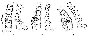
Figure 1: Development process of marginal-type lesions in cervical subcutaneous nodes
Central Type: This type is more common in children and rare in adults. The lesion is located in the center of the vertebral body. In children, the vertebral bodies are small, and the lesion progresses rapidly, affecting the entire ossification center, penetrating the surrounding cartilage shell, and invading the intervertebral disc and adjacent vertebral bodies. In adults, the vertebral bodies are larger, and the lesion progresses slowly. Early lesions may be confined to the center of the vertebral body without invading the intervertebral disc or adjacent vertebral bodies, so early symptoms are not obvious. The lesion is mainly characterized by bone destruction, forming sequestra. In a few cases, after the sequestrum is absorbed, a bone cavity forms, with the cavity wall being grade I dense. The cavity is filled with pus or caseous material. In advanced stages, the entire vertebral body may be destroyed, leading to pathological fracture, compression of the vertebral body into a wedge shape, and cervical kyphosis (Figure 2).
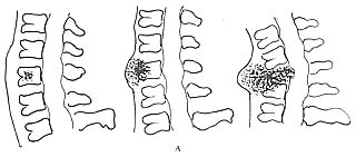
A: Children
B: Adults
Figure 2: Development process of central-type lesions in cervical subcutaneous nodes (top: children; bottom: adults)
Subperiosteal Type: Clinically rare. The lesions are mostly located at the anterior edge of the vertebral body, primarily characterized by bone destruction, often without sequestrum formation, presenting as an ice-melting-like change. This type of lesion may also result from erosion by subcutaneous node lesions outside the vertebral body.
Cervical subcutaneous nodes often form cold abscesses. The subcutaneous nodular granulation tissue, inflammatory exudate, and necrotic tissue from cervical vertebral lesions form pus that penetrates the vertebral cortex and accumulates beneath the periosteum on one side of the vertebra, forming a localized paravertebral abscess. As the disease progresses and pus accumulates, it may break through the anterior vertebral periosteum and anterior longitudinal ligament, collecting anterior to the vertebral periosteum and posterior to the longus colli muscle. For lesions above C4, the abscess is usually located posterior to the pharyngeal cavity, hence also called a retropharyngeal abscess. For lesions below C5, the abscess is typically situated posterior to the esophagus. A large retropharyngeal abscess can push the posterior pharyngeal wall forward, bringing it close to the base of the tongue, causing loud snoring during sleep and even difficulty breathing and swallowing. Pus from lower cervical spondylosis lesions may descend along the longus colli muscle to both sides of the superior mediastinum, enlarging the mediastinal shadow and resembling a tumor. Retropharyngeal and retroesophageal abscesses may rupture into the pharyngeal cavity or esophagus, forming internal fistulas, allowing pus and bone fragments to be swallowed or expelled through the mouth.
Pus from lateral lesions of the vertebral body can also form abscesses on both sides of the neck or spread along the prevertebral fascia and scalene muscles to the supraclavicular fossa, forming multiple abscesses. These abscesses may rupture externally, forming a sinus. Once a sinus forms, it often persists for a long time and becomes difficult to manage, especially when mixed infections occur.
Severe destruction of the affected vertebral body can lead to collapse under pressure. Invasion of the intervertebral disc and cartilage endplate results in narrowing of the intervertebral space. Destruction of the secondary ossification centers of the vertebral body hinders longitudinal growth. Consequently, the physiological curvature of the cervical spine may disappear, and even kyphotic deformity can develop. However, unlike the thoracic or thoracolumbar spine, kyphotic deformity is less common in the cervical spine unless two or more vertebrae are involved. This is mainly due to the cervical spine's inherent physiological lordosis and the fact that the weight of the head is primarily transmitted through the articular processes rather than the vertebral bodies.
Pus, granulation tissue, caseous material, sequestra, and necrotic intervertebral discs from cervical spine tuberculosis may protrude into the spinal canal, compressing nerve roots and the spinal cord. Dislocation or subluxation of the affected vertebrae can also compress the spinal cord. Statistics show that the incidence of paraplegia in cervical spine tuberculosis is approximately 22%.
bubble_chart Clinical Manifestations
The disease has an insidious onset and progresses slowly. Some patients have a history of subcutaneous node disease or contact with subcutaneous node disease. Early symptoms are mild and difficult to detect. Adult patients are often misdiagnosed with wind-dampness or strain and treated with anti-wind-dampness or other symptomatic therapies. Mild symptoms in children are even more easily overlooked. Some patients have no subjective symptoms in the early stages and may be incidentally discovered during physical examinations. Some cases are not diagnosed until the discovery of cold abscesses, cervical deformities, or even paraplegia. Only a few patients experience a relatively acute onset with obvious systemic and local symptoms.
Symptoms and signs
Systemic symptoms: Patients often experience general malaise, fatigue, lack of strength, loss of appetite, weight loss, low-grade fever in the afternoon, night sweats, increased pulse rate, flusteredness, palpitation, and menstrual irregularities, among other grade I toxic and autonomic nervous system dysfunction symptoms. If the abscess becomes secondarily infected, high fever may occur. Fever in pediatric patients may be more pronounced, often accompanied by irritability, reluctance to play, crying when held, and nighttime screaming. Most patients exhibit malnutrition and anemia. If the patient also has pulmonary subcutaneous node, symptoms such as cough, expectoration, hemoptysis, or difficulty breathing may occur. If there is involvement of the urinary system subcutaneous node, symptoms such as frequent urination, urgency, dysuria, and hematuria may appear.
Local symptoms: Persistent mild dull pain in the neck, exacerbated by extension or exertion and relieved by bed rest. Nighttime pain is not significant, and patients generally sleep well, which differs from malignant tumors. If the condition worsens and irritates or compresses nerve roots, the pain may radiate to the shoulders, upper limbs, or the occipital region. The affected spinous process may exhibit tenderness and percussion pain.
Neck stiffness with restricted movement in all directions; patients may turn their entire torso to look downward, often due to protective muscle spasms around the affected vertebrae. Some patients exhibit torticollis deformities; others may have a forward-tilted head, shortened neck, and a tendency to support their chin with both hands to avoid exacerbating pain during movement. This is also known as Rust's sign. The atlantoaxial joint, responsible for head rotation, loses most of its function when affected. Kyphotic deformity is usually not prominent, often presenting as a flattened physiological curvature.
Some patients develop anterior cervical abscesses, which may cause throat discomfort, changes in vocal tone, loud snoring during sleep, or, in severe cases, difficulty breathing and swallowing. A few patients may expel pus, sequestra, or caseous material from the mouth, resulting from the rupture of retropharyngeal or retroesophageal abscesses into the pharynx or esophagus. Physical examination may reveal visible or palpable abscesses in the retropharyngeal area or on both sides of the neck. Fluctuant abscesses in the posterior cervical triangle often indicate cold abscesses but must be differentiated from lymph node subcutaneous node.
When cervical subcutaneous node compresses the spinal cord, patients may develop spastic paralysis. Mild compression may cause incomplete paraplegia, presenting as either motor dysfunction alone or combined with sensory and sphincter dysfunction. Severe compression may lead to complete paraplegia with a distinct sensory level. Tendon reflexes in the limbs are hyperactive, and pathological reflexes such as the Babinski sign are often positive.
bubble_chart Auxiliary Examination
Routine examinations include blood routine, urine routine, stool routine, and liver and kidney function tests. Hemoglobin is usually low, white blood cells are generally not elevated, but they increase significantly when combined with other bacterial infections. The proportion of lymphocytes is generally higher than normal. Urine and stool routine tests can determine whether there is a combined subcutaneous node infection in the urinary system or intestines. Liver function often shows grade I damage, usually with hypoalbuminemia and an inverted albumin-globulin ratio. Serum electrophoresis reveals that as the condition becomes chronic, albumin decreases while α and γ globulins may increase. Anti-tuberculosis drugs can alter this situation, but they are ineffective for drug-resistant cases.
subcutaneous node test: As a diagnostic method, it has only limited reference value. It is helpful for early diagnosis in children under 5 years old who have not received the BCG vaccine. A negative result indicates no subcutaneous node infection, while a positive result indicates prior infection. A change from negative to positive suggests a recent subcutaneous node infection. For children over 5 years old and adults, most already test positive, so this test offers little diagnostic value. However, a strongly positive reaction should be taken seriously.
subcutaneous node culture: This process takes a long time, with a general positive rate of 50–60%. Therefore, relying on pus culture to confirm cervical subcutaneous node diagnosis has a low detection rate.
Animal inoculation test: This has a higher positive rate and aids diagnosis. However, the procedure is complex, time-consuming, and costly, so it should only be used when necessary and feasible.
Pathological biopsy: This is highly valuable for definitive diagnosis. Methods include needle aspiration biopsy and surgical exploration biopsy. Needle aspiration biopsy often yields insufficient material, making diagnosis difficult. If pus or caseous material is found during surgical exploration, subcutaneous node disease can usually be confirmed. If doubts remain, a pathological diagnosis can provide the final determination.
The diagnosis of this disease should be based on a comprehensive analysis of the combination of disease history, symptoms, signs, laboratory tests, and imaging findings. When the lesion progresses to a certain extent, with obvious symptoms and signs, and typical imaging findings, the diagnosis is generally not difficult. However, confirmation still relies on bacteriological and pathological examinations.
Imaging Examination
Plain X-ray films: Including chest X-rays and cervical spine X-rays.
Chest X-rays are used to determine whether there are subcutaneous nodular lesions in the lungs. If subcutaneous nodular lesions are present, their extent and activity should be observed.
In cervical spine subcutaneous nodular lesions, X-ray findings are often inconspicuous at the onset, and positive findings typically appear several months to a year after the disease begins. For central lesions: - Early-stage manifestations include one or two adjacent vertebral bodies showing destructive lucent areas in the central cancellous bone, with blurred edges and generally no significant surrounding bone hyperplasia. - As the lesion progresses, the destructive area gradually expands, either equally upward and downward or predominantly in one direction. Severe destruction may lead to vertebral collapse or flattening. - When the subcutaneous nodular lesion invades the nearby intervertebral disc, the intervertebral space narrows. If it penetrates the disc and invades the adjacent vertebral body, bone destruction can be observed on the affected side. For marginal lesions: - Early-stage manifestations include bone destruction at the anterior, superior, or inferior edges of the vertebral body, often accompanied by narrowing of the adjacent intervertebral space. - This type of lesion is more common in adults, progresses slowly, and tends to be confined to two vertebral bodies, with significant narrowing of the intervening disc and varying degrees of destruction on both sides. Sometimes, localized defects resembling Schmorl’s nodes may appear. - If one side of a vertebral body is severely destroyed, the adjacent vertebral body with minimal destruction may protrude into the destroyed area. For subperiosteal lesions: - The typical manifestation is the formation of a significant abscess near one or two vertebral bodies, with no significant vertebral destruction, only unclear or irregular edges adjacent to the abscess. The intervertebral disc remains normal. - Later, bone erosion and depression appear at the anterior edge of the vertebral body, often requiring tomography for clear visualization. Subcutaneous nodular lesions localized to the vertebral arch, spinous process, or transverse process are rare, presenting as bone destruction in the affected area with adjacent soft tissue swelling shadows. For atlantoaxial joint subcutaneous nodular lesions, an open-mouth cervical spine X-ray is required. - Early-stage findings may only show dislocation or subluxation of the atlantoaxial joint without bone destruction. - In the late stage (third stage), destruction of the lateral mass and odontoid process may be observed, and even odontoid fractures may occur.
The prevertebral soft tissue shadow may widen, with the trachea pushed forward or to one side. If the abscess ruptures, an air-fluid level may be visible. - In advanced stages, calcification may be seen in the abscess.
During the healing phase of cervical spine subcutaneous nodular lesions: - The first sign is the cessation of bone destruction progression, with the edges of the destroyed area becoming clear and denser. - Bone sclerosis gradually appears within the destroyed area. - Severely collapsed vertebral bodies cannot return to normal. If the intervertebral disc is destroyed, healing may occur through bony ankylosis.
Computed Tomography (CT) Cervical spine CT can reveal minute lesions in the vertebral bodies and even the appendages. Transverse scanning is commonly employed. Non-contrast CT with bone windows can display bone destruction in the vertebral bodies as uneven vertebral density, with patchy high-density shadows visible within, sometimes resembling crushed cookie crumbs. It can also show severe bone destruction leading to vertebral collapse, posterior protrusion, and spinal canal stenosis. Abscesses from cervical spine subcutaneous nodes often appear anterior to the vertebrae. Non-contrast CT reveals slightly hypodense prevertebral masses, with CT values indicating fluid density, which is uneven. Post-contrast enhancement shows ring-like enhancement around the abscess margins. Chronic abscesses may exhibit high-density calcifications. CT can also detect epidural abscesses within the spinal canal, which become clearer after intrathecal contrast-enhanced CT. Patients with atlantoaxial subcutaneous nodes often present with torticollis deformity. Plain X-rays may struggle to show the lesions, but non-contrast CT with bone windows can reveal bone destruction in the anterior arch and lateral masses of the atlas, as well as in the odontoid process and vertebral body of the axis. It can also demonstrate anterior atlantoaxial dislocation and paravertebral retropharyngeal abscesses. CT three-dimensional reconstruction is more advantageous for observing odontoid rotational dislocation caused by subcutaneous nodes, lateral atlantoaxial subluxation, and spinal canal stenosis. CT three-dimensional reconstruction also facilitates the assessment of changes in the adjacent relationship between the occipital bone foramen magnum and the atlantoaxial complex due to destruction by subcutaneous node lesions.
Magnetic Resonance Imaging (MRI) can detect cervical spine subcutaneous node lesions earlier than other examination techniques, thereby reducing the occurrence of bone destruction, kyphosis, and paraplegia. Currently, cervical spine MRI examinations mostly use the spin-echo sequence (SE sequence), with sagittal and axial imaging. The vertebral bodies, intervertebral discs, paravertebral soft tissues, and paravertebral abscesses invaded by subcutaneous node lesions show reduced signal intensity on T1-weighted images. On T2-weighted images, the signal intensity increases, and the bone cortex appears blurred. Sagittal imaging can clearly display the smooth boundaries of prevertebral abscesses, which are mostly located anterior to the anterior longitudinal ligament leukorrheal disease. The abscess may rupture posteriorly to the posterior longitudinal ligament leukorrheal disease, causing compression of the dural sac, nerve roots, or even the spinal cord. Severe progression of the disease can lead to pathological fracture, and dislocation may also cause spinal canal stenosis and spinal cord compression. When spinal cord compression is severe, edema and softening degeneration may occur. Therefore, localized high-signal areas within the spinal cord can be observed on T2-weighted images. Axial imaging can reveal the extent of vertebral destruction and the proximity of paravertebral abscesses to the trachea, esophagus, and posterior spinal canal. Gd-DTPA-enhanced MRI images can more clearly display paravertebral abscesses.
Radionuclide Scanning: Areas invaded by subcutaneous nodes show radionuclide concentration, which can help identify lesions in other locations. This examination has good sensitivity but low specificity, so it should be combined with other tests for reference.
Ultrasound Examination: Neck B-mode ultrasound can help determine the nature and approximate extent of cold abscesses, especially those deep in the neck that cannot be palpated during physical examination. Ultrasound-guided needle aspiration biopsy of cold abscesses can also be performed.
bubble_chart Treatment Measures
The history of spinal subcutaneous node treatment dates back to the 17th century, when effective treatments were limited to prolonged bed rest and recuperation. With the clinical application of anti-subcutaneous node drugs and the further development of surgical techniques, especially the improvement in cervical subcutaneous node treatment methods since the 1960s, the cure rate for cervical subcutaneous nodes has significantly increased. Cervical subcutaneous nodes are also a local manifestation of systemic subcutaneous node infection, so systemic treatment should not be overlooked when treating this condition. While emphasizing surgical treatment, effective non-surgical therapy should not be neglected.
Non-surgical therapy
The cervical spine, with its rich blood supply, not only has a low incidence of disease but also exhibits rapid lesion absorption and strong repair capabilities. Therefore, many cases can be cured through non-surgical therapy.
General treatment
Cervical subcutaneous nodes often present with symptoms such as loss of appetite, weight loss, anemia, or hypoproteinemia. The overall condition of the patient is closely related to the improvement or deterioration of the lesions. Rest and nutrition, as crucial steps in improving the patient's general condition, are indispensable in the treatment of cervical subcutaneous nodes. Rest reduces metabolic activity, decreases energy consumption, lowers body temperature, and promotes weight gain, which aids in physical recovery. Therefore, patients must have adequate rest and sleep. Simultaneously, improving nutritional status is equally important. Active nutritional supplementation should include palatable, easily digestible, and nutrient-rich foods. Patients with poor nutritional status may benefit from supplements such as cod liver oil, vitamins B and C. Those with anemia may require iron supplements, vitamin B12, and folic acid. Patients with severe anemia may need intermittent blood transfusions, once or twice a week, with 100–200 ml per session. Patients with impaired liver function require hepatoprotective therapy. Those with concurrent infections should be treated with broad-spectrum antibiotics or sensitive drugs based on drug sensitivity tests. Paraplegic patients require enhanced nursing care to prevent bedsores and to guard against pulmonary and urinary tract infections.
Local immobilization
To alleviate symptoms, prevent worsening deformities, avoid lesion spread, and reduce physical exhaustion, timely rest and cervical immobilization are crucial. Patients with severe conditions may use cervical collars, braces, or plaster casts for protection. Those with more severe conditions or existing paraplegia should remain on strict bed rest. When necessary, occipitomandibular traction or skull traction may be employed. Occipitomandibular traction is suitable for children and patients with shorter disease duration or weaker muscle strength, with a traction weight of 1–2 kg. The traction can be temporarily removed during meals to allow jaw movement. Skull traction is safer and more comfortable, allowing for greater traction weights—up to 5 kg for adults, adjusted for children. After deformity correction, a maintenance weight of 2 kg may be used. During traction, patients should lie supine with a thick cushion under the body and the occiput resting on the bed to position the cervical spine in hyperextension. For patients undergoing long-term traction therapy, precautions should be taken to prevent bedsores at the occipital bone tuberosity. Preventive measures include regular repositioning, placing an air ring under the occipital bone tuberosity, and periodic massage with alcohol application.
Anti-subcutaneous node drug therapy
The use of anti-subcutaneous node drugs plays a vital role in the treatment of cervical subcutaneous nodes, enhancing efficacy and promoting lesion healing. Currently, first-line drugs include isoniazid, rifampicin, pyrazinamide, ethambutol, and streptomycin. Second-line drugs include amikacin, capreomycin, kanamycin, cycloserine, ethionamide, and para-aminosalicylic acid.
Isoniazid (INH) has the strongest early bactericidal effect and is the best at preventing drug resistance. It is rapidly absorbed orally, easily penetrates the pleural and abdominal cavities, cerebrospinal fluid, and joint fluid, and can enter cells, thereby killing intracellular subcutaneous node bacilli.
The adult dosage is 300 mg per day, divided into three doses. For children, the dosage is 10–20 mg per kg of body weight per day. Isoniazid can impair liver function and may cause neuritis or psychiatric symptoms. Regular liver function tests are necessary during treatment. High doses may require supplemental vitamin B6.
Rifampicin (RFP) has the strongest sterilizing effect. After oral administration, it is absorbed through the intestines and can maintain a high concentration in the blood for a relatively long time, allowing it to cross the blood-brain barrier and enter the cerebrospinal fluid. Rifampicin is highly effective in treating subcutaneous node disease.
The adult dose is 450-600mg per day, which can be taken on an empty stomach in the morning or divided into two doses. The general dosage for children is 20mg per kilogram of body weight per day. Rifampin has side effects such as liver function impairment, gastrointestinal reactions, skin reactions, and flu-like reactions. Therefore, it is contraindicated in patients with severe liver function impairment and biliary obstruction, and should be used with caution in the elderly, children, and malnourished individuals.
Pyrazinamide (PZA) has a unique sterilizing effect on intracellular Mycobacterium tuberculosis in acidic environments. The combination of PZA and RFP has the strongest sterilizing effect. The adult daily dosage is 1-1.5g, taken orally in 2-3 divided doses. Toxic effects include liver function impairment and joint pain.
Ethambutol (EMB) has a strong anti-tuberculosis effect and can diffuse into all tissues of the body. The adult dosage is 750mg per day, taken in a single dose to achieve peak blood concentration. Side effects include visual disturbances. The medication should be discontinued immediately if early signs of color vision impairment appear.
Streptomycin (SM) is a bacteriostatic drug that only kills extracellular Mycobacterium tuberculosis. It is poorly absorbed when taken orally. Intramuscular injection allows it to penetrate various tissues, but it cannot or rarely crosses the blood-brain barrier. Long-term use may cause auditory nerve damage and kidney function impairment, so regular kidney function tests are necessary. The adult dose is 1g per day, administered intramuscularly in two divided doses. The dosage for children is 15-30mg per kilogram of body weight per day.
The principles for using anti-tuberculosis drugs are early, adequate, combined, and regular administration. Currently, there are many combination regimens in clinical practice. Some studies have shown that the combined use of INH, RFP, and PZA can exert their individual and synergistic effects, targeting three different metabolic bacterial populations and both intracellular and extracellular bacteria. The drugs achieve bactericidal and sterilizing effects under different pH conditions, significantly shortening the treatment duration. The treatment course is generally 6-9 months. During medication, close monitoring for toxic and side effects is essential, with regular examinations and timely adjustments.
Surgical Treatment
Under the control of anti-tuberculosis drugs, timely and thorough removal of tuberculosis lesions can significantly shorten the treatment course, prevent deformities or paraplegia, and greatly improve the cure rate of cervical spine tuberculosis. At the same time, surgical indications should be emphasized, and surgery should not be overused.
Surgical Indications
1. Presence of a large cold abscess.
2. Imaging shows sequestra and cavity formation within the lesion.
3. Symptoms of spinal cord compression.
4. Persistent sinus tracts.
5. The local lesion is stable, and the patient's overall condition permits surgery.
Preoperative Preparation
In addition to routine preoperative preparations, anti-tuberculosis drugs should be systematically administered to stabilize the lesion, with body temperature and ESR approaching normal. For patients in poor general condition, nutritional support should be strengthened to correct anemia and hypoalbuminemia as much as possible. Blood transfusions or human albumin may be administered if necessary. For patients with atlantoaxial tuberculosis who have dislocation or severe deformity, preoperative traction therapy should be performed to reduce the dislocation and correct the deformity.
Anesthesia and Precautions
For cervical spine tuberculosis patients undergoing surgery, anesthesia is usually performed using tracheal intubation with intravenous compound anesthesia. If necessary, tracheostomy may be performed before intubation. Cervical spine tuberculosis patients often have weak constitutions, especially those with high paraplegia or retropharyngeal abscesses, which pose certain challenges for anesthesia. Limited cervical spine mobility makes it difficult to expose the glottis; destruction of cervical vertebral bone, especially in cases with dislocation, may lead to spinal cord transection and life-threatening complications if excessive force is applied; forceful tracheal intubation or laryngoscopy may rupture a retropharyngeal abscess, causing suffocation and death; patients with high paraplegia have poor cardiopulmonary compensatory function and low tolerance to anesthetic drugs. Therefore, intubation should be performed carefully. For patients with retropharyngeal abscesses or paraplegia, awake intubation is recommended. For large retropharyngeal abscesses, pus should be aspirated before intubation.
Surgical Methods
The surgical treatment for cervical subcutaneous nodes primarily involves the excision of the subcutaneous node lesion. Depending on the specific condition, procedures such as lesion excision with bone grafting, lesion excision with spinal canal exploration, simple abscess incision and pus drainage, or occipitocervical fusion may be performed.
Atlantoaxial subcutaneous node lesion debridement surgery. Atlantoaxial subcutaneous node lesions are mostly located in the anterior arch of the atlas and the odontoid process of the axis. Most cases can resolve with traction, rest, and anti-subcutaneous node medication. If conservative treatment fails, the lesion can be debrided via a transoral approach. Oral hygiene should begin three days before surgery, along with broad-spectrum antibiotic throat sprays. During surgery, the patient is placed in a supine position with the neck hyperextended. First, under local anesthesia, a tracheotomy is performed for general anesthesia. A mouth gag is used to keep the mouth wide open. The oral cavity and posterior pharyngeal wall mucosa are disinfected with thimerosal solution. The uvula is sutured to the soft palate with silk thread, and a tongue depressor is used to press the tongue root downward. Before incision, long gauze strips are used to block the esophagus and tracheal entrance to prevent pus and blood from flowing in. A vertical incision about 4 cm long is made at the most prominent part of the posterior pharyngeal wall abscess, with minimal bleeding. After incising the abscess wall, the pus is immediately suctioned. A small curette is inserted through this incision to remove caseous necrotic material, sequestra, and granulation tissue. When debriding lesions on both sides, care must be taken to avoid injuring the vertebral arteries and veins. After debridement, the wound is irrigated, anti-subcutaneous node medication is injected, and the incision is closed in two layers.
Cervical 2–7 vertebral body lesion debridement surgery. The procedure is generally performed via an anterior approach. After successful anesthesia, the patient is placed in a supine position with the neck hyperextended. A transverse anterior cervical incision or an incision along the anterior border of the sternocleidomastoid muscle is chosen. For cold abscesses in the posterior cervical triangle, a supraclavicular incision may be used. After exposing the abscess via the anterior cervical approach, the skin and normal tissues are protected. The prevertebral soft tissues are palpated to determine the abscess location and extent, and needle aspiration may be performed if necessary. The abscess is incised at its midline to avoid injuring the adjacent cervical sympathetic nerves and phrenic nerves. After incising the abscess wall, the pus is suctioned, and sequestra, necrotic intervertebral discs, granulation tissue, and caseous material are thoroughly removed. The abscess wall should be excised as completely as possible. The vertebral lesions should be debrided until healthy bleeding bone is encountered. If multiple vertebrae are affected, unaffected intervertebral discs between them should also be removed. After irrigation, anti-subcutaneous node medication is placed. Autologous iliac or rib bone grafts may be implanted if necessary. A rubber half-tube drain is placed, and the wound is closed in layers. The drain is removed 24–48 hours postoperatively.
Other procedures. After atlantoaxial subcutaneous node lesion debridement, most patients undergo cervical 1–2 or occipitocervical fusion six months later to maintain cervical stability. For large cold abscesses where lesion debridement is difficult or patients who cannot tolerate prolonged surgery, simple abscess incision and drainage may be performed. For severe lesions causing spinal canal stenosis, vertebral lesion debridement and spinal canal exploration may be conducted. Posterior approach lesion debridement and fusion are currently not recommended.
Postoperative care and rehabilitation
Patients generally need to rest on a hard bed postoperatively. Pediatric patients may require gypsum immobilization. This typically lasts about one month, preferably until X-rays confirm lesion stability. Patients are allowed to ambulate only after bone grafts have fused and erythrocyte sedimentation rate (ESR) has normalized. A cervical collar or brace should be worn for 10–16 weeks during ambulation. Anti-tuberculosis medication should be continued postoperatively, with an appropriate chemotherapy regimen and duration tailored to the patient's systemic condition and lesion stability. To prevent infection, antibiotics may be administered for 7–10 days postoperatively. Nutritional support and systemic care are essential. Liver and kidney function, ESR, and X-rays should be reviewed every three months to monitor lesion healing and stability. Patients are encouraged to build confidence in overcoming the disease and to engage in functional exercises. With comprehensive treatment, cervical subcutaneous node patients achieve a high cure rate of approximately 95%.
The prevention of cervical subcutaneous nodes first involves thoroughly treating the primary disease to prevent subcutaneous node bacteria from spreading from the primary lesion to the cervical region or to rapidly eliminate any bacteria that have already spread to the cervical area, preventing them from developing into a lesion. For cervical subcutaneous nodes that have already formed, early diagnosis and treatment are essential to shorten the course of the disease, reduce disability, and prevent deformities. After the lesion is cured, attention should also be paid to nutrition, avoiding overexertion, and preventing a decline in the body's resistance to reduce the recurrence rate.
Since cervical subcutaneous nodes are secondary lesions, with the vast majority originating from subcutaneous node diseases of the respiratory and digestive systems, the key to prevention lies in preventing the primary disease.
Strengthening prevention and treatment institutions
In recent years, the incidence of subcutaneous node disease has shown an upward trend, reminding people not to slacken their efforts in prevention and treatment. Efforts should be made to strengthen publicity and education, popularize prevention and treatment methods for subcutaneous node disease, establish and improve prevention and treatment institutions at all levels, and ensure sufficient human and material resources to detect subcutaneous node disease promptly and implement standardized and normalized treatment.
Enhancing exercise to protect susceptible individuals
Strengthen physical exercise, improve constitution, and actively enhance the body's resistance. Special attention should be paid to physical exercise for the elderly, children, and patients with various immune impairments to prevent infection or recurrence of subcutaneous node disease. Promote BCG vaccination to transform susceptible individuals into resistant ones.
Eliminating sources of infection
Early detection and thorough treatment of patients with open pulmonary, intestinal, bone and joint, renal, or lymph node subcutaneous nodes are necessary to cure these lesions and prevent patients from shedding bacteria.
Blocking transmission routes
Strengthen disinfection and isolation to block transmission routes. Thoroughly disinfect and handle the excreta of subcutaneous node patients. Ensure proper isolation of subcutaneous node patients and minimize contact between healthy individuals and patients.
In the early stages when bone destruction is not obvious or symptoms are not yet typical, diagnosis can often be challenging and should be differentiated from the following diseases:
**Cervical Pyogenic Osteomyelitis**
The onset is usually acute, with a rapid rise in body temperature, obvious toxic symptoms, and white blood cell counts increasing to 10–20×109/L or higher. Severe neck pain and restricted movement are common. Local swelling and tenderness are often pronounced. However, subacute and chronic cases typically do not present with high fever, making differentiation from subcutaneous node difficult. X-rays may show early sequestrum formation, and in advanced stages, significant osteoproliferation and sclerosis of the vertebral body are visible, often forming thick bony bridges between vertebrae. In contrast, cervical subcutaneous node shows less new bone formation. On MRI, the spread pattern of abscesses also differs: abscesses in this condition have irregular borders and tend to destroy paravertebral ligaments and facet joints, whereas abscesses in cervical subcutaneous node usually display smooth borders with little ligament invasion.
**Cervical Tumor**
Vertebral tumors are often malignant, with benign cases being rare. Among malignant tumors, metastatic carcinoma is the most common, typically occurring in middle-aged and elderly patients. Neck pain is usually severe and worsens at night. Paravertebral shadows are often round. The intervertebral discs remain unaffected. Symptoms do not improve with rest or anti-subcutaneous node treatment and progressively worsen. Sometimes, a primary carcinoma may be identified.
**Spontaneous Atlantoaxial Joint Dislocation**
Patients are mostly children under 10 years old, often following pharyngeal infections. The child may hold their jaw with their hand, present with torticollis, and have restricted neck movement, making it easily misdiagnosed as atlantoaxial joint subcutaneous node. X-rays show anterior dislocation of the atlas and lateral or posterior displacement of the odontoid process, but no bone destruction or swelling of the prevertebral soft tissues.
**Cervical Spondylosis**
Caused by degenerative changes in the cervical intervertebral discs and their secondary effects. It mostly occurs after the age of 30, presenting with neck or neck-shoulder pain, restricted neck movement, and sometimes symptoms of nerve root or spinal cord compression. Patients generally do not exhibit high fever, night sweats, or cervical kyphosis. Although X-rays may show narrowed intervertebral spaces and osteoproliferation at vertebral edges, there is no bone destruction or thickening of prevertebral soft tissues. MRI may reveal posterior disc protrusion and reduced signal intensity, but cold abscesses are rarely observed.


