| disease | Pericoronitis of Wisdom Teeth |
| alias | Pericoronitis |
Inflammation of the soft tissue around the crown of the wisdom tooth (third molar) is called pericoronitis. It commonly occurs in young adults aged 18 to 25 and is one of the prevalent oral diseases.
bubble_chart Etiology
During the eruption process of the third molar or when eruption is difficult, part of the crown is covered by the free gingiva, forming a blind pocket (gingival pocket) between the crown and the gingival flap. The blind pocket Neijing often retains food debris and bacteria. This local condition makes it easy for bacteria to grow and multiply. If factors such as the common cold, fatigue, or other reasons lead to a decrease in the body's resistance, or due to local trauma (such as a bite from the opposing tooth), pericoronitis of the wisdom tooth may be induced. Because the mandibular third molar often lacks sufficient space for eruption and is prone to impaction, this condition is commonly seen in this tooth. Clinically common types of impaction include mesial impaction, horizontal impaction, and vertical impaction.
bubble_chart Clinical Manifestations
The main symptoms of acute pericoronitis of the wisdom tooth are swelling and pain in the soft tissues around the tooth crown. If the inflammation affects the masticatory muscles, it can cause varying degrees of restricted mouth opening. If it spreads to the lateral pharynx, swallowing pain may occur, leading to difficulties in chewing, eating, and swallowing for the patient. In severe cases, systemic symptoms such as general discomfort, headache, elevated body temperature, and loss of appetite may also be present.
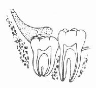
Figure 2: The blind pocket around the crown and the swollen gingival flap.
bubble_chart Auxiliary Examination1. Examination reveals partial eruption of the mandibular third molar, with gingival flap coverage and formation of a blind pouch. The surrounding soft tissue of the crown is red and swollen, the edge of the gingival flap is eroded, and there is purulent discharge in the blind pouch (Figure 3-15). Sometimes a pericoronal abscess may form, accompanied by facial swelling, ipsilateral submandibular lymph node enlargement, and tenderness.
2. X-ray dental examination can detect the presence of an impacted wisdom tooth, as well as its impacted form and position.
3. Laboratory tests: During the acute suppurative pericoronitis phase, there is often an increase in the total white blood cell count to varying degrees, along with a rise in the proportion of neutrophils.
1. It mostly occurs in young people, especially those aged 18 to 25. There are systemic predisposing factors or a history of recurrent episodes.
2. In the early stage of acute pericoronitis, there are generally no obvious systemic reactions. The patient feels distending pain and discomfort in the affected area, with aggravated pain during chewing, swallowing, and mouth-opening movements. Examination may reveal impacted teeth, swelling in the retromolar area, and purulent discharge in the pericoronal pocket.
3. As the inflammation progresses further, involving the masseter and medial pterygoid muscles, swelling in the mandibular angle area occurs, accompanied by varying degrees of limited mouth opening or even complete inability to open the mouth. Systemic symptoms become evident, often with submandibular lymph node enlargement and tenderness. Without timely and appropriate treatment, it may develop into pericoronal abscess, maxillofacial cellulitis, or even osteomyelitis.

bubble_chart Treatment Measures
The treatment of pericoronitis of the wisdom tooth primarily involves enhancing the patient's immune resistance, controlling infection, and promoting the resolution of inflammation. After the acute phase, surgical treatment of the causative tooth should be considered to prevent recurrence.
1. Systemic Treatment
Depending on the condition, antibiotics or oral administration of heat-clearing and toxin-removing Chinese herbal medicine may be selected for treatment.
2. Local Treatment
Local treatment for pericoronitis of the wisdom tooth is crucial. The blind pouch can be irrigated daily with a 1–3% hydrogen peroxide solution and normal saline or other sterile solutions, followed by the application of 3% iodine glycerin. Additionally, a compound borax solution or nitrofurazone solution can be used for gargling multiple times a day. In the early stages, local physiotherapy and topical application of Chinese herbal medicine can also aid in the absorption of inflammation. Acupuncture therapy may help alleviate pain and improve mouth opening. If a pus cavity forms, incision and drainage may be performed.
3. Management of the Causative Tooth
After the acute inflammation subsides, further treatment of the causative tooth should be undertaken to prevent recurrence. If the tooth is properly positioned, can erupt normally, and has an opposing tooth for masticatory function, a wedge-shaped excision of the pericoronal gingival flap may be performed (Figure 1). Otherwise, the tooth should be extracted.

① Incision ② Exposing the entire crown after gingival excision ③ Suturing
Figure 1 Wedge-shaped excision of the pericoronal gingival flap of the mandibular third molar
If acute pericoronitis is not thoroughly treated, it can become chronic, leading to recurrent episodes or even the formation of fistulas. If the inflammation continues to spread, the following complications may occur. For example, it may extend beneath the periosteum to form a subperiosteal abscess, or pus may spread along the outer surface of the mandible, forming multiple abscesses. This can result in an abscess or gingival fistula near the buccal side of the mandibular first or second molar (Figure 1). Alternatively, the infection may spread outward, forming a subcutaneous abscess in the cheek or perforating the skin to create a cutaneous fistula. Clinically, if a patient presents with a cheek cutaneous fistula, the possibility of pericoronitis should be considered to avoid misdiagnosis. Severe pericoronitis may also lead to complications such as perimandibular cellulitis, mandibular osteomyelitis, or even systemic infection.
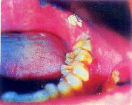
Figure 1: Gingival fistula between teeth 6 and 7 caused by pericoronitis of the mandibular wisdom tooth.
1. Pericoronitis of the mandibular wisdom tooth combined with a gingival fistula in the buccal vestibule of the mandibular first molar should be differentiated from a buccal fistula caused by periapical lesions of the mandibular first molar. In the former case, clinical examination of the first molar reveals no definitive lesions, and periapical radiographs show no periapical pathology, but there is an impacted wisdom tooth and a history of redness and swelling. In the latter case, the first molar exhibits caries, pulp disease, and periapical destruction.
2. Malignant tumors in the third molar region Although malignancies in this area are often accompanied by inflammation, they are primarily proliferative masses with solid infiltrative growth. Radiographic examination may reveal localized osteolytic destruction.


