| disease | Tooth Crack |
| alias | Tooth Microcrack, Incomplete Tooth Fracture, Cracked Tooth |
Also known as incomplete tooth fracture or microcrack. It refers to non-physiological fine cracks on the surface of the tooth crown, often difficult to detect. The cracks of a cracked tooth usually extend into the dentin structure and are one of the causes of toothache. Due to its relatively common occurrence in clinical practice and the tendency for these cracks to be overlooked, clinicians should pay sufficient attention. Cracked teeth most frequently occur in maxillary molars, followed by mandibular molars and maxillary premolars. The first molar is significantly more affected than the second molar, especially the mesiopalatal cusp, as it is the primary working cusp during mastication, bearing the greatest occlusal force and having the most optimal cusp-fossa relationship with the central fossa of the mandibular molar. Although maxillary molars have oblique ridges, unevenly worn high and steep cusps and tight occlusal relationships can also lead to cracks in the mesial or distal fossae of the occlusal surface, or in the fissures between the buccal or lingual cusps.
bubble_chart Etiology
1. The weak points in tooth structure are predisposing factors for the occurrence of cracked teeth. These weak areas not only have low resistance to cracking themselves but are also sites of stress concentration when the tooth is subjected to normal occlusal forces.
2. The larger the cuspal incline, the greater the horizontal component force generated, and the higher the chance of crack formation.
3. Traumatic occlusal forces—when pathological wear leads to steep cusps, the cuspal inclination also significantly increases. The horizontal component force produced during normal occlusion also rises, creating traumatic occlusal forces that deepen and widen the enamel lamellae at the base of the fissures toward the dentin. This marks the beginning of crack formation. Under continued occlusal force, the cracks gradually deepen toward the pulp. Thus, traumatic occlusal forces are the causative factors of tooth cracks.
bubble_chart Clinical ManifestationsThe locations of hidden cracks all overlap with certain pits and fissures on the occlusal surface and extend toward one or both marginal ridges. Hidden cracks in maxillary molars often overlap with the mesiolingual groove on the occlusal surface (Figure 1); hidden crack lines in mandibular molars frequently overlap with the mesiodistal developmental grooves on the occlusal surface and extend beyond the marginal ridge to reach the proximal surface. However, there are also buccolingual hidden cracks that overlap with the buccolingual grooves on the occlusal surface, while hidden cracks in premolars often run in a mesiodistal direction.
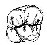
Figure 1 Hidden cracks in maxillary molars
Superficial hidden cracks often show no obvious symptoms, but when deeper, they may become sensitive to hot or cold stimuli or cause discomfort during occlusion. Deep hidden cracks, as they reach the deep layer of dentin, often exhibit symptoms of chronic pulpitis, sometimes acutely, accompanied by localized severe pain during chewing. If the above symptoms are present but no deep caries or periodontal pockets are found in the affected tooth, and no sensitive spots are detected on the tooth surface, the possibility of hidden cracks should be considered. Generally, a sharp probe can be used for examination. If the hidden crack is not obvious, iodine tincture can be applied to stain and reveal it clearly. Sometimes, placing the probe on the crack and applying pressure or prying may cause pain. Grinding along the crack may reveal that it has reached the deep layer of dentin. Placing a cotton swab on the cusp of the suspected tooth and asking the patient to bite down may elicit a brief tearing-like pain, indicating the likely presence of a hidden crack.
bubble_chart Diagnosis1. Medical History Often presents with symptoms of masticatory discomfort or occlusal pain.
2. Clinical Manifestations Most commonly occurs in premolars and molars, with the maxillary first molar being the most frequently affected. Careful examination may reveal shallow black or deep brown fissure lines, which may traverse the occlusal surface of the tooth or only be visible near the marginal area. Pain is often elicited when biting on a cotton swab or during percussion at the site of the fissure.
3. Auxiliary Examination The application of iodine tincture or Chinese Gentian violet can make the fissure lines more distinct. Cold testing tends to be more sensitive at the site of the fissure.
bubble_chart Treatment Measures
1. For superficial cracks with no obvious symptoms and normal pulp vitality, occlusal adjustment can be performed to reduce lateral splitting forces and prevent the crack from deepening. Alternatively, a cavity can be prepared to grind away the crack as much as possible, followed by preventive filling.
2. For deeper cracks or cases with existing pulp pathology, it is crucial to extensively adjust the cusp inclines during pulp treatment, completely eliminate the cracking forces on the affected tooth, and promptly restore it with a full crown after treatment. During pulp therapy, if the occlusal surface is perforated, the tooth's resistance to occlusal forces is significantly reduced. Even if the bite has been reduced during treatment, the tooth is highly prone to splitting along the crack due to chewing or other reasons during the course of treatment. Therefore, a band can be bonded to protect the crown at the beginning of pulp therapy, and a full crown restoration should be performed promptly after completing the pulp treatment.


