| disease | Tricuspid Atresia |
Tricuspid atresia is a cyanotic congenital heart disease, accounting for approximately 1-5% of all congenital heart defects. Among cyanotic congenital heart diseases, it ranks third after tetralogy of Fallot and transposition of the great arteries. The main pathological features include tricuspid valve atresia or absence of the tricuspid orifice, patent foramen ovale or atrial septal defect, mitral valve and left ventricular hypertrophy, and right ventricular hypoplasia.
bubble_chart Pathological Changes
Under normal embryonic development, the endocardial cushions fuse, evenly dividing the atrioventricular canal into left and right orifices and participating in the formation of the membranous part of the ventricular septum and the closure of the first atrial septal foramen. It is generally believed that during the embryonic stage, the fusion site of the anterior and posterior endocardial cushions is biased toward the right side. The rightward shift of the ventricular septum causes unequal division of the atrioventricular orifice, and the occlusion of the right atrioventricular canal orifice leads to the future development of tricuspid atresia.
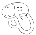
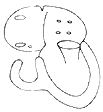
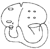
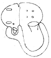
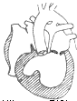
**Pathological Anatomy:** In tricuspid atresia, the right atrium does not directly communicate with the right ventricle, while the left atrium connects to the left ventricle via the mitral valve. No tricuspid valve tissue or orifice is observed in the right atrium. At the base of the right atrium, the original tricuspid valve site is most commonly replaced by muscular tissue (approximately 76%). A thin membranous structure is present in 12% of cases, while fused valve leaflets forming a membranous structure account for 6%, with chordae-like tissue possibly attached to the ventricular side of the fused leaflets. In another 6%, the atrioventricular orifice is obstructed by valve leaflet tissue attached to the right ventricular wall, which Vanpragh termed the Ebstein type (Figure 2).
 |  |
(1) Muscular type | (2) Membranous type |
 |  |
(3) Valvular type | (4) Ebstein type |
**Figure 2: Anatomical Classification of Tricuspid Atresia**
Vanpragh classified this condition into three types: ① **Muscular type (84%)**: Presents as a fibrous depression. Microscopic examination reveals radially arranged muscle fibers. ② **Membranous type (8%)**: Accompanied by juxtaposed atrial appendages, displaying transparent fibrous tissue. ③ **Ebstein type (8%)**: The atrialized right ventricle forms a blind-ended pouch below the right atrium. The right atrial wall is thickened and enlarged, the left atrium is dilated, and there may be a patent foramen ovale, atrial septal defect, or even a single atrium. The right ventricle is underdeveloped, particularly in the inflow tract. When pulmonary atresia is present, the right ventricle may be absent or reduced to a small cavity only a few millimeters in diameter, with rudimentary papillary muscles possibly present in the lower part of the right ventricular cavity. Ventricular septal defects of varying sizes may exist between the left and right ventricles, with larger defects correlating with a larger right ventricular cavity. Occasionally, a narrow, thin-walled right ventricular outflow tract may be observed below the pulmonary valve. In rare cases, the right ventricle is entirely absent or appears as a small fissure within the right ventricular wall below the pulmonary valve. In extremely rare instances, the left and right ventricles are malpositioned, with the mitral valve straddling the displaced ventricular septum, and the right ventricle assuming the primary blood-pumping function. In such cases, the right ventricle on the left side of the heart is better developed, while the left ventricle on the right side is hypoplastic.
 |  |  | |
| (1) Type IA pulmonary stirred pulse atresia | (2) Type IB pulmonary stirred pulse hypoplasia with small ventricular septal defect | (3) Type IC pulmonary stirred pulse normal with large ventricular septal defect | |
 |  |  | |
| (4) Type IIA pulmonary stirred pulse atresia | (5) Type IIB pulmonary stirred pulse valve or subvalvular stenosis | (6) Type IIC pulmonary stirred pulse dilation | |
 |  | ||
| (7) Type IIIA pulmonary stirred pulse or pulmonary stirred pulse subvalvular stenosis | (8) Type IIIB aortic stirred pulse subvalvular stenosis | ||
Figure 3 Keith classification of tricuspid atresia

If the defect or patent foramen ovale enters the left atrium, and if the defect is small, the systemic venous pressure rises, leading to hepatomegaly and right heart failure. ② The complete mixing of systemic venous blood and oxygenated pulmonary venous blood in the left atrium results in varying degrees of reduced blood oxygen saturation in the stirred pulse. Those with high pulmonary blood flow may not exhibit cyanosis or may show grade I cyanosis, while those with narrowed pulmonary stirred pulse outlets may exhibit grade III cyanosis. ③ Due to right ventricular hypoplasia, the ventricular cavity is very small, so the left ventricle takes on the pumping function of both ventricles, often becoming enlarged and leading to left heart failure. Approximately 20% of patients with tricuspid atresia present with cyanosis clinically due to accompanying pulmonary stirred pulse outlet stenosis. Another group of cases with normal or increased pulmonary blood flow may develop heart failure or pulmonary vascular obstructive sexually transmitted disease changes. In cases of major stirred pulse dextro-transposition, especially those with a large pulmonary stirred pulse accompanied by aortic coarctation or hypoplasia, severe heart failure may lead to early death after birth. As pulmonary vascular obstructive sexually transmitted disease worsens and pulmonary blood flow gradually decreases, cyanosis also progressively intensifies.
bubble_chart Clinical Manifestations
(1) Symptoms The survival period of patients with tricuspid atresia is closely related to pulmonary blood flow. Those with near-normal pulmonary blood flow can survive for up to 8 years or more; those with excessive pulmonary blood flow generally survive only about 3 months after birth; those with less than normal pulmonary blood flow have a survival period between the aforementioned two scenarios. Keith et al. reported that 50% of tricuspid atresia patients survive to 6 months, 33% to 1 year, and only 10% to 10 years. Cases with small atrial septal defects clinically present with systemic venous congestion, jugular vein distension, hepatomegaly, and peripheral edema. Due to reduced pulmonary blood flow, most cases exhibit cyanosis from the neonatal period, dyspnea after exertion, and may assume a squatting position or experience hypoxic syncope. Patients over 2 years old often develop clubbing of fingers and toes. In cases with increased pulmonary blood flow, cyanosis is less severe, but dyspnea, tachypnea, and recurrent pulmonary infections are common, often presenting as congestive heart failure.
(2) Signs A systolic blowing murmur caused by pulmonary stenosis or ventricular septal defect is often heard at the left sternal border, and a continuous machinery-like murmur may be audible in cases with patent ductus arteriosus. Patients with increased pulmonary blood flow may exhibit a diastolic rumbling murmur. Additionally, signs such as hepatomegaly, edema, jugular vein distension, and pulmonary edema may be present.
bubble_chart Auxiliary Examination
In 90% of cases, the electrocardiogram shows left axis deviation and malposition of the {|###|} great artery. Patients with thickened pulmonary {|###|} arteries have normal or right axis deviation. All precordial leads exhibit left ventricular hypertrophy and T-wave inversion. In 80% of cases, the P wave is tall or widened with notching.
X-ray examination reveals considerable variability in chest X-ray findings. Cases with reduced pulmonary blood flow show normal heart shadow or grade I enlargement, while those with increased pulmonary blood flow exhibit significant cardiac enlargement. The typical chest X-ray features include a straightened right heart border, rounded left heart border, elevated apex, and a concave waist of the heart, sometimes resembling tetralogy of Fallot. Malposition of the {|###|} great artery may present an egg-shaped heart shadow. Cases with reduced pulmonary blood flow show markedly decreased lung markings, whereas those with pulmonary congestion exhibit increased lung markings.
Cardiac catheterization and {|###|} angiography: A right heart catheter can pass through the atrial septal defect into the left atrium, with right atrial pressure higher than left atrial pressure. The pressure gradient is inversely proportional to the diameter of the atrial defect—smaller defects result in larger gradients. The {|###|} artery shows reduced oxygen content, while the left atrium, left ventricle, pulmonary {|###|} artery, and aorta have identical oxygen levels. Selective right atrial angiography reveals contrast medium flowing from the right atrium into the left atrium and left ventricle, then into the pulmonary {|###|} artery and aorta. An unopacified triangular area, the right ventricular window, is visible below the cardiac shadow, located between the right atrium, left ventricle, and diaphragm
. Occasionally, angiography may reveal a ventricular septal defect, right ventricular cavity, outflow tract, and pulmonary {|###|} artery. Additionally, it can demonstrate the spatial relationship and position of the two great {|###|} arteries. Left ventricular angiography can assess the presence of mitral regurgitation.
M-mode echocardiography shows the disappearance of the tricuspid valve's double-peaked curve. Four-chamber views fail to detect tricuspid valve echoes, with interrupted echoes in the atrial septum and upper ventricular septum. Echocardiography and Doppler studies also reveal blood flow from the right atrium to the left atrium and then into the left ventricle. The mitral valve exhibits increased motion amplitude, while the right atrium, left atrium, and left ventricular cavity are enlarged, and the right ventricle is small or absent.
Clinically presenting with cyanosis, dyspnea, and lack of strength, along with an electrocardiogram showing left axis deviation and left ventricular hypertrophy, as well as tall and wide P waves, should raise high suspicion for possible tricuspid atresia. Right heart catheterization and cardiac angiography, along with echocardiography, can confirm the diagnosis. Differential diagnoses include tetralogy of Fallot, Ebstein's anomaly, transposition of the great arteries, double outlet right ventricle, and single ventricle.
bubble_chart Treatment Measures
The prognosis of tricuspid atresia is extremely poor, with a very short survival period. Approximately 70% of affected infants die within the first year of life. In cases where pulmonary blood flow is reduced, grade III cyanosis is observed. For patients with a pressure gradient between the right and left atria, the following palliative surgeries can be performed to increase pulmonary circulation blood flow.
(1) Palliative Surgeries
1. Systemic-pulmonary shunt: Commonly used methods include the left subclavian artery-pulmonary artery end-to-side anastomosis (Blalock-Taussig shunt) or connecting a segment of Gortex artificial vessel between the subclavian artery and the pulmonary artery. Alternatively, descending aorta-left pulmonary artery side-to-side anastomosis (Potts shunt) or ascending aorta-main pulmonary artery side-to-side anastomosis (Waterston shunt) can be performed. The latter two procedures may cause pulmonary artery distortion or an overly large anastomosis, leading to excessive pulmonary blood flow.
2. Balloon catheter atrial septal defect enlargement or closed atrial septal resection: In tricuspid atresia, two-thirds of atrial communications are due to patent foramen ovale, and one-third are due to atrial septal defects. If right heart catheterization reveals that right atrial pressure exceeds left atrial pressure by >0.67 kPa (5 mmHg), the atrial communication must be enlarged. A balloon catheter can be passed through the atrial septal defect to expand the defect. This method can be performed during cardiac catheterization and is often used in infants and young children to alleviate symptoms. Alternatively, a closed method can be used to create a defect in the atrial septum, relieving right atrial and caval hypertension and mitigating right heart failure.
3. Superior vena cava-right pulmonary artery anastomosis (Glenn surgery): The Glenn procedure has relatively good outcomes, with the advantages of not increasing left ventricular load or causing pulmonary vascular disease. However, the surgical mortality rate is higher in patients under 6 months of age, and the interruption of left and right pulmonary artery continuity complicates future reconstructive surgeries.
4. Pulmonary artery banding: Excessive pulmonary blood flow can lead to congestive heart failure and pulmonary vascular obstructive disease. If medical therapy fails to control heart failure, pulmonary artery banding can be performed to reduce pulmonary blood flow, improve heart failure, and prevent pulmonary vascular disease.
(2) Corrective Surgery
In 1968, Fontan successfully treated tricuspid atresia by performing a right atrium-pulmonary artery anastomosis while suturing the atrial septal defect. The goal of the Fontan procedure is to direct all systemic venous blood returning to the right atrium into the pulmonary artery for oxygenation without relying on the right ventricle to pump blood. The anatomical anomaly is preserved. Surgical indications include:
① Mean pulmonary artery pressure <2 kPa (<15 mmHg),
② Pulmonary vascular resistance <4 Wood units/m²,
③ Left ventricular ejection fraction >0.6,
④ Left ventricular end-diastolic pressure <1.6 kPa (<12 mmHg),
⑤ No significant mitral valve disease,
⑥ Age >2–3 years,
⑦ Sinus rhythm,
⑧ Aorta-pulmonary artery diameter ratio ≥0.75.
The Fontan procedure includes the following operative methods:
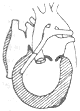
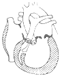
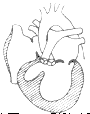
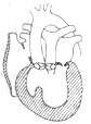
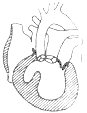
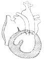
1. Right atrium-pulmonary artery connection: Suitable for tricuspid atresia with transposition of the great arteries or pulmonary stenosis but well-developed left and right pulmonary arteries. During surgery, the main pulmonary artery is transected at its root, the proximal end is closed, and the main pulmonary artery is rerouted behind the aorta to the right side and anastomosed to the roof of the right atrium. The main and branch pulmonary arteries must be fully mobilized to prevent postoperative traction and anastomotic stenosis. When closing the atrial septal defect with a pericardial patch, the roof of the left atrium is incorporated into the right atrium to ensure adequate anastomotic diameter. The anastomotic diameter should not be less than 2 cm in children under 2 years of age and 2.5–3 cm in those over 3 years. Alternatively, a valved external conduit can be placed between the right atrium and pulmonary artery, and the atrial septal defect can be closed with a patch via a right atrial incision. Before chest closure, the external conduit must be checked for compression; if compressed, part of the sternal plate should be resected (Figure 1).
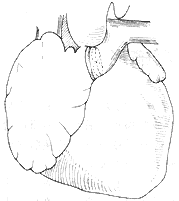
(1) The same type of main stirred pulse is anastomosed between the right atrium and the main pulmonary stirred pulse.
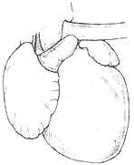
(2) The right ventricle is anastomosed to the right atrium with a Teflon ring support.
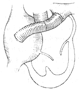
(3) A Daeron conduit with a porcine main stirred pulse valve is anastomosed between the right atrium and the main pulmonary stirred pulse.
Figure 1: Schematic diagram of tricuspid atresia surgery.
2. Right atrium-right ventricular outflow tract anastomosis: Applicable when there is no stenosis in the right ventricular outflow tract, no stenosis in the pulmonary stirred pulse valve ring or main pulmonary stirred pulse, or no gap between the main stirred pulse and the superior vena cava, making it unsuitable for anastomosis between the top of the right atrium and the pulmonary stirred pulse. The surgical method involves making a "门"-shaped incision in the right atrium, flipping the atrial wall to the right ventricular outflow tract incision, and anastomosing it to the lower edge of the incision. The anterior wall is covered with a pericardial patch to form a channel. Alternatively, an external conduit can be placed between the right atrium and the right ventricle. Under extracorporeal circulation, a right ventricular incision is made to remove hypertrophic muscles in the fistula disease cavity. The ventricular septal defect is directly sutured or repaired with a patch. Through the right atrial incision, the atrial septal defect is closed with a patch. Finally, a Dacron patch or Gortex external conduit is used to anastomose the right atrium to the fistula disease cavity of the right ventricle.
3. Disconnection of the superior vena cava, with the distal end of the superior vena cava anastomosed to the right pulmonary stirred pulse and the proximal end anastomosed to the main pulmonary stirred pulse: During surgery, the superior vena cava and the left and right pulmonary stirred pulses are fully mobilized to prevent twisting of the superior vena cava or pulmonary stirred pulse during anastomosis, which could cause anastomotic stenosis. The top of the right atrium is preserved to avoid injury to the sinoatrial node stirred pulse.
During surgery, the following should be noted: ① Maintain the anatomical and functional integrity of the right atrium as much as possible to ensure effective pulmonary circulation dynamics and reduce atrial arrhythmias postoperatively. ② The diameter of the valved or non-valved external conduit should be sufficiently large—approximately 20 mm for children around 6 years old and 22–25 mm for older children. The conduit should be pre-clotted with blood before heparinization to prevent bleeding after heartbeat. ③ The conduit should be placed in an appropriate position to avoid compression by the sternum. ④ Postoperatively, right atrial pressure should be measured. If it exceeds 3.3 kPa (25 mmHg) and right cardiac output is below 2 L/m2, an anastomosis between the superior vena cava and the right pulmonary stirred pulse should be performed to reduce right atrial pressure. A temporary cardiac pacemaker should be installed to control heart rate.
Postoperative management: Monitor cardiopulmonary function postoperatively. Early maintenance of right atrial pressure >2.0 kPa (15 mmHg) is essential; if this cannot be maintained, blood and plasma transfusion should be administered. For low cardiac output syndrome, drugs such as dopamine, isoproterenol, or nitroprusside should be used. If there is significant early postoperative bleeding, fresh blood, platelets, and fibrinogen should be promptly administered. Increased right atrial pressure postoperatively may restrict lymphatic回流, leading to increased drainage, which can be managed with diuretics and/or digitalis Rehmannia. Postoperative anticoagulation should be maintained for 2–3 months.
Postoperative efficacy and prognosis: Shunt surgery: Trusle reported the outcomes of 148 cases of tricuspid atresia shunt surgeries performed between 1947 and 1978, including 52 Potts procedures, 46 Blalock procedures, 22 Glenn procedures, 9 Waterston procedures, and 19 other procedures. The surgical mortality rate was 47.4% for infants under 6 months and 13.9% for those over 6 months. Among the 95 surviving cases followed up, 44 were asymptomatic, 48 had mild to grade II symptoms, and 3 had severe activity limitations. Among these procedures, the Glenn procedure was found to have the best outcomes. Dick reported that a large group of patients survived 10–15 years after shunt surgery. The early mortality rate for the Fontan procedure was 20–30%, but significant improvements have been made, with satisfactory surgical outcomes. Early complications included right heart failure, pleural effusion, hepatomegaly, and ascites, most of which resolved within one week, though persistent pleural effusion remained. Most postoperative patients experienced the disappearance of cyanosis and a significant improvement in activity tolerance. Miller reported that right heart catheterization in postoperative patients showed an average right atrial pressure of 1.87–2.40 kPa (14–18 mmHg) and an arterial oxygen saturation of 87–92%. However, this procedure still presents long-term issues such as hemodynamic abnormalities due to homograft or heterograft valve dysfunction. The long-term increase in right atrial load leads to right atrial enlargement and predisposes to atrial arrhythmias, though most cases show satisfactory early results.


