| disease | Esophagus Cancer |
| alias | Esophageal Cancer, Carcinoma of Esophagus |
Esophageal cancer is a malignant tumor that occurs in the epithelial tissue of the esophagus, accounting for 2% of all malignant tumors. Approximately 200,000 people worldwide die from esophageal cancer each year. China is a high-incidence area for esophageal cancer, with deaths from esophageal cancer ranking second only to stomach cancer. The age of onset is mostly over 40, and it is more common in men than in women. However, in recent years, there has been an increasing trend in the incidence among those under 40 years old. The occurrence of esophageal cancer is related to chronic stimulation by nitrosamines, inflammation and trauma, genetic factors, as well as the content of trace elements in drinking water, grains, and vegetables. However, the exact cause is not well understood and requires further research and exploration.
bubble_chart Epidemiology
(1) Incidence and Mortality Rates The incidence of this disease varies significantly among different countries (or regions), and there can be notable differences even within the same country or among different ethnic groups. In Europe, America, and Oceania, the incidence of esophageal cancer generally ranges from 2 to 5 per 100,000 (with the exception of France, where it reaches 13.6 per 100,000). In the Central Asian region of the former Soviet Union, the rate is as high as over 100 per 100,000. In Asian countries, the incidence ranges from 1.2 to 32 per 100,000, but in the coastal areas of the Black Sea in Iran, it reaches over 100 per 100,000.
According to a survey of over 850 million people in 29 provinces, cities, and autonomous regions in China from 1974 to 1978, approximately 700,000 people died from malignant tumors, of which 157,000 died from esophageal cancer (accounting for 22.4%), second only to stomach cancer (22.8%). Among the mortality rates of various malignant tumors, esophageal cancer ranks first in nine provinces and cities: Henan (40.55%), Jiangsu, Jiangxi, Hebei, Shaanxi, Anhui, Sichuan, Hubei, and Beijing. Currently, China is one of the countries with the highest esophageal cancer mortality rates in the world, with an average annual mortality rate of 14.59 per 100,000. The lowest rate is in Yunnan Province (1.05 per 100,000), and the highest is in Henan Province (32.22 per 100,000).
(2) Gender and Age The reported incidence of this disease varies significantly by gender abroad, with a male-to-female ratio ranging from 1.1 to 17:1. According to survey data from various regions in China, the male-to-female incidence ratio ranges from 1.3 to 2.7:1.The disease primarily affects older age groups. The proportion of cases under 35 years old is very small, and it increases with age after 35. The highest proportion is in the 60-64 age group (17.95%), followed by the 65-69 age group, and it gradually decreases after 70 years old.
The exact cause of esophageal cancer is unknown. Clearly, environmental factors and certain carcinogens are important contributing factors.
(1) Nitrosamines and mycotoxins: It is now known that nearly 30 types of nitrosamines can induce tumors in animals. Domestically, nitrosamines such as methylbenzyl nitrosamine, ethyl nitrosoguanidine, methylamyl nitrosamine, and diethylnitrosamine have been successfully used to induce esophageal cancer in rats. Surveys in China have found that in high-incidence areas, the levels of nitrates, nitrites, and secondary amines in food and water are significantly higher, and these levels are positively correlated with the prevalence of esophageal cancer and grade III esophageal epithelial hyperplasia. These substances are easily synthesized into carcinogenic nitrosamines in the stomach.
(2) Esophageal injury, esophageal diseases, and dietary stimulation: Esophageal injury and certain esophageal diseases can promote the development of esophageal cancer. The incidence of esophageal cancer is higher in patients with corrosive esophageal burns and strictures, achalasia, esophageal diverticula, or reflux esophagitis than in the general population. It is speculated that this is due to long-term chronic inflammation, ulcers, or chronic irritation caused by esophageal retention, leading to epithelial hyperplasia and eventually cancer. Epidemiological surveys have found that residents in high-incidence areas of esophageal cancer have habits of consuming very hot food, drinking strong alcohol, eating large amounts of pepper fruit, chewing areca seeds, or tobacco, all of which can cause chronic physical and chemical irritation of the esophageal mucosa, leading to local epithelial cell hyperplasia. Animal experiments have shown that diffuse or focal epithelial hyperplasia may be a precancerous lesion of esophageal cancer.
(4) Genetic factors: The incidence of esophageal cancer often shows familial clustering. Surveys in provinces such as Shanxi, Shandong, and Henan in China have found that about 1/4 to 1/2 of cases have a positive family history. In high-incidence areas, the proportion of positive family history is higher, with the highest being paternal, followed by maternal, and the lowest being collateral.
bubble_chart Pathological Changes
The site of esophageal cancer lesions varies across different regions in China, but the middle segment is the most common (52.69% to 63.33%), followed by the lower segment (24.95% to 38.92%), and the upper segment is the least common (2.80% to 14.0%). Among the 622 cases in our hospital, 68.90% were in the middle segment, 26.40% in the lower segment, and 4.70% in the upper segment.
(1) Clinical pathological staging and classification
1. Clinical pathological staging The clinical pathology of esophageal cancer is of great significance for the selection of treatment plans and the evaluation of treatment outcomes. The clinical pathological staging criteria established at the 1976 National Esophageal Cancer Conference are shown in Table 18-7.
Table 18-7 Clinical pathological staging of esophageal cancer
| Stage | Lesion length | Lesion extent | Metastasis status |
| 0 | Not specified | Limited to the mucosal layer | No metastasis |
| 1 | <3cm | Invades submucosal layer | No metastasis |
| 2 | 3~5cm | Invades part of the muscular layer | No metastasis |
| 3 | >5cm | Invades through the muscular layer or outer layer | Local lymph node metastasis |
| 4 | >5cm | Significant external invasion | Distant lymph node or organ metastasis |

2. Pathological morphological classification
(1) Pathological morphological classification of early esophageal cancer: Early esophageal cancer can be classified into latent type, erosive type, patch type, and papillary type based on morphology.
(2) Pathological morphological classification of middle and advanced stage esophageal cancer: It can be classified into medullary type, fungating type, ulcerative type, constrictive type, intraluminal type, and unclassified type. Among these, the medullary type has the highest degree of development. A small number of middle and advanced stage esophageal cancers cannot be classified into the above types and are referred to as unclassified.
3. Histological classification
(1) Squamous cell carcinoma: Most common.
(2) Adenocarcinoma: Less common, and can be further divided into simple adenocarcinoma, adenosquamous carcinoma, mucoepidermoid carcinoma, and adenoid cystic carcinoma.
(3) Undifferentiated carcinoma: Less common but highly malignant.
The majority of cancers in the upper and middle segments of the esophagus are squamous cell carcinomas, while cancers in the lower segment are mostly adenocarcinomas.
(2) Spread and metastasis patterns of esophageal cancer
1. Intramural spread of esophageal cancer The cancerous transformation of basal cells in the epithelium adjacent to esophageal cancer or the formation of carcinoma in situ is one of the surface spread patterns of cancer. Cancer cells often infiltrate the lymphatic vessels of the esophageal submucosal layer.
2. Direct invasion of adjacent organs Upper segment esophageal cancer can invade the larynx, trachea, and cervical soft tissues, and even invade the bronchi, forming a bronchial-esophageal fistula; it can also invade the thoracic duct, azygos vein, hilum, and lung tissue, and in some cases, invade the main artery to form an esophageal-aortic fistula, causing massive hemorrhage and death. Lower segment esophageal cancer often involves the cardia and pericardium.
3. Lymphatic metastasis is relatively common, accounting for about two-thirds of the cases. Mid-esophageal cancer often metastasizes to the paraesophageal or hilar lymph nodes, and can also metastasize to the cervical, perigastric, and left gastric artery lymph nodes. Lower esophageal cancer often metastasizes to the paraesophageal, perigastric, left gastric artery, and abdominal lymph nodes, and occasionally to the superior mediastinum and cervical lymph nodes. The order of lymphatic metastasis is the mediastinum, abdomen, trachea and paratracheal, hilar, and bronchial lymph nodes.
4. Hematogenous metastasis is commonly seen in patients with advanced stage. The most common sites of metastasis are the liver (accounting for about 1/4) and the lungs (accounting for about 1/5). Other organs, in order of frequency, include the bones, kidneys, adrenal glands, pleura, peritoneum, pancreas, heart, lungs, thyroid, and brain.
bubble_chart Clinical Manifestations
(I) Early symptoms of esophageal cancer
1. Dysphagia: The most common symptom, which can disappear and recur on its own without affecting eating. It often occurs when the patient is emotionally disturbed, so it is easily mistaken for a functional symptom.
2. Pain behind the sternum and under the xiphoid process: More common. Pain behind the sternum or under the xiphoid process occurs when swallowing food, which can be burning, stabbing, or pulling in nature, especially when swallowing rough, hot, or irritating food. Initially intermittent, the pain can become severe and persistent when the cancer invades nearby tissues or penetrates. The location of the pain often does not completely correspond to the location of the lesion in the esophagus. The pain can often be temporarily relieved by antispasmodics.
3. Food retention infection and foreign body sensation: When swallowing food or water, there is a feeling of slow passage and retention of food, as well as a tightness behind the sternum or a feeling of food sticking to the esophageal wall, which disappears after eating. The location of the symptoms often corresponds to the location of the lesion in the esophagus.
4. Dryness and tightness in the throat: Especially noticeable when swallowing dry and rough food. The occurrence of this symptom is also often related to the patient's emotional fluctuations.
5. Other symptoms: A few patients may experience discomfort, pain, and belching behind the sternum.
(II) Late stage [third stage] symptoms of esophageal cancer
1. Dysphagia: Progressive dysphagia is the main symptom of the vast majority of patients when they seek medical attention, but it is a manifestation of the more advanced stage of the disease. Because the esophageal wall is elastic and has the ability to expand, dysphagia only occurs when about 2/3 of the esophageal circumference is infiltrated by cancer. Therefore, after the above early symptoms appear, the condition gradually worsens over several months, from being unable to swallow solid food to being unable to swallow liquid food. If the cancer is accompanied by inflammation, edema, or spasm of the esophageal wall, it can worsen dysphagia. The location of the obstruction often corresponds to the location of the cancer.
2. Food reflux: Often occurs when dysphagia worsens, with a small amount of reflux containing food and mucus, and may also contain blood and pus.
3. Other symptoms: When the cancer compresses the recurrent laryngeal nerve, it can cause hoarseness; invasion of the phrenic nerve can cause hiccups or phrenic nerve paralysis; compression of the trachea or bronchus can cause shortness of breath and dry cough; erosion of the main stirred pulse can cause fatal bleeding. Complications such as esophageal-tracheal or esophageal-bronchial fistula or cancer located in the upper esophagus can cause cervical sympathetic nerve paralysis syndrome when swallowing liquids.
(III) Signs
Early signs are often absent. In the advanced stage, signs such as weight loss, anemia, malnutrition, dehydration, or cachexia may appear. When the cancer metastasizes, enlarged and hard superficial lymph nodes or an enlarged and nodular liver may be palpable.
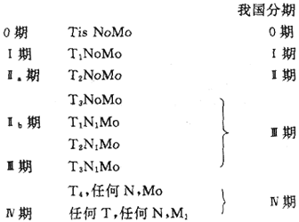
(IV) Clinical staging
In 1987, the International Union Against Cancer (UICC) revised the TNM classification for esophageal cancer.
1. Primary tumor (T) staging: Tx Primary tumor cannot be assessed; To Primary tumor size and location unknown; Tis Carcinoma in situ; T1 Tumor infiltrates the esophageal mucosal layer or submucosal layer; T2 Tumor infiltrates the esophageal muscle layer; T3 Tumor infiltrates the esophageal outer membrane; T4 Tumor invades adjacent structures (organs) of the esophagus.
2. Regional lymph nodes (N) staging: Nx Regional lymph nodes cannot be assessed; No No regional lymph node metastasis; N1 Regional lymph node metastasis.
3. Distant metastasis (M) staging: Mx Distant metastasis status unknown; Mo No distant metastasis; M1 Distant metastasis present.
4. TNM staging
bubble_chart Auxiliary Examination
(1) X-ray Barium Meal Examination Esophageal X-ray barium meal examination can reveal the stagnation of barium at the site of the cancer, with a narrow flow of barium in the diseased segment; the esophageal wall is stiff, peristalsis is weakened, the mucosal pattern becomes thickened and disordered, with rough edges; the esophageal lumen is narrow and irregular, with grade I dilation above the obstruction, and there may be ulcer niches and filling defects. Conventional X-ray barium meal examination often fails to detect superficial and small cancers. The use of sodium methyl cellulose and barium for double-contrast imaging can more clearly display the esophageal mucosa, improving the detection rate of esophageal cancer.
(2) Fiber Esophagogastroscopy This allows direct observation of the morphology of the cancer and enables pathological biopsy under direct vision to confirm the diagnosis.
(3) Esophageal Mucosal Exfoliative Cytology A wire mesh balloon double-lumen tube cell collector is swallowed into the esophagus, inflated after passing the diseased segment, and then slowly withdrawn. The mesh is used to collect cells for cytological examination, with a positive rate of over 90%. This method often detects some early cases and is an important method for large-scale screening of esophageal cancer.
(4) Esophageal CT Scan CT scanning can clearly show the relationship between the esophagus and adjacent mediastinal organs. Normally, the esophagus is clearly demarcated from adjacent organs, with a wall thickness not exceeding 5mm. If the esophageal wall thickens and the boundary with surrounding organs becomes blurred, it indicates the presence of esophageal disease.
(5) Other Examination Methods The use of toluidine blue or iodine staining during endoscopy has certain value in the early diagnosis of esophageal cancer. This method is simple, easy to perform, and has the advantages of accurate localization and determination of the cancer's extent.
For any suspected cases, a barium swallow X-ray examination should be performed. Early signs of esophageal cancer on X-ray include: ① localized thickening and disruption of the mucosal folds. ② localized rigidity of the esophageal wall. ③ localized small filling defects. ④ small niches. In the advanced stage, there are mostly filling defects, luminal narrowing, or obstruction. For those highly suspected but not definitively diagnosed, esophagoscopy and biopsy should be performed. Esophageal cytology examination by balloon cytology and radioactive isotope 32P are helpful for the early diagnosis of cancer.
bubble_chart Treatment Measures
The proliferation cycle of normal esophageal epithelial cells is the longest in the human digestive tract. The process from grade III hyperplasia of esophageal basal cells to carcinogenesis takes approximately 1 to 2 years; the progression from early esophagus cancer (cytological detection of cancer cells with normal X-ray esophageal membrane imaging or only grade I lesions) to advanced stage invasive cancer usually takes 2 to 3 years, or even longer; in some cases, patients can "live with cancer" for more than 6 years. Therefore, the early treatment of esophagus cancer yields good results. Even for advanced stage cases, proper treatment can lead to positive outcomes. Generally, surgical treatment is preferred for earlier stage lesions; for more advanced stage lesions located in the middle or upper segments, especially in older patients or those with surgical contraindications, radiation therapy is more suitable.
(1) Surgical Treatment
Surgical intervention is the primary method for treating esophagus cancer. The resection rate for lower segment cancer is 90%, for middle segment cancer it is 50%, and for upper segment cancer, the average resection rate ranges from 56.3% to 92.9%.
Contraindications for surgery include: ① Clinical and X-ray examinations confirming extensive esophageal lesions involving adjacent organs such as the trachea, lungs, mediastinum, and major arteries. ② Severe cardiac, pulmonary, hepatic, or renal insufficiency or cachexia that makes the patient unable to tolerate surgery. Apart from these conditions, surgical treatment should be pursued as soon as the diagnosis is confirmed and the patient's physical condition permits. Additionally, depending on the condition, surgeries can be categorized into palliative and radical surgeries. Palliative surgeries are mainly for advanced stage patients who cannot undergo radical surgery or post-radiotherapy, and include procedures like esophageal-gastric bypass, gastrostomy, and intraluminal esophageal stenting to alleviate difficulty in eating. Radical surgeries are determined based on the lesion location and the patient's specific condition. In principle, the majority of the esophagus should be removed, with the resection margin at least 5 cm away from the tumor.
(2) Radiation Therapy
Radiation therapy for esophagus cancer includes both radical and palliative types. For cervical and upper thoracic esophagus cancer, surgery is associated with significant trauma and high complication rates, whereas radiotherapy causes less injury and is more effective than surgery, making it the preferred choice. Patients in relatively good general condition, able to consume semi-liquid or liquid diets, with thoracic esophagus cancer without supraclavicular lymph node metastasis or distant metastasis, no tracheal invasion, no signs of esophageal perforation or bleeding, and lesion length <7-8 cm without medical contraindications, are suitable for radical radiotherapy. Other patients may undergo palliative radiotherapy aimed at relieving esophageal obstruction, improving difficulty in eating, reducing pain, enhancing quality of life, and prolonging survival.
(3) Drug Treatment
1. The cell proliferation cycle of chemotherapy for esophageal cancer is approximately 7 days, slightly longer than the cycle of normal esophageal epithelial cells. Theoretical calculations suggest a doubling time of about 10 days, indicating fewer proliferating cells and more non-proliferating cells. Therefore, although many chemotherapeutic drugs are currently used for this disease, few have proven efficacy. The most commonly used drugs include Bleomycin (BLM), Mitomycin C (MMC), Adriamycin (ADM), 5-Fluorouracil (5-Fu), Methotrexate (MTX), Lomustine (CCNU), Mitoguazone (MGAG), Vindesine (VDS), Etoposide (VP-16), and Cisplatin (DDP). The remission rate of single-agent chemotherapy is 15-20%, with a remission stage of 1-4 months. Combination chemotherapy mostly adopts regimens based on DDP and BLM, with an efficacy rate mostly exceeding 30%, and a remission stage of about 6 months. Combination chemotherapy is not only used for intermediate and advanced stage esophageal cancer but also for comprehensive treatment with surgery and radiotherapy. Currently, commonly used combination chemotherapy regimens in clinical practice include DDP-BLM, BLM-ADM, DDP-VDS-BML, and DDP-ADM-5-Fu. Clinical observations have shown that chemotherapeutic drugs such as DDP, 5-Fu, and BLM have radiosensitizing effects. Over the past decade, these chemotherapeutic drugs have been used as sensitizers in combination with radiotherapy for the treatment of esophageal cancer, achieving encouraging therapeutic outcomes.
2. Chinese medicinals treatment Currently, a combination of the main formula with pattern identification and treatment, as well as strengthening the body and promoting blood circulation to remove stasis, is commonly used. In North China, Rabdosia rubescens and its active component, rubescensin, have been applied. Experimental studies have shown that they have a significant cytotoxic effect on the human esophageal squamous carcinoma cell line CaEs-17 and exhibit inhibitory effects on various animal transplanted tumors. Clinical applications have also demonstrated certain therapeutic efficacy.
The prognosis for esophageal cancer patients is generally better for squamous cell carcinoma than for adenocarcinoma; the constrictive and fungating types have a better prognosis than the ulcerative and medullary types. The 5-year survival rate for early-stage esophageal cancer without metastasis or invasion is 60%, while for those with invasion or metastasis or mid-esophageal cancer, the 5-year survival rate is less than 25%. The average 5-year survival rate ranges from 18.1% to 40.8%. However, foreign reports indicate a very poor prognosis for esophageal cancer, with a 5-year survival rate of less than 5%.
1. Change unhealthy eating habits, avoid eating moldy food, and reduce or eliminate the consumption of pickled vegetables.
2. Improve water quality and reduce the content of nitrites in drinking water.
3. Promote the use of trace element fertilizers to correct soil deficiencies in molybdenum and other trace elements.
4. Use Chinese and Western medicines and vitamin B2 to treat esophageal epithelial hyperplasia to block the cancerization process. Actively treat diseases related to esophageal cancer, such as esophagitis, esophageal leukoplakia, achalasia, and esophageal diverticulum.
5. Monitor high-risk populations, popularize cancer prevention knowledge, and raise awareness of cancer prevention.
This disease should be differentiated from the following conditions:
(1) Esophageal achalasia: Patients are mostly young women with a long course of illness, and symptoms vary in severity. Barium swallow examination shows a smooth funnel-shaped narrowing at the lower end of the esophagus, which can be dilated with the use of antispasmodics.
(2) Benign esophageal stricture: This can be caused by scars resulting from accidental ingestion of corrosive agents, esophageal burns, foreign body injury, chronic ulcers, etc. The course is long, and dysphagia does not worsen beyond a certain point. Detailed history taking and X-ray barium swallow examination can help differentiate.
(3) Benign esophageal tumors: Mainly rare leiomyomas, with a long course and intermittent dysphagia. X-ray barium swallow examination may show round, oval, or lobulated filling defects in the esophagus with smooth edges and normal surrounding mucosal folds.
(4) Globus hystericus: Mostly seen in young women, with a sensation of a ball-like foreign body in the throat that disappears during eating, often triggered by psychological factors. There is actually no organic esophageal lesion, and it is not difficult to differentiate from esophageal cancer.
(5) Iron deficiency pseudomembranous esophagitis: Mostly in women, besides dysphagia, there may also be microcytic hypochromic anemia, glossitis, achlorhydria, and koilonychia.
(6) Lesions of surrounding organs: Such as mediastinal tumors, aortic aneurysm, thyroid enlargement, and cardiac enlargement. Except for mediastinal tumors invading the esophagus, X-ray barium swallow examination may show smooth indentations in the esophagus with normal mucosal folds.


