| disease | Oral Candidiasis |
| alias | Oral Candidiasis |
Oral candidiasis is a mucosal disease of the oral cavity caused by infection with the fungus Candida. In recent years, due to the widespread clinical use of antibiotics and immunosuppressants, the occurrence of dysbiosis or reduced immunity has led to an increasing number of infections of internal organs, skin, and mucous membranes by fungi, and the incidence of oral mucosal candidiasis has also increased accordingly. Gruby (1842) isolated yeast-like fungi from the lesions of aphtha patients; Berkhont (1923) identified these fungi as belonging to the Cryptococcaceae family, including Candida, Candida glabrata, and Candida pseudotropicalis. Among these, Candida albicans is the most significant pathogen. Thrush is the most common form of oral candidiasis.
bubble_chart Etiology
25-50% of healthy individuals may carry Candida in their oral cavity, vagina, and digestive tract without developing symptoms; however, under certain conditions, non-pathogenic Candida can transform into pathogenic forms, which is why Candida is often referred to as an opportunistic pathogen. In conditions such as infant thrush, denture stomatitis, angular cheilitis, candidal leukoplakia, and chronic mucocutaneous candidiasis, the detection rates of Candida albicans are 84%, 69%, 77%, 84%, and 100%, respectively.
1. Virulence and types of pathogenic fungi: Candida albicans is an oval, budding yeast-like fungus that can produce pseudohyphae in culture media, tissues, and secretions. It is Gram-positive, measuring 2-3μm × 4-6μm, with elongated budding cells resembling hyphae, hence the name pseudohyphae. Pseudohyphae form budding spores at the nodes and sometimes produce thick-walled spores at the ends. Yeasts do not form true hyphae.
The virulence of Candida depends on the metabolic products of toxic substances. In the digestive tract or vagina, the yeast form of Candida is non-pathogenic, but when it develops into the hyphal form, it becomes pathogenic. The toxins of Candida albicans exhibit phospholipase-A-type activity similar to viruses, and injecting a suspension of this fungus into animal veins can be lethal. Therefore, the virulence and type of the pathogen are closely related to its disease-causing potential. In healthy carriers, the bacterial count in saliva is below 200/ml, making it difficult to detect the pathogen directly using standard microscopy.Candida albicans has strong adhesion to oral mucosal epithelium, which serves as its "foothold" for pathogenesis. This adhesion relies on the mannoside protein portion of the epithelial cell surface, acting as a cell surface receptor. Therefore, substances that disrupt mannoside proteins or similar structures can inhibit adhesion, providing a pathway for exploring new therapeutic drugs.
2. Host defense mechanisms: Human serum contains an antifungal component (serum factor) that inhibits the growth of Candida albicans. This factor is present in newborns (1-3 months) but at lower levels than in mothers, reaching adult levels by 6-12 months. Thus, infants under six months, especially those under one month, are most susceptible to oral mucosal candidiasis. Additionally, neutrophils, monocytes, and eosinophils in the human body also have the ability to digest and kill Candida albicans.
3. Impact of drugs and other factors on host defense: The misuse of corticosteroids (SH) often leads to Candida infections. SH can weaken the reticuloendothelial system, reduce inflammatory responses, and decrease antibody formation. On the other hand, SH can enhance fungal activity and toxicity. Immunosuppressants and antimetabolic drugs share these properties, creating conditions conducive to fungal proliferation and spread.
4. Systemic diseases of the host: Congenital immunodeficiency (e.g., thymic atrophy), exposure to high doses of X-rays, agammaglobulinemia, and diseases affecting the immune system such as lymphoma, Hodgkin's disease, and leukemia are all prone to concurrent candidiasis. Abnormal iron metabolism is considered one of the causes of candidiasis, possibly due to iron deficiency leading to abnormalities in the body's enzyme systems (iron is a component of enzymes involved in cellular redox processes, such as peroxidases and cytochromes), resulting in immune dysfunction.
Endocrine dysfunction, such as hypothyroidism, Addison's disease, and hypopituitarism, increases susceptibility to candidiasis.
The skin surface pH of diabetic patients is low, and the sugar content is high, which is conducive to the growth and invasion of Candida albicans. It is also believed that the reduced ability to inhibit fungi is due to the lower fatty acid content in the stratum corneum of diabetic patients.
Severe immunodeficiency diseases are often associated with oral candidiasis.
5. Other factors Environmental factors and working conditions are also related to the onset of Candida albicans, such as working in high temperature and humid conditions, which can easily lead to cutaneous candidiasis. Chronic local irritations, such as dentures, orthodontic appliances, excessive smoking, etc., can also be factors for Candida albicans infection. Contact with pestilence is also an important pathogenic factor. In the nursery of maternity hospitals, pathogens can originate from the mother's vagina, causing thrush in newborns. Due to maternal vaginal infections, the incidence of cutaneous candidiasis is highest in newborns within 20 days.
bubble_chart Clinical Manifestations

Oral candidiasis can be classified according to the main lesion sites into: candidal stomatitis, candidal cheilitis and angular cheilitis, and chronic mucocutaneous candidiasis.
Other oral diseases related to Candida albicans infection include: lichen planus, hairy tongue, and median rhomboid glossitis.
1. Candidal stomatitis
(1) Acute pseudomembranous type (thrush): Acute pseudomembranous candidal stomatitis can occur in people of any age, but is most common in newborn infants, with an incidence rate of 4%, also known as neonatal thrush or thrush disease.
Neonatal thrush mostly occurs within 2 to 8 days after birth, with the common sites being the cheeks, tongue, soft palate, and lips. The affected mucosal areas are congested, with scattered soft white spots like snow, the size of a pinhead, "white debris all over the tongue at birth" ("Complete Book of Sores and Wounds"); soon they merge into white or bluish-white velvety patches, which can continue to expand and spread. In severe cases, the tonsils, pharynx, and gums may be affected, leading to: "the entire mouth is covered with white snow-like patches, even the throat is swollen and layered" (Waike Zhengzong). Early mucosal congestion is more obvious, hence the contrast between bright red and snow white. In older lesions, mucosal congestion decreases, and the white patches take on a pale yellow hue. The patches are not very tightly attached and can be wiped off with slight force, exposing red mucosal erosions and grade I bleeding. The child may be dysphoric, restless, crying, and have difficulty breastfeeding, sometimes with grade I fever, and the systemic reaction is generally mild; but in a few cases, it may spread to the esophagus and bronchi, causing candidal esophagitis or pulmonary candidiasis. A few patients may also develop generalized cutaneous candidiasis in infants, chronic mucocutaneous candidiasis. (Color Figure 1)
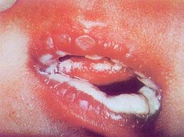
Color Figure 1
(2) Acute atrophic type: Acute atrophic candidal stomatitis is more common in adults, often due to long-term use of broad-spectrum antibiotics, and most patients originally suffer from debilitating diseases, such as leukemia, malnutrition, endocrine disorders, post-chemotherapy for tumors, etc. Certain skin diseases such as systemic lupus erythematosus, psoriasis, pemphigus, etc., during the extensive use of penicillin and streptomycin, can also develop candidal stomatitis, hence this type is also known as antibiotic stomatitis. It should be noted that this adult acute candidal stomatitis may have pseudomembranes and be accompanied by angular cheilitis, but sometimes it mainly manifests as mucosal congestion and erosion and clumped atrophy of the tongue papillae, with thickened tongue coating around. Patients often first experience abnormal taste or loss of taste, dry mouth, and mucosal burning pain.
(3) Chronic hypertrophic type: This type, also known as proliferative candidal stomatitis, can be seen on the buccal mucosa, tongue dorsum, and palate. Due to the deep penetration of hyphae into the mucosa or skin, causing parakeratosis, acanthosis, epithelial hyperplasia, microabscess formation, and inflammatory cell infiltration of the lamina propria papillae, the superficial pseudomembrane is tightly attached to the epithelial layer and is not easy to peel off. Histological examination can reveal grade I to grade II epithelial dysplasia, and some believe that candidal leukoplakia has a malignant transformation rate of more than 4%, especially in elderly patients, who should be vigilant and strive for early biopsy to clarify the diagnosis. (See the section on oral leukoplakia)
The buccal mucosal lesions of this type are often symmetrically located in the triangular area inside the corners of the mouth, presenting as nodular or granular hyperplasia, or as tightly fixed white keratinized patches, similar to general mucosal leukoplakia. Palatal lesions can develop from denture stomatitis, with the mucosa showing papillary hyperplasia; tongue dorsum lesions can manifest as filiform papillary proliferation, gray-black in color, known as hairy tongue, hence hairy tongue also belongs to this type (see diseases of the lips and tongue for details).
Hypertrophic candidal stomatitis can be a component of the symptoms of chronic mucocutaneous candidiasis, and can also be seen in patients with immunodeficiency syndromes and endocrine dysfunction.
(4) Chronic Atrophic Type: This type is also known as denture stomatitis. The affected area is often the palatal and gingival mucosa in contact with the palatal side of the upper denture, and it is more common in female patients (some statistics show that the incidence rate is 1/4 for females wearing upper dentures, while it is 1/10 for males). The mucosa appears bright red with edema, or with yellow-white streaks or patchy pseudomembranes. In 90% of patients, Candida albicans can be detected in the patches or pseudomembranes. Among patients with candidal cheilitis or angular cheilitis, 80% have denture stomatitis. Conversely, this type of lesion can often occur alone and does not necessarily involve concurrent damage to the lips and corners of the mouth.
Denture stomatitis often occurs simultaneously with papillary hyperplasia of the palate. Before considering surgical removal, antifungal treatment should be administered first, which can significantly reduce the degree of hyperplasia and narrow the scope of surgery required.
The fungi attached to the denture are the main pathogenic cause. Regular cleaning with 2% chlorhexidine or nystatin can inhibit the fungi. A flexible denture base made of silicone rubber seems to be more prone to retaining and adsorbing fungi, thus increasing the chance of denture stomatitis.
Fungal stomatitis caused by mandibular dentures is rare, possibly due to the strong negative pressure adsorption of maxillary dentures, which displaces antibodies in saliva from this area. The base surface is in wide and tight contact with the mucous membrane, allowing a large number of pathogenic fungi to be retained.
2. Candidal cheilitis: This is a chronic cheilitis caused by Candida infection, mostly occurring in elderly patients (over 50 years old). It usually occurs on the lower lip and may be accompanied by candidal stomatitis or angular cheilitis.
Gansen classified this disease into two types. The erosive type has a long-standing bright red erosion in the middle of the lower lip vermilion, with surrounding hyperkeratosis and surface desquamation, making it easily confused with discoid lupus erythematosus lesions and similar to actinic cheilitis. The granular type presents with swelling of the lower lip, with scattered small granules at the vermilion-skin border, very similar to glandular cheilitis. Therefore, for candidal cheilitis, scales and small granular tissues from the edge of the erosion should be scraped and examined microscopically for fungi. Only when budding spores and pseudohyphae are repeatedly found and culture confirms Candida albicans can the diagnosis be confirmed.
3. Candidal angular cheilitis: This disease is characterized by bilateral involvement, with rhagades in the skin and mucous membrane of the angular area, adjacent skin and mucous membrane congestion, and often erosion and exudate or thin crusts at the rhagades, causing pain or bleeding when opening the mouth. This type of angular cheilitis, characterized by moist white erosion, should be distinguished from vitamin B2 deficiency or bacterial angular cheilitis. The former is often accompanied by glossitis, cheilitis, scrotitis, or vulvitis, while the latter usually occurs unilaterally and is positive for bacterial culture (mainly streptococci). Candidal angular cheilitis mostly occurs in children, debilitated patients, and patients with blood diseases.
Angular cheilitis in elderly patients is often related to the shortening of the occlusal vertical dimension, causing the skin of the angular area to collapse into a groove, leading to saliva spilling into the groove, thus keeping it moist and conducive to fungal growth. A report of 150 denture wearers found 75 cases of angular cheilitis, with causes including vertical dimension shortening, certain systemic factors, and local irritation and infection from denture ulcers.
In children, angular cheilitis secondary to Candida infection due to chapped lips in dry winters is also common.
4. Chronic mucocutaneous candidiasis: This is a special type of Candida albicans infection involving the oral mucosa, skin, and nail beds. It usually starts in childhood and lasts for years to decades, often accompanied by endocrine or immune dysfunction and low cellular immune function, making it a manifestation of a syndrome. Lehner classified this group of diseases into the following types:
(1) Multiple endocrine disorder type: Often occurs around puberty, with initial stages showing hypoparathyroidism or adrenal cortical insufficiency and chronic keratoconjunctivitis, but candidal stomatitis may be the earliest manifestation.
(2) T-lymphocyte deficiency type: This disease can be seen in patients with hypergammaglobulinemia and malignant lymphoreticular tumors.
(3) Familial Chronic Mucocutaneous Candidiasis: This type can be seen in children and may also initially occur in adults after the age of 35 (late-onset type). Both are related to abnormalities in iron absorption and metabolism, possibly due to a reduction in the inhibitory factors for Candida albicans when iron is deficient, leading to the proliferation and invasion of pathogenic bacteria.
Various chronic mucocutaneous candidiasis often first manifest as persistent or recurrent thrush and angular cheilitis; subsequently, erythematous desquamative rashes may appear on the head, face, and limbs, along with thickening of the nail plates. Hair loss and horn-like lesions on the forehead and nose may also occur.
The most reliable laboratory diagnostic method for Candida albicans is currently considered to be the formation of thick-walled spores on cornmeal agar, while the simplest method is direct microscopic examination of specimens.
Dentists often take samples of oral mucosal pseudomembranes, exfoliated epithelium, and scabs, place them on a slide, add a few drops of 10% potassium hydroxide solution, cover with a coverslip, and gently heat to dissolve keratin, then immediately examine under a microscope. If pseudohyphae or spores are found, a fungal infection can be confirmed, but a culture is still necessary to definitively diagnose Candida albicans.
Acute pseudomembranous candidal stomatitis should be differentiated from acute coccus stomatitis (membranous stomatitis). Membranous stomatitis is caused by infections of coccus such as Staphylococcus aureus, hemolytic streptococcus, and pneumococcus, and is more common in children and the elderly. It can occur in any part of the oral mucosa, with obvious congestion and edema in the affected area, and a large amount of fibrinogen exudes from blood vessels, coagulating into gray-white or gray-yellow pseudomembranes that are smooth and dense, slightly raised above the mucosal surface. The pseudomembranes are easily wiped off, leaving an eroded surface with oozing blood. Regional lymph nodes may be swollen, and systemic reactions may occur. Smear examination or bacterial culture can identify the main pathogenic bacteria.
bubble_chart Treatment Measures
Oral candidiasis is primarily treated with local therapy, but severe cases and chronic candida infections often require systemic treatment to be effective.
1. Local drug treatment
(1) 2-4% sodium bicarbonate (baking soda) solution: This drug is commonly used to treat thrush in infants and young children. It is used to rinse the mouth before and after feeding to eliminate residual curd or sugars that can decompose to produce acid, making the oral environment alkaline, which can inhibit the growth and reproduction of Candida albicans. Mild cases may not require other medications, and lesions can disappear within 2-3 days, but medication should be continued for a few more days to prevent recurrence. This solution can also be used to clean the nipples before and after feeding to avoid cross-infection or reinfection.
(2) Chinese Gentian violet solution: At a concentration of 1:100,000, Chinese Gentian violet can still inhibit the growth of Candida. A concentration of 1/2000 (0.05%) is suitable for oral mucosa, applied three times daily to treat thrush and angular cheilitis in infants and young children. However, after staining, it is not suitable for observing changes in lesions. Commercially available 1% Chinese Gentian violet alcohol solution is too irritating for direct use on the oral mucosa of infants and young children but can be used for skin lesions.
(3) Chlorhexidine: Chlorhexidine has antifungal properties and can be used as a 0.2% solution or 1% gel for local application, rinsing, or gargling. It can also be combined with nystatin to form an ointment or cream, which may include a suitable amount of triamcinolone acetonide to treat angular cheilitis and denture stomatitis (the cream can be applied to the tissue surface of the denture and worn in the mouth). Alternating rinses with chlorhexidine solution and sodium bicarbonate solution can eliminate the synergistic pathogenic bacteria of Candida albicans—Gram-negative bacteria.
2. Antifungal drug treatment
(1) Nystatin (mycostatin): This drug belongs to the tetraene class of antibiotics, with 1mg equivalent to 2000U, and should be stored at low temperatures. It is poorly absorbed by the intestines, so it is mostly used to treat skin, mucosa, and digestive tract infections of Candida albicans. Locally, a water suspension of 50,000-100,000 U/ml can be applied every 2-3 hours and can be swallowed after application. It can also be used as a mouthwash or made into lozenges, emulsions, etc. Children (1-2 years old) can take 100,000 U orally each time, three times a day; adults can take 500,000-1,000,000 U orally each time, three times a day. The bacteriostatic effect of this drug may be due to the destruction of the cell membrane releasing potassium, leading to the cessation of glycogen decomposition within the cell and loss of vitality. Oral side effects are minimal, occasionally causing nausea, diarrhea, or loss of appetite. The treatment course is 7-10 days.
(2) Miconazole: This is a synthetic broad-spectrum antifungal drug, with the domestic trade name of Daktarin for topical use of miconazole nitrate. In addition to its antifungal properties, this drug also has antibacterial effects against Gram-positive bacteria. Powder can be used for oral mucosa, and cream is suitable for glossitis and angular cheilitis, with a general treatment course of 10 days.
(3) Clotrimazole: A synthetic broad-spectrum antifungal agent with relatively high toxicity. It is rapidly absorbed after oral administration, reaching peak blood concentration in 4-5 hours, and can enter the mucosa and saliva. This drug can affect the synthesis of ergosterol, leading to defects in the fungal cell membrane, causing the contents to leak out and resulting in fungal death. Adults can take 0.5g orally three times a day, with a maximum daily dose of 3g. The main side effects of this drug are gastrointestinal reactions; long-term use can affect liver function and cause leukopenia, so local preparations are mostly used now.
(4) Ketoconazole: A new antifungal drug recommended abroad in the 1970s for the treatment of Candida albicans. It inhibits fungal cell membrane DNA and RNA, with rapid efficacy. Oral absorption reaches its peak 2 hours after administration, and it reaches the affected area through blood circulation. The dose is 200mg orally once daily, with a treatment course of 2 to 4 weeks. It can also be used in combination with other topical antifungal drugs for better efficacy. It also has significant therapeutic effects on fungal infections outside the oral cavity, such as skin and digestive tract infections, and has currently replaced amphotericin abroad. This drug should not be taken with antacids or anticholinergic drugs to avoid affecting absorption.
3. Comprehensive Treatment
For symptoms such as obvious congestion and edema of the mucous membrane, red tongue texture, fetid mouth odor, yellow urine, constipation, etc., which are indicative of up-flaming of heart fire or stomach heat with dampness, "Kouyan Infusion Granule" can be taken (refer to the chapter on herpes simplex).
In addition to antifungal medications, patients who are physically weak, have immunodeficiency diseases, or related systemic diseases and chronic candidiasis infections often require comprehensive treatment measures to enhance the body's immunity, such as injections of transfer factor, thymosin, lipopolysaccharides, etc., supplementation with iron and vitamin A; as well as multiple small blood transfusions.
The treatment duration for oral candidiasis should be appropriately extended, generally for 14 days, as stopping medication too early can easily lead to recurrence of the disease. The course of treatment for the hypertrophic (proliferative) type should be even longer, with reports suggesting up to 3-4 months. For candidal leukoplakia with insignificant efficacy, surgical removal should be considered as early as possible.
4. Prevention
Avoid cross-infection in the delivery room; during childbirth, attention should be paid to the disinfection of the perineum, birth canal, the hands of the delivery personnel, and all delivery equipment.
Frequently clean the baby's mouth with warm boiled water, boil and disinfect feeding utensils, and keep them dry. Before breastfeeding, it is best to clean the mother's nipples with a 1/5000 chlorhexidine hydrochloride solution and then wipe them clean with cold boiled water.
Children should protect their lips from dryness and cracking in winter and correct the bad habit of licking lips and sucking tongues.
Patients who have been using antibiotics and immunosuppressants for a long time, or those suffering from chronic debilitating diseases, should be vigilant about the occurrence of candidiasis infections, especially paying attention to the easily overlooked deep (visceral) candidiasis complications.


