| disease | Duodenal Diverticulum |
The exact incidence of duodenal diverticula is difficult to determine, as many diverticula do not produce clinical symptoms and are not easily detected in a timely manner. Reports indicate that the detection rate of duodenal diverticula during gastrointestinal barium meal examinations is 1%, while the discovery rate during autopsies can be as high as 22%. 90% of the diverticula are single, and 80% are located in the second part of the duodenum, especially on the medial wall or concave surface. This condition predominantly occurs in patients aged 40 to 60, and is rare in those under 30. There is no difference in incidence between genders.
bubble_chart Clinical Manifestations
Duodenal diverticula do not have typical clinical manifestations, and the symptoms that occur are mostly caused by complications. Epigastric fullness is a relatively common symptom, caused by diverticulitis, accompanied by belching and dull pain. The pain is irregular and cannot be relieved by antacid medications. Nausea or vomiting is also common. When the diverticulum is filled with food and becomes distended, it can compress the duodenum and cause symptoms of partial obstruction. Initially, the vomitus consists of gastric contents, followed by bile, and may even be mixed with blood; symptoms may be relieved after vomiting. When the diverticulum is complicated by ulcer or bleeding, symptoms similar to ulcer disease or hematochezia may appear respectively. When the diverticulum compresses the common bile duct or the pancreatic duct opening, it can further cause cholangitis, pancreatitis, or obstructive jaundice. After perforation of the diverticulum, symptoms of peritonitis may appear.
bubble_chart DiagnosisSome smaller and concealed diverticula can only be detected during hypotonic duodenography with the aid of gastrointestinal barium meal examination.
bubble_chart Treatment Measures
(1) Treatment Principles: Asymptomatic duodenal diverticula do not require treatment. When there are certain clinical symptoms without other pathological changes, medical treatment should be initiated first, including dietary adjustments, antacids, antispasmodics, etc. Additionally, adopting a lateral position or changing various postures can help empty the accumulated food in the diverticulum. Since diverticula are mostly located on the medial wall of the second part of the duodenum or even embedded within pancreatic tissue, surgical resection is relatively difficult. Therefore, surgery is considered only when medical treatment is ineffective and there are recurrent complications such as diverticulitis, bleeding, or compression of adjacent organs.
(2) Surgical Methods: In principle, the most ideal surgical method is diverticulectomy. For smaller diverticula, simple inversion may be performed. When multiple diverticula are present and there are technical difficulties in resection, a bypass surgery may be adopted, such as performing a Billroth II partial gastrectomy and selective vagotomy.
If it is difficult to locate the diverticulum during surgery, the duodenum can be incised to find the opening of the diverticulum from within the lumen, and its base can be inverted into the intestinal lumen for resection. After diverticulectomy, the intestinal wall incision should be sutured in an inverted manner perpendicular to the long axis of the intestinal loop (as shown in Figure 1) to avoid intestinal stenosis.
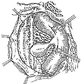
(1) Incise the abdominal membrane on the lateral side of the duodenum, free the duodenum and retract it medially to expose the diverticulum
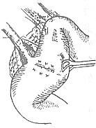
(2) After diverticulectomy, transversely (i.e., perpendicular to the long axis of the intestinal loop) invert and suture the intestinal wall incision
Figure 1: Diverticulectomy of the descending part of the duodenum.


