| disease | Femoral Head Osteoepiphysiochondropathy |
| alias | Perthes Disease |
Legg-Calve-Perthes disease (LCP), commonly referred to as Perthes disease, is the most common osteochondrosis. It predominantly affects children aged 3 to 10 years, with a higher incidence in boys than in girls, at a ratio of 4-6:1. Occasionally, it may occur in children younger than 2 years or older than 10 years. Bilateral involvement is seen in about 10% of cases, and the disease course typically lasts 2 to 4 years. This condition is characterized by ischemic necrosis of the epiphysis, primarily affecting the epiphysis of the femoral head and the metaphysis of the femur, and in rare cases, it may also involve the acetabulum.
bubble_chart Clinical Manifestations
Initial stage [first stage]: Symptoms are relatively vague, with a feeling of easy fatigue in the limbs often being the first symptom. There is grade I pain in the hip during weight-bearing, but it disappears after rest, accompanied by an indistinct limp. Hip abduction and rotation are affected early on, and pain can be felt when tapping along the long axis of the femur. There is tenderness in the front of the hip joint, and the pain radiates to the knee joint, leading to misdiagnosis as a knee condition.
As the condition progresses, the pain becomes persistent. The child's limp becomes noticeable, and the gluteal and thigh muscles undergo disuse atrophy. The Trendelenburg test is positive, and hip flexion with inversion causes relative shortening of the affected limb. With the formation of a flat hip, the absolute length of the limb also becomes shorter compared to the healthy side. This leads to early-onset osteoarthritis in adulthood.
In the initial stage of the lesion, X-rays show no positive findings. The early X-ray manifestation of femoral head osteoepiphysio-chondropathy is the outward displacement of the ossification center of the femoral head and the widening of the medial joint space (a widening of 1-2 mm has diagnostic value and can be compared with the healthy side). This is due to increased intra-articular pressure caused by synovitis, leading to swelling of the joint capsule fat pad. Some believe that the outward displacement of the femoral head is due to the displacement of the ossification center rather than the entire femoral head. After the femoral head is displaced outward, the anterior-superior quarter of the head inevitably bears excessive load, and the acetabular edge continuously compresses the femoral head, causing marginal compression fractures. This is more clearly visible on X-rays taken in the hip abduction position (frog-leg position), hence some emphasize the routine use of abduction position films. Joint contrast imaging may show thickening of the medial cartilage of the femoral head, possibly due to the cessation of ossification center development and proliferation of medial cartilage cells. The ossification center shown on X-rays is also smaller, with increased epiphyseal density and temporary cessation of development. Later, a dome-like subchondral lucency appears in the femoral head, visible only on abduction X-rays, indicating the fracture line of the necrotic area. When the lower limb is abducted, the volume between the edge of the ossification center and the epiphyseal cartilage suddenly increases, possibly slightly separating bone fragments, allowing air to enter the fracture line and become visible, known as the intra-epiphyseal gas sign. The anterolateral part of the epiphysis is the first to necrotize (and also the first to show repair), visible only in the abduction position, while on anteroposterior X-rays it often appears as if the entire femoral head is affected. The femoral head begins to flatten, the metaphysis thickens, and disuse osteoporosis occurs in the hip bones. Subsequently, the repair process begins, and X-rays show coexisting bone necrosis, bone absorption, and new bone deposition. The femoral head gradually restores its smooth outer edge, but if not treated promptly and effectively, it becomes mushroom-shaped. To adapt to the deformation of the femoral head, the acetabulum also flattens and becomes shallow with an irregular shape. The acetabulum cannot fully contain the femoral head, leading to semi-dislocation, and the femoral neck becomes wide and short. Due to changes in the weight-bearing line, early signs of osteoarthritis can be seen in adulthood. In recent years, CT and MRI (magnetic resonance imaging) have been widely used clinically, greatly aiding in the early diagnosis of this condition.
bubble_chart Treatment MeasuresAt the onset of the disease, due to the high sensitivity of the child's hip joint, skin traction can be applied for 1-2 weeks initially. Further treatment should be considered only after the acute symptoms have subsided.
(1) Non-surgical treatment: In the past, long-term hip spica cast fixation was used, but due to its significant impact on the child's development and joint function, it is now rarely or no longer used. Various types of abduction braces are currently the commonly adopted treatment methods, aiming to: ① deeply position the femoral head within the acetabulum; ② avoid compression of the femoral head by the acetabular labrum; ③ ensure even pressure distribution on the femoral head; ④ maintain good mobility of the hip joint; and ⑤ preserve the round shape of the femoral head and the normal acetabulum as much as possible. A group of 68 children treated with abduction and internal rotation brace fixation achieved an excellent and good rate of 91%, but the average fixation time was 19 months, which is still too long.
(2) Surgical treatment: The idea is to alter the circulation of the ossification center of the femoral head through surgical methods, enabling blood supply communication between the femoral head and neck, using techniques such as drilling, bone grafting, or blood supply communication, but these have not yielded effective results. Some advocate for the use of hip joint synovial membrane resection to treat this disease, which has shown certain effects, but the mechanism of the surgery's effect is still unclear. Others use pedicled (muscle flap or vascular) bone grafting, vascular implantation, etc. In recent years, subtrochanteric or intertrochanteric osteotomy has been relatively recognized abroad. The advantages of this surgery include: ① treatment can be completed within 6-8 weeks post-surgery; ② no further use of braces or other measures to limit activity and weight-bearing is needed after surgery; ③ osteotomy can cause hyperemia in the upper segment of the femur; ④ its efficacy is not inferior to long-term abduction brace fixation. Complications of osteotomy include limb shortening deformity, residual hip bone flipping, non-union at the osteotomy site, and limited joint mobility. It is generally believed that surgery under the age of 7 yields good results, and the effects continue to improve in the years following surgery. The healing process of femoral head epiphyseal necrosis can be shortened. The average limb shortening post-surgery is 1.4cm.
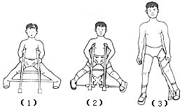
Figure 1: Brace treatment
(1) Petrie abduction cast (2) Bobechko abduction brace
(3) Tachdjian abduction brace. The common feature of these braces is to maintain the hip joint in an abducted and internally rotated position, but the hip joint can flex and extend freely, and the child is required to walk on the ground.
It is related to the age of onset, the duration of the disease history, and the correctness of the treatment method. Generally, those who develop the disease before the age of 5 have a good prognosis and may not even require any treatment.
To determine the prognosis and decide on the treatment method, Catterall classified the disease into four grades based on X-ray findings (anteroposterior and abduction views). Grade I: Only the anterior part of the epiphysis is affected, the joint surface is not collapsed, the affected bone can be completely absorbed, there is no necrotic bone formation, no metaphyseal bone changes, and the regeneration process can reach a complete degree (Figure 2). Grade II: The anterior and lateral parts of the epiphysis are affected to varying degrees, the femoral head is collapsed but can still maintain the original height of the epiphysis, necrotic bone is formed but can be absorbed, cystic changes appear in the metaphysis and will disappear later (Figure 3). Grade III: Only a small part of the epiphysis is not necrotic, due to new bone covering the necrotic bone, the phenomenon of "head within a head" appears on the anteroposterior X-ray, the femoral head is collapsed and can no longer maintain its original height, and the metaphysis has widened (Figure 4). Grade IV: The entire epiphysis has become necrotic, the femoral head has taken on a mushroom shape, the metaphysis has significant widening and other bone changes, and remodeling has occurred on the femoral head, but it is difficult to restore its original shape.


Figure 2 Catterall Grade I
(1) Anteroposterior view showing the lesion only within the epiphysis (2) Frog-leg lateral view

Figure 3 Catterall Grade II
(1) Both the epiphysis and metaphysis are affected (2) Lateral view showing two connected lesions
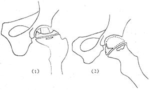
Figure 4 Catterall Grade III
(1) "Head within a head" appearance, metaphysis thickened
(2) Lateral view showing the head has lost its original appearance
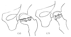
Figure 5 Osteochondrosis
(1) The entire femoral head and neck are affected, accompanied by bone sclerosis, joint space narrowing
(2) Lateral view
There is no doubt that Grades I and II have a good prognosis, while Grades III and IV have a poor prognosis. After further research, Catterall also found that some children recover without treatment, while others require timely and correct treatment, otherwise the consequences are severe. He proposed a concept called "femoral head at risk signs." "At risk signs" clinically include: obese children, limited hip movement with adduction contracture; on X-ray, they include: Gage sign, lateral subluxation of the femoral head, calcification on the lateral side of the epiphysis, and horizontal growth plate of the epiphysis. If these signs appear, timely treatment is necessary, and intertrochanteric or subtrochanteric osteotomy is the most effective treatment method. If there are no "at risk signs," regardless of the group, no treatment is necessary.
At the start of treatment, if the femoral head remains round, the outcome is good; if the head has already flattened, it may not necessarily return to its original shape. Once the healing process begins, the femoral head will not continue to deform. Some believe that if the disease course has lasted more than 20 months, even osteotomy may not yield good results.
Femoral head osteoepiphysio-chondropathy needs to be differentiated from the following diseases.
(1) Hip joint subcutaneous node: It is often difficult to distinguish, especially in the early stages. The hip joint subcutaneous node is a localized, progressive, destructive, inflammatory sexually transmitted disease that can affect the femoral head, acetabulum, and femoral neck. The X-ray sign of joint capsule swelling due to joint effusion can persist for a considerable period. In contrast, femoral head osteoepiphysio-chondropathy is a subchondral aseptic necrosis sexually transmitted disease, characterized by increased bone density, deformation, and secondary hip osteoarthritis on X-ray, without significant joint effusion or abscess formation.
(2) Transient (temporary) synovitis of the hip joint: Both conditions are similar in terms of age of onset and synovitis manifestations, but the course of the disease differs. Transient synovitis shows no abnormal X-ray findings. In recent years, 99mTc scanning has been helpful: femoral head osteoepiphysio-chondropathy shows reduced 99Tc uptake.
(3) Cretinism: The epiphyseal changes in cretinism can manifest as irregular calcification points, but their appearance and fusion times are delayed compared to normal children. The long diameter of the bone is shortened due to endochondral ossification disorder. Additionally, the child may have intellectual disabilities, which can help differentiate.
(4) Slipped capital femoral epiphysis: The clinical symptoms of both conditions are similar. However, slipped capital femoral epiphysis occurs at an older age, and limited internal rotation and adduction of the hip joint (Drehman's sign) are characteristic.


