| disease | Tricuspid Valve Insufficiency (Surgical) |
Tricuspid valve insufficiency can be either relative or organic. In the relative type, the valve membrane itself is not diseased, but rather the right ventricular hypertrophy and corresponding dilation of the atrioventricular ring cause poor coaptation of the tricuspid valve leaflets, leading to insufficiency. Patients with grade III rheumatic heart disease and mitral stenosis or insufficiency often have relative tricuspid insufficiency. Organic tricuspid insufficiency is a sequela of wind-dampness heat and is relatively rare in clinical practice, usually accompanied by mitral and aortic valve lesions. Pathological changes include fibrous thickening and shrinkage of the valve membrane, shortening of the chordae tendineae, and dilation of the valve ring, preventing complete coaptation of the valve membrane during cardiac contraction. This condition is often combined with fusion of the valve commissures, resulting in concurrent stenosis.
bubble_chart Pathological Changes
The pathophysiology of tricuspid insufficiency is the result of tricuspid regurgitation, where blood flows back from the right ventricle into the right atrium during systole, leading to significant enlargement of the right atrium, increased pressure, and impaired venous return. Due to the increased load on the right ventricle, it compensates by thickening, making it prone to right heart failure.
bubble_chart Clinical Manifestations
The symptoms and signs of tricuspid valve insufficiency are related to the degree of valve membrane insufficiency. Grade I insufficiency is clinically difficult to detect. More severe cases may present with fatigue, poor appetite, distending pain in the liver area, abdominal distension, and lower limb edema.
Typical signs include: jugular vein distension with pulsation; hepatomegaly with palpable pulsation; and a holosystolic blowing murmur at the fourth intercostal space along the left sternal border, which intensifies at the end of deep inspiration (Carvallo's sign). Typical signs may be absent in severe cases of tricuspid regurgitation. If the liver becomes cirrhotic due to long-term congestion, pulsation may no longer be present; once the right heart's volume load reaches its peak, the murmur no longer intensifies with inspiration, so Carvallo's sign may be negative.X-ray images show hypertrophy of the right atrium and right ventricle, with a bulging right heart border, along with changes caused by other valve membrane lesions.
The electrocardiogram shows atrial hypertrophy with tall and wide P waves; and right bundle branch block or right ventricular hypertrophy, even myocardial strain. Atrial fibrillation is common.
Echocardiography and Doppler examination: cross-sectional ultrasound can detect the size of the tricuspid valve ring and understand the thickening of the valve membrane, which helps distinguish between relative and organic sexually transmitted disease changes. In tricuspid valve insufficiency, contrast echocardiography can show microbubbles moving back and forth across the tricuspid valve; Doppler can directly monitor abnormal signals from the right ventricle to the right atrium and estimate the degree of regurgitation.
Cardiac catheterization shows a prominent V wave in the right atrial pressure waveform, with a steep y descent, more pronounced during inspiration. The right atrial pressure waveform is similar to the right ventricular pressure waveform, only with a smaller amplitude, known as right ventricularized right atrial pressure, which is a manifestation of grade III tricuspid regurgitation.
Heart blood vessel angiography: right ventricular angiography and right anterior oblique cineangiography can show tricuspid regurgitation and its degree. However, due to the catheter crossing the tricuspid valve, there is a potential for false positives.The diagnosis of tricuspid valve insufficiency should include an understanding of the degree of insufficiency. Typical clinical signs are of some value in diagnosing grade III tricuspid valve insufficiency. In the past, right ventricular angiography was used as a means to diagnose suspected cases and estimate the degree of regurgitation. In recent years, ultrasound and Doppler examinations have gradually replaced invasive tests.
bubble_chart Treatment Measures
In cases of mild relative tricuspid insufficiency, after the correction of primary lesions in other valve membranes and a period of recovery, the degree of insufficiency often decreases or even disappears due to the reduction in right ventricular pressure and the shrinking of the right heart. However, in some severe cases of Rheumatic heart disease, the hemodynamic disturbances caused by tricuspid regurgitation within a few days post-operation can contribute to low cardiac output, which is one of the factors leading to surgical mortality. Additionally, some patients with pulmonary stirred pulse hypertension do not achieve the expected results and remain in a state of right heart failure for a long time post-operation. Therefore, in recent years, it has been advocated to perform tricuspid annuloplasty concurrently with other valve membrane surgeries for moderate or greater tricuspid insufficiency to achieve more satisfactory results.
Organic tricuspid insufficiency generally requires surgical treatment. For mild cases, direct incision of the fused commissures followed by annuloplasty can be performed; for more severe cases, valve membrane replacement should be considered.
Tricuspid annuloplasty: There are three common methods: ① Annular plication (Figure 1), where 1-2 mattress sutures are made along the commissure of the anterior and posterior leaflets and the posterior annulus using double-armed non-traumatic sutures, with pledgets on both sides, to shorten the annulus after tying. ② De Vega procedure (Figure 2), where a double-armed non-traumatic suture is used to make a double-layer cross-stitch along the anterior and posterior annulus, with pledgets at both ends, and the suture is tightened and tied to shorten the enlarged posterior and anterior annulus. ③ Carpentier ring fixation (Figure 3), where the Carpentier ring, an oval semi-circular ring made of stainless steel and wrapped in polyester fabric, is used. The ring is placed along the tricuspid annulus with mattress sutures and then sutured to the appropriately sized Carpentier ring. After fixation, the enlarged anterior and posterior annulus can be reduced, restoring good coaptation of the tricuspid valve. The modified Carpentier ring, made of elastic material, can adapt to the movement of the atrioventricular ring during the cardiac cycle, reducing the stress on the suture line and thus the likelihood of dehiscence.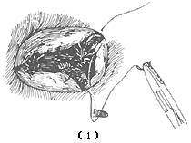
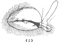
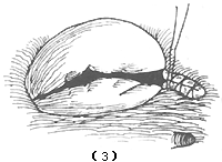
Figure 1 Tricuspid insufficiency annular plication
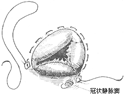
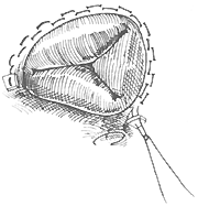
Figure 2 De Vega tricuspid annuloplasty
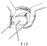
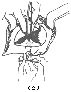
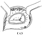
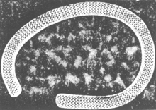
Figure 3 Tricuspid insufficiency Carpentier ring fixation
(1)~(4) show the surgical steps (5) shows the modified Carpentier tricuspid ring


