| disease | Cervical Spondylolysis |
Cervical spondylolysis with spondylolisthesis is relatively rare, most commonly occurring at the C6 level, with slippage generally not exceeding 1°. In 1951, Perlman and Hawes first reported a case of C6 spondylolysis with cervical spondylolisthesis in a young male. To date, the total number of reported cases worldwide does not exceed one hundred.
bubble_chart Etiology
Most authors consider this condition to be a congenital malformation. It is often associated with other congenital cervical spine anomalies, such as spina bifida, congenital absence of the pedicle, and articular process dysplasia. Some authors believe this condition is related to heredity, with reports of a pair of twins both affected by the disease.
bubble_chart Pathological Changes
The part between the superior and inferior articular processes of the vertebral arch is called the pars interarticularis. In the lumbar spine, this area is relatively narrow and elongated, referred to as the isthmus, while in the cervical spine, it appears columnar. Cervical spondylolysis refers to the non-union of this part on either one or both sides, which can lead to anterior slippage of the cervical vertebra (Figure 1), causing cervical instability and symptoms of spinal cord or nerve root irritation. The forward-slipped vertebral body may compress the esophagus anterior to the vertebrae. Sometimes, this part may become elongated without complete fracture, and dynamic flexion-extension X-rays reveal instability.
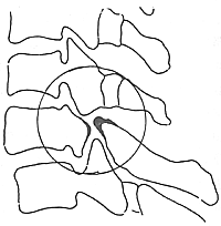
Figure 1 Cervical spondylolysis with slippage.
bubble_chart Clinical Manifestations
This condition is more common in young males, with a male-to-female ratio of 2–3:1. The affected segments can involve the cervical spine from C2
to C7, with C6 being the most frequently involved, accounting for more than 70%. Bilateral vertebral arch defects are more common than unilateral ones.In cases of cervical spondylolysis without spondylolisthesis, clinical symptoms of cervical instability may occur, primarily manifesting as pain in the occipital region and shoulders. Patients with concurrent cervical spondylolisthesis may exhibit symptoms of nerve root compression and dysphagia. Spinal cord compression symptoms are less common, but if cervical spondylolisthesis is accompanied by herniation of intervertebral disc or cervical spinal canal stenosis, spinal cord compression symptoms are more likely to occur. Some cases may present no clinical symptoms and are only discovered incidentally on X-ray examination, revealing cervical spondylolysis with spondylolisthesis.
Physical examination may reveal restricted cervical spine movement, and cervical motion can trigger or exacerbate clinical manifestations. In patients with cervical spondylolisthesis, a "step-like" deformity may be palpated in the neck. Occasionally, the neck may exhibit a torticollis-like deformity. For those with concurrent spina bifida, clinical symptoms and signs of spina bifida may also be present.
Clinical manifestations and X-ray examinations are sufficient to confirm the diagnosis of this condition. When necessary, tomography, flexion-extension dynamic radiography, myelography, CT, and MRI can be performed. X-ray examinations include anteroposterior, lateral, and oblique views of the cervical spine. When diagnosing this condition, it is important to differentiate it from cervical vertebral arch destruction caused by trauma, tumors, or other factors.
Non-surgical treatment
For asymptomatic individuals, cervical spondylolysis discovered only during physical examination or X-ray imaging generally does not require special intervention. For those without neurological symptoms, conservative treatments such as local immobilization, physical therapy, and oral analgesics usually achieve satisfactory outcomes.
Surgical treatment
For patients with significant nerve root compression symptoms, conservative treatment may be attempted first. Cervical fusion surgery can be considered once neurological symptoms alleviate or disappear. For patients with concomitant herniation of intervertebral disc or spinal canal stenosis presenting spinal cord compression symptoms, decompression combined with cervical fusion surgery may be considered.


