| disease | Cervical Facet Traumatic Degenerative Arthritis |
Chronic neck pain is a common yet often overlooked clinical symptom. Clinicians tend to associate it with cervical herniation of intervertebral discs or neck muscle strain. However, as research on facet joints deepens, increasing evidence suggests that cervical facet joint lesions are one of the significant causes of neck pain. When degenerative changes affect part or all of the posterior cervical facet joints, leading to traumatic arthritis reactions and a series of clinical symptoms, it is termed cervical facet traumatic degenerative arthritis. This condition is relatively common in clinical practice and is one of the major causes of chronic neck pain, cervicogenic headache, and radicular cervical spondylosis.
bubble_chart Etiology
The specific pathogenesis and pathological changes of traumatic degenerative arthritis of the cervical facet joints remain unclear due to a lack of in-depth research. However, trauma and degeneration are two definitive factors contributing to the onset of this disease.
Anatomical Basis
The cervical facet joints, like various large joints throughout the body, are synovial joints composed of superior and inferior articular processes. The articular processes form an angle of approximately 40°–45° with the vertebral body plane, with smooth articular surfaces covered by hyaline cartilage and relatively loose joint capsules. The cervical facet joints play a role in guiding and limiting the direction of motion in the spinal segments. For example, during cervical flexion, the inferior articular process of the upper vertebra slides forward over the superior articular process of the lower vertebra, but the joint capsule restricts excessive flexion. Although the arrangement of cervical facet joints facilitates flexion and extension, they are relatively unstable and prone to injury. These anatomical characteristics form the basis for pathological changes in the facet joints.
TraumaIn daily life, activities such as flexion, extension, and rotation of the head and neck rely heavily on the coordination of intervertebral joints and bilateral facet joints. Poor working postures, such as those of typists or household laborers who work with their heads bent forward for prolonged periods, generate shear forces on the facet joints, placing the joint capsules under tension. Excessive tension can lead to traumatic inflammatory reactions in the joint capsules, causing them to gradually thicken and harden. In car accidents, the whiplash effect suddenly applies acceleration-deceleration forces to the facet joints, resulting in injuries such as articular cartilage and subchondral fractures, joint capsule tears, and intra-articular hematomas. Traumatic inflammatory changes in the joint capsules activate synovial cells, increase synovial fluid secretion, and may also involve hematomas, leading to joint swelling and elevated intra-articular pressure. This stimulates the C-type nerve fibers in the capsule wall, causing symptoms such as neck pain. Some studies have shown that injecting saline into the facet joint capsule can induce neck pain. Trauma to the joint capsule can also dilate microvessels, allowing formed elements such as red and white blood cells to exude into the joint fluid, where they precipitate and organize, leading to joint adhesions. Injury to the articular cartilage roughens the joint surfaces, increasing the likelihood of further trauma and resulting in gradual cartilage atrophy, joint space narrowing, and other arthritic changes. Trauma to the bone and joints is a significant factor in the development of degeneration.
Degeneration
With aging, degeneration is an inevitable process. After wear or trauma to the facet joints, the joint capsules may thicken and form scars. Repeated damage to the joint capsules impairs synovial fluid secretion, reducing lubrication. The articular cartilage, deprived of nutrients, undergoes degenerative changes, gradually thinning and becoming rough. Cracks may appear on the cartilage surface, extending into the cartilage or even forming small fragments that detach into the synovial fluid. Due to wear, the interlocking articular surfaces not only sustain injury but also, according to Wolff's law, gradually develop subchondral bone densification. Increasing roughness and hardening of the joint surfaces lead to hypertrophy of the facet processes and the formation of osteophytes at the margins. Changes in mineral concentration in the soft tissues can also calcify the joint capsules and alter their structure, making the facet joints more susceptible to injury during movement and accelerating degenerative changes.Relationship Between Intervertebral Disc Degeneration and Facet Joint Pathology
After intervertebral disc degeneration, the facet joints are often prone to severe injury due to changes in the pivot points of spinal flexion and extension. During spinal flexion and extension, vertebral bodies move over the incompressible nucleus pulposus, with the facet joints limiting motion. If disc degeneration occurs, movement becomes unbalanced and irregular. Experiments have shown that during normal full flexion and extension, the pivot point passes behind the nucleus pulposus. However, with disc degeneration, the pivot shifts backward and, in severe cases, may even pass through the facet joints, subjecting them to significant damage during flexion and extension. This irregular motion accelerates facet joint degeneration. After intervertebral disc degeneration, the upper and lower vertebral bodies may move closer together, loosening the facet joint capsules. At this stage, the facet joints may progress from uneven misalignment to subluxation, causing injury to the joint capsules and surrounding soft tissues. However, some studies suggest that there is no clear correlation between cervical facet joint degeneration and intervertebral disc degeneration.
There is no clear boundary between trauma and degeneration. As causative factors of disease, they have a mutually reinforcing effect: trauma accelerates the progression of degeneration, while degeneration increases the likelihood of trauma.bubble_chart Clinical Manifestations
History of neck trauma Patients usually have a history of head or neck trauma or long-term poor working postures such as typing or car accidents. It is particularly common in those with a history of whiplash injuries from motorcycle accidents or car collisions. After whiplash injuries caused by car accidents, the incidence of chronic neck pain due to facet joints is quite high, with Barnsley's survey suggesting it can be as high as 54%.
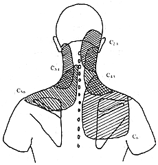
Neck pain Chronic neck pain is a characteristic manifestation of this condition, often presenting as persistent dull pain that can be triggered or worsened by movement. The C2–3 or C5–C6 facet joints are highly susceptible to trauma and strain, and the corresponding incidence rates are also high. Facet joint lesions at different segments can cause pain in distinct regions, with specific distribution patterns: (Figure 1)

Figure 1 Schematic diagram of pain distribution caused by traumatic degenerative arthritis in cervical facet joints
1. C2–3 facet joints: Pain is located in the upper cervical region and extends to the occipital area. In severe cases, the pain may spread to the ear, vertex, forehead, or eyes.
2. C3–4 facet joints: Pain occurs in the posterolateral cervical region, extending upward to the suboccipital area but not beyond the occipital region, and downward not exceeding the shoulder girdle. The distribution resembles that of the levator scapulae muscle.
3. C4–5 facet joints: Pain is distributed in a triangular area, bounded by the posterior midline and the posterolateral cervical border. The base is an imaginary line parallel to the scapular spine, passing through the junction of the outer and middle thirds of the clavicle.
4. C5–6 facet joints: Pain is distributed in a triangular, sleeve-like pattern, with the apex pointing toward the posterior midline of the neck and enveloping the anterior, posterior, and superior aspects of the shoulder girdle. The baseline is the scapular spine.
5. C6–7 facet joints: Pain is distributed in a quadrilateral shape covering the supraspinous and infraspinous fossae.
6. C7–T1 facet joints: Pain is generally concentrated in the lower half of the C6–7 region.
The above pain distribution patterns represent the primary regions affected by facet joint lesions at each segment. In severe cases, the pain may spread more widely, but these areas are often the most severely affected. Pain may be bilateral if both facet joints are involved. Overlapping pain regions due to multiple facet joint involvement can complicate diagnosis.
Referred pain Due to the extensive distribution of cervical nerve roots in the head, neck, chest, and upper limbs, referred pain is common in addition to local pain. Headache, primarily caused by referred pain from C2–3 facet joint involvement, is frequent and easily misdiagnosed. It may present as chronic persistent dull pain or even mimic typical migraine or forehead pain. Shoulder pain, often caused by C5–6 facet joint involvement, can be confused with scapulohumeral periarthritis. Additionally, chest pain and upper limb pain may also occur.
Local signs Traumatic degenerative arthritis of the facet joints often exhibits obvious fixed tenderness, exacerbated by movement. Neck mobility may be reduced due to pain, and the neck may even assume a forced posture. Blockade of the medial branches of the posterior rami of the spinal nerves innervating the affected facet joints can alleviate the pain.
bubble_chart Auxiliary Examination
X-ray films must include anteroposterior, lateral, and left/right oblique views. Early stages often show no significant changes, but later stages reveal narrowing of the joint space and loosening; gradual proliferation at the joint protrusions, forming sharp bone spurs; in the late stage [third stage], the joint exhibits hypertrophic changes, with obvious osteophyte formation at the periphery, causing the intervertebral foramen to shrink and deform.
Tomography can more clearly display the narrowing, osteophytes, and deformities of the joint in the advanced stage.
CT scans can clearly show the extent of facet joint lesions and their relationship with the spinal canal and root canal in cross-sectional views. Common signs include: (1) formation of bone spurs at the facet joint margins; (2) hypertrophy of the facet joints; (3) narrowing of the joint space; (4) thinning of the articular cartilage; (5) "vacuum phenomenon" within the facet joints; (6) calcification of the joint capsule; and (7) subchondral bone sclerosis of the facet joints. However, in early stages, CT scans are less effective than X-rays. One advantage of CT scans is the ability to simultaneously observe intervertebral discs and rule out disc diseases.
SPECT: Conventional ECT images are not very helpful for observing this condition, but high-spatial-resolution ECT images can clearly show increased radionuclide uptake in the facet joints. This method has the advantage of high sensitivity and can serve as a screening tool before facet joint arthrography.
Facet joint arthrography is highly useful for the early diagnosis of traumatic degenerative arthritis of the facet joints. It must be performed under fluoroscopic guidance, using either a lateral or posterior approach, similar to facet joint injection. After confirming the needle's entry into the facet joint cavity under anteroposterior and lateral fluoroscopy, a water-soluble contrast agent is injected. In early stages, joint capsule laxity or rupture and signs of facet joint inflammation can often be observed. In the advanced stage, arthrography is often not performed due to the difficulty of puncture and obvious imaging changes. After the procedure, injecting a mixture of local anesthetic and steroids can also provide therapeutic benefits.
Other tests: To rule out possible concurrent intraspinal lesions, disc diseases, or other conditions, magnetic resonance imaging (MRI), myelography, or discography may be performed when conditions permit to clarify the diagnosis.
Cervical traumatic degenerative facet arthritis in the early stages primarily manifests as chronic neck pain, often mistaken for muscle strain and overlooked due to the lack of characteristic symptoms and radiographic abnormalities, leading to misdiagnosis and confusion with fistula disease. Diagnosis is relatively easier for advanced-stage patients. The diagnostic criteria include:
History of head or neck trauma—Patients with a history of trauma, especially from car accidents, should be highly suspected, though some may not have a clear traumatic history.
Pain—The pain distribution follows specific patterns. Patients with significant tenderness should undergo a nerve block test, which serves both diagnostic and therapeutic purposes. Since the facet joints are innervated by the medial branches of the dorsal rami, which lie close to the bone at the waist of the articular process, pain relief after blocking these nerves suggests facet joint involvement. Pain relief within about 10 minutes after local anesthetic injection, lasting over 2 hours, is considered a positive response. This test is one of the characteristic diagnostic features in the early stages. Compared to intra-articular blocks, medial branch nerve blocks are simpler, less invasive, and safer, and can be successfully performed even in cases of narrowed joint spaces. However, due to the significant influence of psychological factors on pain perception, the false-positive rate is relatively high.
Comparative block test—Since simple nerve block tests are prone to psychological interference and high false-positive rates, a comparative block test can be performed for patients suspected of psychological influence. The principle is that bupivacaine's duration of action is about 2–3 times that of lidocaine. By comparing the duration of symptom relief after blocking with each drug, the test can determine whether the response aligns with their pharmacological profiles, significantly reducing false positives. Note that the second block should only be performed after the effects of the first block have completely worn off.
Intra-articular facet block test—This involves injecting local anesthetics into the facet joint under fluoroscopy, providing more accurate diagnostic results. However, due to its complexity and higher risk of complications, it is often replaced by medial branch nerve blocks during the diagnostic phase.
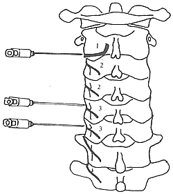
Medial branch nerve block technique—Must be performed under fluoroscopic guidance. First, identify the affected facet joints based on pain distribution and tenderness points. Then, using a 22-gauge (20–24 gauge) 90mm spinal needle, perform a lateral approach puncture to the dorsal side of the corresponding facet joint. Confirm needle placement based on bony resistance and fluoroscopic imaging, then inject 0.5ml of 2% lidocaine or 0.5ml of 0.75% bupivacaine at the waist of the articular process (lateral view). Since each facet joint is innervated by the medial branches of two adjacent spinal nerves, blocks must be performed at both the superior and inferior articular processes. The C2–3 facet joint is unique, and medial branch blocks are unreliable here. Instead, a three-point block of the third occipital nerve is required: (1) below the superior articular cartilage, (2) below the inferior articular cartilage, and (3) between the superior and inferior articular processes. Successful blockade is confirmed by loss of sensation in the occipital region (its cutaneous distribution). Pain relief starting within 10 minutes and lasting over 2 hours is considered a positive response. However, this test has a high false-positive rate (27%, Figure 1) and should be interpreted in clinical context.

Figure 1: Schematic diagram of medial branch nerve block
1. Third occipital nerve; 2. Medial branch; 3. Articular branch
Radicular symptoms—Early stages present as irritative symptoms, while late-stage (third-stage) symptoms may result from direct compression due to articular process hypertrophy, osteophyte formation, etc. Compression of the C3 and C6 spinal nerve roots is common and may manifest as clinical features of cervical spondylotic radiculopathy. Imaging characteristics The diagnosis of advanced stage patients is not difficult, mainly based on findings from plain radiographs, tomography, and CT scans. However, early-stage patients often show no abnormal manifestations. Although SPECT and small joint arthrography can aid in the early diagnosis of this disease, they are not as sensitive and reliable as nerve block tests. bubble_chart Treatment Measures Currently, although there are various treatment options for traumatic degenerative facet arthritis of the cervical spine, there is still a lack of particularly effective therapeutic methods. Conservative treatment should be selected based on the condition and managed with different approaches. During the acute exacerbation phase, treatment primarily focuses on rest, heat therapy, and analgesia. Local tuina, acupuncture, oral anti-inflammatory and analgesic medications, as well as block therapy, can all be effective. Resting on a wooden board bed and using a cervical collar for protection when getting out of bed clinically are recommended. After the acute phase, physical therapy and self-tuina exercises can be appropriately initiated to strengthen the neck muscles. Moderate exercise not only prevents continuous and firm compression of the opposing cartilage surfaces but also allows the articular cartilage to receive nutrients from the synovial fluid. Therefore, a balance between activity and rest should be maintained. For intractable pain, when conservative treatment is ineffective, episodes are frequent, and work and daily life are affected, injection therapy and surgical therapy should be considered.
**Nerve Block Therapy**
This method has both significant diagnostic value and therapeutic effects, such as pain relief and alleviation of local muscular rigidity. Whether during the acute exacerbation phase or the chronic phase, it is an effective means of pain relief.
**Facet Joint Injection**
This is not only an effective diagnostic method but also a highly effective treatment. It is suitable for patients who test positive in nerve block tests.
**Contraindications**
(1) Active bleeding disorders or patients undergoing anticoagulant therapy;
(2) Local infection at the puncture site;
(3) Patients with cerebrovascular diseases: to avoid ischemia caused by stimulation of hardened blood vessels;
(4) Cervical instability is a relative contraindication, as the puncture process may lead to nerve injury.
**Method**
The procedure must be performed under fluoroscopic guidance. The patient is placed in a prone position, with sponges placed under the chin and chest. The degree of head flexion is adjusted to ensure clear visualization of the facet joints in both anteroposterior and lateral views under fluoroscopy. The skin entry point is selected two segments below the target. After disinfecting the skin and draping, local anesthesia is administered using 2% lidocaine. A 22-gauge spinal needle is inserted via a posterior or lateral approach, with continuous adjustment of direction until the joint space is directly punctured. A loss of resistance is felt upon penetrating the joint capsule, at which point the needle is advanced to the cartilage surface. Under fluoroscopy, the needle is slightly withdrawn after confirming its entry into the joint cavity. If joint arthrography is performed, contrast medium can be injected. A mixture of 0.5 ml of 2% lidocaine and 2 mg of betamethasone is injected, with the total volume not exceeding 1 ml. The needle is then removed, and the patient is observed for 20 minutes before leaving. The local anesthetic provides temporary pain relief, while the steroid offers long-term anti-inflammatory and analgesic effects, alleviating symptoms for 2 weeks to 4 months (Figure 1).
**Figure 1** Schematic diagram of facet joint injection and joint puncture in anteroposterior and lateral views
**Radiofrequency Thermal Coagulation of the Medial Branch of the Posterior Spinal Nerve**
**Indications**
(1) Patients with a clear diagnosis and positive nerve block test results;
(2) Patients for whom conservative treatment and intra-articular injection therapy are ineffective.
**Method**
Under X-ray fluoroscopy, a spinal needle is inserted near the lower half of the lateral aspect of the facet joint. After confirming the position, the stylet is removed, and an electrode is inserted for treatment. To enhance efficacy, Bogduk suggests that the needle should be inserted obliquely from above downward, positioning the electrode tangentially to the joint and parallel to the nerve, with a recommended temperature of 90°C.
This treatment method is simple to perform and minimally invasive, but its long-term efficacy is relatively poor, with a cure rate of about 40%. It also requires specialized equipment. Some have raised concerns that it may accelerate degeneration or lead to osteophyte formation, which is why this method is not widely used at present.
**Direct Visual Medial Branch Rhizotomy**
**Indications**
(1) Clear diagnosis with positive nerve block test results;
(2) Ineffective conservative treatment, with persistent pain episodes affecting the patient's work and daily life;
(3) No evidence of osteophyte formation or abnormal imaging findings.
**Method** Under local anesthesia, the affected segment's facet joint is exposed via a posterior approach. First, identify the mamillary process of the superior articular process and the accessory process at the root of the transverse process on one side. Between the two mamillary processes lies a tubular structure formed by fibrous connective tissue. Incising this tube reveals the medial branch of the dorsal ramus and the articular branch, which are then severed and extracted. The joint capsule can also be excised simultaneously. Postoperatively, bed rest is required for 4–7 days. The procedure must involve resection of both the superior and inferior medial branches of the dorsal rami corresponding to the affected facet joint.
Since a single facet joint is innervated not only by the medial branches of the two adjacent dorsal rami but also potentially by communicating branches from other spinal nerves, complete visualization and resection are challenging. Consequently, the outcomes are less favorable than anticipated. After nerve resection, the formation of a neuroma due to nerve regeneration may exacerbate pain, necessitating dorsal rhizotomy. For these reasons, this surgical approach is now rarely adopted. Posterior Cervical Facet Decompression
This procedure is indicated for patients with clear facet joint hyperplasia and osteophyte formation compressing the spinal nerve roots, resulting in radicular symptoms.
Method: Under local anesthesia, a posterior cervical approach is used. After exposing the bilateral facet joints, a drill bit approximately 3mm in diameter is used to create holes in the posterior facet joints from superficial to deep layers. When approaching the root canal, the patient may experience pain, and a thin nerve dissector is used for separation and release. The decompression area can then be further expanded using a drill or curette. If excessive joint resection risks instability, bone grafting may be performed between the laminae or spinous processes on the same or contralateral side to maintain spinal stability.
Due to the proximity of the facet joints to critical structures such as the spinal canal, vertebral artery, and spinal nerve roots, this procedure carries significant risks and requires advanced technical skills. Surgeons must possess extensive clinical knowledge and surgical expertise to perform it, while strictly adhering to indications.
Rehabilitation
Traumatic degenerative arthritis of the cervical facet joints is often caused by neck trauma or chronic strain. However, many cases are not suitable for surgical intervention, making rehabilitation therapy particularly important for patients.
First, healthcare providers should help patients build confidence in overcoming the condition. A key clinical feature of traumatic degenerative arthritis of the cervical facet joints is chronic neck pain, which is a subjective symptom heavily influenced by psychological factors. Therefore, addressing the patient’s mental state is crucial for both treatment and recovery. Emphasis should be placed on: (1) Eliminating pessimistic attitudes by providing scientific explanations to patients, reassuring them that symptoms can be alleviated with proper treatment and that excessive worry is unnecessary; (2) Managing impatience by helping patients understand that recovery is a long-term process, encouraging active cooperation with various therapies.
Second, available resources should be utilized to implement the most beneficial rehabilitation measures for patients. The use of braces can help maintain the head and neck in an optimal position. Infrared therapy and hot compresses may also aid in symptom relief. Natural factors such as seawater and mineral springs may offer benefits and should be utilized. Additionally, patients should be taught self-rehabilitation techniques, including self-traction therapy, workplace exercises, and occupational sports therapy. Therapeutic physical exercises may be particularly beneficial for patients. Pay attention to good sleeping and working postures. People spend 6 to 9 hours sleeping every day, so placing the head and neck in an appropriate position is of great significance in preventing small joint diseases caused by strain. Generally, it is considered ideal to keep the head and neck in a naturally extended position, and one must avoid the misconception that "a high pillow ensures sound sleep." At work, it is necessary to avoid maintaining the same posture for too long, improve working conditions, balance work and rest, and adhere to workplace exercises. If necessary, a change of job may be required. Practice self-protection to prevent injuries. Although the saying goes, "You can't avoid misfortune," using seat belts in cars and airplanes can reduce the severity of trauma and slow the progression of diseases. Treat acute injuries promptly. During the acute injury phase, it is important to rest in bed, immobilize and protect the neck with a cervical brace, and, if necessary, take oral medications such as ibuprofen to reduce inflammation and relieve pain. This minimizes the traumatic response of the injured small joints. Complications of small joint injection: (1) Excessive forward insertion of the needle may injure the vertebral artery; (2) Excessive inward insertion may enter the epidural space or even mistakenly enter the subarachnoid space, potentially causing high epidural block or even total spinal anesthesia symptoms; (3) Transient headache, vertigo, or grade I balance disorder symptoms may occur, possibly due to local proprioceptor block, which usually resolves on its own shortly after. 1. Lateral cervical herniation of intervertebral disc: Caused by sudden excessive movement of the neck or degenerative changes in the intervertebral disc, it is also one of the common disease causes of chronic neck pain. It can occur acutely or chronically. The main symptoms include neck pain, limited movement, resembling a "stiff neck," with pain radiating to the shoulders and occiput. Often, there is pain and numbness in one upper limb. It rarely occurs bilaterally simultaneously, and there may be no symptoms between episodes. Physical examination reveals the head and neck often in a rigid position, with tenderness in the lower cervical spinous processes and scapular region. Tilting the head backward and sideways toward the affected side or applying pressure to the top of the head can induce pain, radiating to the hand. Pulling the affected upper limb can also cause pain. X-ray plain films may show narrowing of the intervertebral space, indicating degenerative changes. Myelography + CT scans and MRI can reveal the herniated disc. Simple herniation of intervertebral disc can be easily distinguished from traumatic degenerative changes in the facet joints. However, if both coexist, misdiagnosis or fistula disease may occur. Therefore, if chronic neck pain persists after effective treatment for disc disease, the possibility of traumatic degenerative arthritis in the facet joints should be considered.
2. Neck soft tissue strain: Primarily manifests as simple muscle soreness and discomfort in the neck without conduction or radiation to other areas, mostly due to work fatigue. Symptoms can quickly resolve with rest or counteractive muscle movements (e.g., switching from prolonged forward head posture to backward extension). The degree of pain and the effectiveness of muscle activity adjustments are often proportional to the duration of the condition. Most patients experience strain symptoms in the posterior neck muscles and ligaments. Sometimes, these symptoms overlap with bone and joint disorders, requiring careful differentiation. Treatment should focus on correcting work and lifestyle habits, posture, and balance. Appropriate physiotherapy, exercise therapy, and tuina can be beneficial. Acute exacerbations may require neck immobilization, oral analgesics, and sedatives. If necessary, 1% procaine injection at tender points can be performed.
3. Tension headache: The headache is usually bilateral, involving the entire head. The pain is dull, with a typical tightening or pressing sensation, graded as I or II in severity, and does not worsen with routine activities. Headaches recur but do not exceed 15 days per month, and may be accompanied by one of three major symptoms: nausea, photophobia, or phonophobia. Physical examination generally shows no positive signs, and third occipital nerve block tests are ineffective.
4. Scapulohumeral periarthritis: This condition commonly occurs between the ages of 50 and 60. The onset is slow, with gradually worsening shoulder joint pain and restricted movement. It often follows a unique clinical course: after progressing to a certain point, the condition stabilizes, followed by gradual pain reduction or disappearance and restoration of joint mobility. The entire course is prolonged, often lasting months or years. There are many tender points around the shoulder joint, but X-rays show no positive findings. Pain relief can be achieved with peri-shoulder tender point blocks or axillary nerve blocks.
5. Neurosis: More common in young women, symptoms are exaggerated without positive signs. Distracting the patient during history-taking or physical examination may significantly alleviate symptoms. Suggestive therapy can be notably effective, and permanent pain relief may be achieved with medial branch blocks of the spinal nerves.
Additionally, since car accidents involve legal, financial compensation, and other social issues, special attention should be paid to distinguishing whether chronic neck pain is caused by psychological factors or organic facet joint disorders.


