| disease | Malignant Glaucoma |
| alias | Ciliary Ring Block Angle Closure Glaucoma, Ciliary Block Glaucoma, Malignant Glaucoma |
Malignant glaucoma, also known as ciliary block glaucoma.
bubble_chart Etiology
1. Internal factors: Anatomical and physiological factors.
(1) Variations within the normal range of anatomical structure and genetic defects: such as microphthalmos, small angle membrane, hyperopia, shallow anterior chamber, and high-rolled red membrane, which result in a shallow anterior chamber and narrow angle, leading to impaired aqueous humor drainage.
(2) Physiological changes: Pupillary block, shallow anterior chamber, narrow angle, and grade II pupillary dilation are key conditions. Additionally, with advancing age, the lens grows progressively, gradually pressing against the pupillary margin, creating pupillary block between the iris and lens. This causes the posterior chamber pressure to exceed the anterior chamber pressure. Combined with weakened elasticity of the corneal-scleral membrane, which lacks compensatory capacity for sudden pressure increases, the peripheral iris is pushed forward, resulting in iris bombe and angle closure, leading to increased intraocular pressure.
2. External factors
(1) Emotional hormones: Dysfunction of the central nervous system, imbalance of excitation and inhibition in the cerebral cortex, and impairment of the diencephalic intraocular pressure regulation center. Vascular motor nerve dysfunction causes congestion and edema of the pigment membrane, while sympathetic nerve excitation leads to pupillary dilation, both of which can push the iris root toward the periphery, obstructing the angle.
(2) The use of mydriatic drops, darkroom testing, or prolonged exposure to movies or television can cause pupillary dilation, angle obstruction, and subsequent elevation of intraocular pressure.
Schematic diagram of angle-closure glaucoma onset: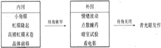
During surgery, due to the sudden and massive outflow of aqueous humor, the high intraocular pressure drops abruptly. This causes the vitreous body, which had been edematous due to the previously high intraocular pressure, to suddenly expand and impact the lens, leading to zonule rupture. Sometimes, the zonules are injured during the procedure, and combined with their inherent fragility, the lens shifts forward, resulting in pupillary block, which obstructs the angle or the surgical filtration pathway.
The cause induced by miotics is the contraction of the ciliary muscle, leading to ciliary block. The zonules of the lens become lax, and the ciliary body adheres to the equatorial region of the lens. Aqueous humor accumulates behind the lens, causing both the lens and the iris to move forward. The iris becomes highly bulged, the anterior chamber generally shallowens, and aqueous humor drainage is obstructed. At this point, the fluid can only be diverted posteriorly, leading to anterior displacement of the vitreous (aqueous humor accumulates behind the vitreous). This further pushes the lens forward, making the anterior chamber even shallower and causing the angle to close again, forming a vicious cycle. Thus, it manifests as ciliary block angle-closure glaucoma.
bubble_chart Clinical Manifestations
Malignant glaucoma is a type of refractory glaucoma that is difficult to diagnose and has intraocular pressure (IOP) that is hard to control. It is generally considered a severe complication following anti-glaucoma surgery, characterized by postoperative elevation of IOP, forward displacement of the lens-iris diaphragm, and significant shallowing or even disappearance of the entire anterior chamber. Typical cases often occur within hours, days, or even months after surgery. However, in some cases, malignant glaucoma may develop without prior anti-glaucoma surgery, instead being triggered by the topical use of miotic agents, trauma, or uveitis. These factors lead to ciliary muscle contraction and ciliary block due to transparent mechanisms.
bubble_chart Treatment Measures
For malignant glaucoma, emergency measures should be taken as early as possible to reduce the pressure in the posterior part of the eyeball and break the ciliary block. At the same time, drug treatment including hyperosmotic agents, carbonic anhydrase inhibitors, and cycloplegics should be administered, supplemented with corticosteroid drugs to reduce inflammatory reactions and ciliary body edema. Commonly used eye drops include: 1% atropine and 10% phenylephrine solution or 0.5% tropicamide, applied alternately several times a day. Pupil dilation can cause the ciliary body to move backward. If the anterior chamber forms and intraocular pressure normalizes after drug treatment, various medications can be gradually discontinued, starting with the hyperosmotic agents, followed by the carbonic anhydrase inhibitors, while the cycloplegics should be maintained for a longer period. If drug treatment proves ineffective after 4–5 days, surgical intervention may be considered.
Surgical treatments include vitreous puncture and drainage or anterior chamber air injection. If these are ineffective, lens extraction or vitrectomy may be performed. After the ciliary block is relieved, if intraocular pressure remains high, oral acetazolamide and timolol eye drops may be administered.


