| disease | Cervical Vertebral Fracture Dislocation without Spinal Cord Injury |
Violence-induced fracture of cervical vertebrae dislocation is often accompanied by varying degrees and types of spinal cord injury. However, some very severe fracture dislocations may not be associated with or only have minor spinal cord or nerve root injuries. This particular type of injury is fortunate for the injured.
bubble_chart Pathogenesis
Flexion violence is the primary cause of fracture of cervical vertebrae dislocation. Fracture dislocation leads to localized narrowing of the cervical spinal canal, making it highly susceptible to spinal cord injury, especially in cases of fracture dislocation at the cervical enlargement (C5–C7), where spinal cord injury is more likely to occur. Under normal anatomy, the sagittal diameter of the spinal canal at C3–C7 is approximately 14mm. If it measures 12mm or less, the spinal cord will be compressed. Therefore, when fracture dislocation causes anterior displacement of the vertebral body by one-third to one-half of its sagittal diameter, spinal cord compression is almost inevitable. Based on the injury history and X-ray features, cases with obvious fracture dislocation but fortunate absence of spinal cord injury are primarily due to the formation of a fulcrum between the affected vertebrae during cervical vertebrae dislocation. This creates tensile stress in the posterior structures, causing the spinous processes and laminae to separate, tearing the interlaminar ligaments, and often accompanied by vertebral arch fractures. The separation of the anterior and posterior structures of the cervical spinal canal creates sufficient safe space, allowing the spinal cord to retract posteriorly in a flexed position and avoid compression. After the injury, the patient's neck muscles tense to stabilize the cervical spine in a flexed position, preventing spinal cord injury.
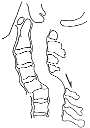
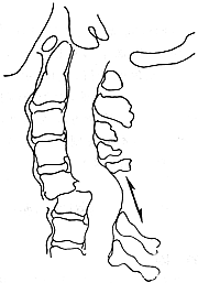
bubble_chart Pathological ChangesThere are two variations in this type of cervical vertebrae fracture dislocation. In one type, which is combined with a vertebral arch fracture, the vertebral body shifts forward, while the vertebral arch, lamina, and spinous process remain at the lower spinal level and maintain a normal alignment (Figure 1). In the other type, where the vertebral arch is fractured, the vertebral arch and other structures maintain a normal connection sequence with the posterior structures of the upper spine. The vertebral arches, laminae, and spinous processes of the two dislocated vertebrae separate and spread apart upward and downward (Figure 2). In cases with a vertebral arch fracture, the posterior structures spread apart between the vertebral arches above the dislocated vertebral body. The spinal cord at the fracture dislocation segment does not angle synchronously with the dislocated vertebra but moves backward away from the bony protrusion on the anterior wall of the spinal canal. The authors performed myelography on such patients and clearly visualized the appearance of the displaced spinal cord. Blothman also observed this type of dislocation. Although it causes a considerable number of spinal cord injuries, some patients with such injuries show no damage to the neural tissue. It is suggested that this may be related to the destruction of the posterior structures at the injury segment and their separation toward the lateral side of the spinal canal.

Figure 1: Fracture dislocation combined with a spinous process fracture, aligned with the lower vertebral segment

Figure 2: Separation and displacement of the spinous process at the fracture dislocation segment
This type of cervical spine injury without spinal cord compression presents with normal sensory and motor function in the limbs and trunk, as well as normal urination and defecation functions, or only symptoms of nerve root irritation. Local symptoms at the injury site are the prominent clinical manifestations of this type of injury.
1. Neck pain: The pain is mostly located at the injured segment, but it can also involve the entire neck and may be accompanied by tenderness.
2. The cervical spine exhibits a forward flexion deformity or stiffness: This manifests as a forced forward tilting of the head and neck with a stiff deformity.
3. Restricted movement: The range of motion in the neck is reduced, and sometimes rotation is also limited. This is related to pain at the injured segment and muscular rigidity, and the patient's own sense of insecurity is also one of the reasons for the restricted movement.
4. Limbs: Sensation in the limbs and trunk is normal, movement is unrestricted, tendon reflexes are normal, and there are no pyramidal tract signs.
If there is only a clear history of cervical spine {|###|}injury{|###|}, and local symptoms are still significant but a final diagnosis cannot be made, X-ray examination is the primary basis for confirmation. Routine anteroposterior cervical spine radiographs are taken to show whether the spinous processes are laterally displaced and changes in the intervertebral joints; left and right oblique views are used to assess changes in the intervertebral foramina and posterolateral structures; flexion and extension lateral views (should be performed under physician supervision) mainly reveal the following:
2. Anterior {|###|}dislocation{|###|} of one facet joint, anterolateral {|###|}dislocation{|###|} of the vertebral body, with fractures of the vertebral arch and facet processes.
3. Anterior {|###|}dislocation{|###|} of the vertebral body combined with a fracture of the anterior vertebral margin.
4. If there is no fracture in the posterior cervical structures, the spinous processes of the vertebrae above and below the {|###|}dislocation{|###|} are splayed apart; in cases with vertebral arch fractures, the lamina and spinous processes remain normally aligned with the lower vertebrae.
Tomography and CT scans of the injured segment can reveal subtle or occult bone and joint {|###|}injuries{|###|}, while MRI imaging can display the morphology of the spinal cord and its relationship with the spinal canal.
bubble_chart Treatment Measures
Non-surgical treatment
Emergency management should pay close attention to the mechanism of injury. Once diagnosed, cervical spine extension should be avoided to prevent spinal cord injury. Occipitomandibular traction or skull traction should be applied, with the direction of traction slightly flexing the cervical spine. The traction weight should start at 2–3 kg, similar to that for fracture-dislocation combined with spinal cord injury, and gradually increase under close observation. Once reduction is achieved, the cervical spine should be maintained in a physiological position. After 3 weeks, a head-neck-thorax gypsum cast should be applied for 3 months. During traction, if neurological irritation or compression symptoms occur, the traction weight and direction should be adjusted. If symptoms persist or worsen, traction should be discontinued immediately to avoid spinal cord injury for the sake of reduction. Maintain traction weight for 3 weeks, followed by a gypsum cervical collar fixation, and proceed to late-stage [third-stage] surgical fixation.
Surgical treatment
The goal of surgery is to restore cervical spine stability and repair or expand the spinal canal at the injured segment to prevent future chronic compression. The anterior approach is used to remove the posterior edge of the vertebral body protruding into the spinal canal at the injured segment. The procedure must be meticulous to avoid spinal cord injury. Autologous iliac bone is grafted into the intervertebral space to ensure bony fusion of the anterior cervical structure for stability. Postoperatively, a gypsum cervical collar should be applied for 3 months.


