| disease | Parapharyngeal Space Infection |
Parapharyngeal space infection refers to an acute suppurative infection in the parapharyngeal space, with the main clinical manifestations being redness and swelling of the lateral pharyngeal wall and protrusion of the palatine tonsil.
bubble_chart Etiology
The parapharyngeal space is located lateral to the pharyngeal cavity, between the superior pharyngeal constrictor muscle and the medial pterygoid muscle and the deep lobe of the parotid gland. Anteriorly, it is bounded by the pterygomandibular ligament and the upper edge of the submandibular gland; posteriorly, by the prevertebral fascia. The space is shaped like an inverted pyramid, with its base at the top formed by the temporal and sphenoid bones of the skull base, and its apex extending downward to the hyoid bone. The styloid process and the muscles attached to it divide the space into anterior and posterior compartments: the anterior compartment is called the anterior parapharyngeal space, and the posterior compartment is the posterior parapharyngeal space. The anterior compartment is small and contains the ascending pharyngeal artery and vein, lymphatics, and loose connective tissue. The posterior compartment is larger and contains the internal carotid artery and vein, the 9th to 12th cranial nerves, and the superior deep cervical lymph nodes. The parapharyngeal space communicates with the pterygomandibular, infratemporal, sublingual, submandibular, and retropharyngeal spaces. The neurovascular bundle connects superiorly to the intracranial space and inferiorly to the mediastinum, serving as a potential pathway for the spread of infection (Figure 2).
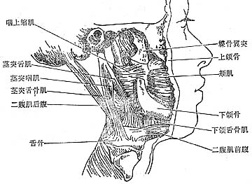
Figure 2 Anatomical location of the parapharyngeal space
Most cases are odontogenic in origin, particularly pericoronitis of the mandibular wisdom tooth, as well as the spread of palatine tonsillitis and infections from adjacent spaces. Occasionally, it may be secondary to parotitis, otogenic infections, or inflammation of the superior deep cervical lymph nodes.bubble_chart Clinical Manifestations
The local symptoms of parapharyngeal space infection mainly manifest as redness and swelling of the lateral pharyngeal wall, protrusion of the palatine tonsil, and swelling that may extend to the ipsilateral soft palate, palatoglossal arch, and palatopharyngeal arch, with the uvula being pushed to the unaffected side. If accompanied by inflammation of the pterygomandibular space or submandibular space, the swelling in the lateral pharyngeal and upper neck regions becomes more extensive and pronounced.
Patients experience subjective symptoms such as pain during swallowing, difficulty eating, and limited mouth opening. If accompanied by laryngeal edema, hoarseness, varying degrees of dyspnea, and coughing during eating may occur. If the parapharyngeal space infection is inadequately managed, it can lead to severe complications such as pulmonary infection, sepsis, and thrombophlebitis of the internal jugular vein.
Clinically, it is important to differentiate this condition from diseases with similar local manifestations, such as rapidly progressing malignant tumors in the lateral pharyngeal region or secondary infections of cystic sexually transmitted diseases.
1. Medical History A history of acute pericoronitis of the mandibular wisdom tooth, acute tonsillitis, or infections in adjacent spaces such as the pterygomandibular, buccal, submandibular, or sublingual spaces.
2. Clinical Manifestations More common in children and adolescents. In addition to severe systemic signs of infection and toxicity, the local presentation often exhibits the following three characteristic features.(1) Pharyngeal Signs Unilateral redness, swelling, and tenderness in the pharynx within the oral cavity. The swollen area includes the pterygomandibular ligament region, soft palate, and uvula, which deviates to the unaffected side. The patient experiences pain during swallowing and difficulty eating. Pus can be aspirated from the most prominent area of pharyngeal swelling.
(2) Cervical Signs Swelling and tenderness at the level of the greater horn of the hyoid bone, slightly below the angle of the mandible on the affected side.
(3) Restricted Mouth Opening Due to inflammatory stimulation causing spasm of the medial pterygoid muscle (the lateral boundary of this space), the patient exhibits varying degrees of restricted mouth opening.
bubble_chart Treatment Measures
The parapharyngeal space is deeply located, and the presence of an abscess is usually confirmed by puncture. The puncture is performed by entering between the superior pharyngeal constrictor muscle and the medial pterygoid muscle through the medial side of the pterygomandibular fold in the oral cavity. Once pus is aspirated, incision and drainage should be performed immediately.
Intraoral approach for incision and drainage: For patients without significant mouth-opening limitations, a longitudinal incision can be made slightly medial to the pterygomandibular fold through the mucosal layer. A hemostat is then used to bluntly dissect submucosally along the medial side of the pterygoid muscle to enter the abscess cavity. The mucosal incision should not be too deep to avoid accidental injury to major blood vessels and nerves (Figure 1).
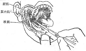
Figure 1 Intraoral incision and drainage for parapharyngeal space abscess.


