| disease | Bile Reflux Gastritis |
| alias | Alkaline Reflux Gastritis |
Reflux gastritis, also known as alkaline reflux gastritis, refers to a syndrome characterized by a series of symptoms such as upper abdominal pain, vomiting bile, abdominal distension and fullness, and weight loss, caused by the reflux of bile into the stomach. It commonly occurs after gastrectomy or gastrointestinal anastomosis, with an overall incidence of about 5%. The incidence after Billroth II gastrectomy is 2 to 3 times higher than that after Billroth I. Given its distinct symptoms, pathological changes, and treatment response compared to other postgastrectomy syndromes, Roberts et al. classified this condition as a separate disease entity, distinct from other postgastrectomy complications.
bubble_chart Pathogenesis
The occurrence of this syndrome first requires the fundamental condition of pyloric dysfunction or pyloric insufficiency, such as after gastrectomy or gastrointestinal anastomosis, where {|###|}gall fel{|###|} can directly reflux into the stomach. Some patients have no surgical history, yet duodenal contents can reflux into the stomach through an incompetent pylorus, causing reflux gastritis. After cholecystectomy, the function of storing {|###|}gall fel{|###|} is lost, and {|###|}gall fel{|###|} continuously flows into the duodenum. If it refluxes into the stomach through an incompetent pylorus, it can similarly cause reflux gastritis.
Pure {|###|}gall fel{|###|} coming into direct contact with the gastric mucosa generally does not cause damage. However, by stimulating gastric acid secretion, bile salts combined with gastric acid can enhance the activity of acidic hydrolases, disrupt lysosomal membranes, and dissolve lipoproteins, thereby impairing the barrier function of the gastric mucosa. H
+ reverse diffusion increases, entering the mucosa and submucosa, which can stimulate mast cells to release histamine. The latter further stimulates the secretion of gastric acid and pepsin, ultimately leading to gastric mucosal inflammation, erosion, and hemorrhage. When {|###|}gall fel{|###|} mixes with pancreatic juice, the lecithin in {|###|}gall fel{|###|} interacts with phospholipase A in pancreatic juice to convert into lysolecithin. If this refluxes into the stomach, it can also damage the gastric mucosal barrier.Gastrin can stimulate the proliferation of gastric mucosal cells to strengthen their barrier function and prevent the reverse diffusion of H+. However, after Billroth II gastrectomy, gastrin secretion decreases by approximately 50–75%, which may be one of the important pathogenic factors of this syndrome.
Post-gastrectomy reflux of {|###|}gall fel{|###|} into the stomach is a common phenomenon, but not everyone develops symptoms. The pathogenesis may also be related to the following factors: 1. **Gastric emptying disorder**: Prolonged retention of reflux fluid in the stomach raises the pH, making it easier for aerobic and anaerobic bacteria to grow in the residual stomach. These bacteria can release bile salts, causing gastric mucosal inflammation and symptoms. 2. **Changes in bile acid composition**: Gadacz found that those with normal bile acid composition do not develop symptoms, while those with significantly increased deoxycholic acid often experience symptoms. 3. **Presence of bacteria in gastric juice**: Symptomatic patients have Gram-negative bacilli or Pseudomonas in their gastric juice. Treatment with doxycycline can alleviate symptoms, whereas asymptomatic individuals have no bacteria in their gastric juice. 4. **Sodium concentration in gastric juice**: Those with sodium concentrations exceeding 15 mmol/L are prone to gastritis, while those with concentrations below 15 mmol/L do not develop gastritis.
Most patients complain of persistent burning pain in the upper middle abdomen, with increased pain after meals, which is not relieved by alkaline medications or may even worsen. A few patients may experience retrosternal pain or a sensation of indigestion. Gallbladder vomiting is a characteristic manifestation. Due to impaired gastric emptying, vomiting often occurs in the evening or at night, and the vomitus may contain food, occasionally with small amounts of blood. Fearing symptom aggravation after eating, patients reduce their food intake, leading to anemia, weight loss, malnutrition, and diarrhea.
After gastric surgery, if the aforementioned characteristic symptoms are discovered, the following examinations should be performed:
(1) Endoscopy Direct visualization of bile reflux, gastric mucosal congestion, edema, or erosion can be observed. Biopsy indicates gastritis. Although bile reflux is a common occurrence after gastrectomy, if endoscopy reveals atrophic gastritis, bile reflux gastritis can be confirmed.
(2) Gastric Aspirate Measurement After inserting a gastric tube, fasting and postprandial gastric fluids are aspirated to measure bile acid content. If the basal acid output (BAO) is <3.5 mmol/h and bile acid exceeds 30 μg/ml, bile reflux gastritis can be diagnosed.
(3) Isotope Measurement Intravenously inject 2 mCi 99mTc-diethylenetriamine pentaacetic acid (DTPA), and observe the liver and biliary tract every 5 minutes for 1 hour. After 1 hour, the patient drinks 100 ml of water containing 0.3 mCi 99mTc to accurately determine the position of the stomach. Subsequently, over the next 2 hours, the liver, gallbladder, and stomach areas are examined every 15 minutes to determine the gastrointestinal reflux index. The normal value is 8.6 ± 6.0, while in cases of reflux gastritis, it increases to 86.3 ± 7.1. Alternatively, a 99m

bubble_chart Treatment Measures
Drug therapy should be attempted first, and surgical treatment should only be considered for those who fail drug therapy and have severe symptoms.
(1) Drug Therapy
Cholestyramine is a basic anion exchange resin that binds to bile salts in the stomach and accelerates their excretion. Initially, take 4g one hour after meals and add another dose before bedtime. It usually takes effect within 1 to 2 weeks, after which the dosage can be gradually reduced. Fat-soluble vitamins should be supplemented simultaneously. If there is no effect after 3 months of treatment, it is considered a treatment failure.
Metoclopramide (also known as gastric motility stimulant) can promote contractions of the gastric cardia and proximal small intestine, accelerating gastric emptying. This can alleviate symptoms in patients with gastric emptying disorders and may also reduce the secretion of bile and pancreatic juice, thereby relieving symptoms of reflux gastritis.
(2) Surgical Treatment There are essentially four surgical methods.
1. Conversion to Billroth I Procedure For patients who originally underwent Billroth II gastrectomy, converting to Billroth I can improve symptoms in approximately half of the patients.
2. Roux-en-Y Procedure For patients who originally underwent Billroth II surgery, the afferent limb at the anastomosis is transected, and the proximal cut end is anastomosed to the efferent limb (see Figure 1).
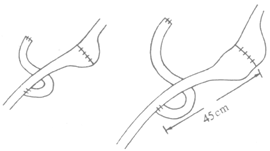
Figure 1 Roux-en-Y Gastrojejunostomy
3. Jejunal Interposition For patients who originally underwent Billroth I gastrectomy, a segment of jejunum approximately 20 cm long is interposed between the gastroduodenal anastomosis, with an efficacy rate of 75%.
4. Tanner Procedure Suitable for patients who originally underwent Billroth II surgery, the jejunal afferent limb is transected, the distal cut end is anastomosed to the jejunal efferent limb to form a loop, and the proximal cut end is anastomosed to the jejunum 50 cm distal to the original gastrojejunal anastomosis (see Figure 2).
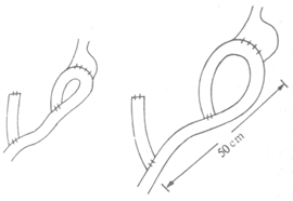
Figure 2 Tanner Procedure
In addition, to prevent anastomotic ulceration, vagotomy may be considered.


