| disease | Pubic Tuberculosis |
The pubic subcutaneous node is relatively rare, with only 18 cases (0.43%) out of 4,140 cases. The part of the pubis near the pubic symphysis is called the pubic body, which branches into the superior ramus and inferior ramus. The distal end of the superior ramus forms part of the acetabulum, while the distal end of the inferior ramus connects with the ischial ramus. The junction of the two pubic bones is called the pubic symphysis. The anterior surface of the pubic body is the perineal surface, and its posterior surface is the pelvic surface.
bubble_chart Pathological Changes
Subcutaneous nodes of the pubis often present as localized bone destruction, with the lesion extending upward to the superior pubic ramus and downward to the inferior pubic ramus. Crossing the midline may destroy the pubic symphysis and involve the contralateral pubic body.
The lesions are mostly osteolytic, with possible local sequestra. When the lesion affects the pubic rami, periosteal reactions are often observed.
Cold abscesses may spread along the pubis and adductor muscles, forming multiple abscesses in the groin or medial thigh. Abscesses from combined lesions of the pubic symphysis may flow into the pyramidalis muscle or rectus sheath, where granulation tissue or small sequestra can be seen. Lesions on the posterior aspect (pelvic surface) of the pubic body may form abscesses between the bladder and pubis, and cold abscesses often rupture to form sinuses.
bubble_chart Clinical Manifestations
This disease is commonly seen in women of childbearing age. Without subcutaneous nodules in other areas, systemic symptoms are usually absent, and the onset is generally slow with mild local pain. Severe bone destruction often leads to limping. Local swelling is common, with noticeable tenderness, and abscesses or sinus tracts often form by the time medical attention is sought. Apart from slight abduction limitation in the affected hip joint, there is no functional impairment.
bubble_chart Auxiliary ExaminationX-ray imaging reveals localized bone destruction in the pubis, often with sequestra. When the lesion involves the pubic symphysis, widening or dislocation of the symphysis can be observed.
Based on medical history, symptoms, signs, and X-ray findings, the diagnosis is usually not difficult.
bubble_chart Treatment Measures
1. For cases without dead bone and without sinus involvement, simple treatment with anti-subcutaneous node medication can achieve cure.
2. For cases where non-surgical treatment is ineffective, lesion debridement therapy is adopted.
(1) Pubic Lesion Debridement
1. Anesthesia: Local anesthesia, epidural block, or general anesthesia.
2. Position: The patient lies supine with the hips slightly elevated and legs apart. A urinary catheter is placed preoperatively to clarify the position of the urethra and avoid injury during surgery.
3. Incision: A curved incision as shown in Figure 1.
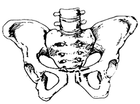
Figure 1: Two curved incisions for pubic symphysis lesion debridement
4. Exposure of the lesion: For female patients, the mons pubis and labia majora are turned downward; for male patients, the spermatic cord is retracted bilaterally. The pubic periosteum and its ligaments are incised, followed by subperiosteal dissection to expose and debride the lesion. If exposure is unsatisfactory, subperiosteal dissection is performed to reveal the pelvic surface. Care must be taken to avoid injury to the urethra and bladder during surgery.
Hemostasis should be carefully managed during the operation to prevent extensive postoperative perineal swelling.
Postoperatively, the patient should remain in bed for 1–2 months, gradually resuming activity.
(2) Ischial (Bursal) Subcutaneous Node Lesion Debridement1. Anesthesia: Continuous epidural anesthesia or general anesthesia.
2. Position: The patient lies in the lateral position, with the torso at a 60° angle to the operating table, maintained by sandbags. The affected hip and knee joints are flexed at 45°, while the unaffected leg is extended, secured with restraints.
3. Procedure:
(1) Incision: Starts 2 cm medial to the ischial tuberosity, follows the gluteal fold along the lower edge of the gluteus maximus, and ends posteromedial to the greater trochanter.
(2) Exposure of the lesion: After incising the skin, subcutaneous tissue, and superficial and deep gluteal fascia, the gluteal muscle is reflected upward to immediately expose the ischial (or bursal) subcutaneous node lesion.
(3) Debridement of the lesion: For subcutaneous node bursitis, complete dissection and removal are required. During ischial subcutaneous node debridement, special attention must be paid to thoroughly clearing lesions on the medial side of the ischium.
(4) Suturing: The area is irrigated, hemostasis is ensured, and the wound is closed in layers. A closed silicone tube drainage is placed. Due to the proximity of the incision to the anus, precautions against bacterial infection are necessary.
4. Postoperative care: Anti-subcutaneous node medication is continued as before. The patient should rest in bed for 3–6 weeks postoperatively, gradually resuming activity.
1. Suppurative pubic osteitis presents with high fever, elevated white blood cell count, and obvious local symptoms, with diagnosis confirmed by bacteriological examination.
2. Non-suppurative pubic osteitis occurs in women during pregnancy and after childbirth, and in men after prostate surgery, with local pain. X-ray shows narrowing or widening of the pubic symphysis, symmetrical grade I destruction of bilateral pubic bodies, and sclerotic changes. The bone destruction of the pubic subcutaneous node is often unilateral, frequently accompanied by sequestrum formation.


