| disease | Unilateral Facet Joint Dislocation |
Unilateral facet joint dislocation is a relatively common injury, usually caused by the combined action of flexion and rotational forces.
bubble_chart Pathogenesis
Similar to flexion injury, when the head and neck are subjected to forces such as flexion impact, the neck not only flexes but also rotates to one side. When flexion and rotational forces act simultaneously on the cervical spine, the injured segment forms a forward and downward torsional force. With the posterior central part of the intervertebral disc as the pivot, the inferior articular process of the upper cervical vertebra on one side rotates backward, while the inferior articular process on the other side slides forward, surpassing the superior articular process of the lower vertebra and locking in front of it, creating a "locked" state (Figure 1). During the mutual impact of the superior and inferior articular processes, joint fracture may also occur.
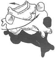
Figure 1 Mechanism of unilateral facet dislocation under flexion and rotational forces.
bubble_chart Pathological Changes
Bilateral facet joint capsule tears may involve damage to the anterior longitudinal ligament, posterior longitudinal ligament, intervertebral disc, and posterior ligamentous complex. Due to the dislocation shifting the facet joint anteriorly, the intervertebral foramen becomes deformed or narrowed, making the nerve roots prone to injury. This dislocation is considered a relatively "stable" state of cervical spine injury, but the articular surfaces of the facet joints on the non-dislocated side remain separated. The annulus fibrosus of the intervertebral disc is subjected to continuous rotational stress. This asymmetric dislocation causes deformation and narrowing of the spinal canal at the injury level, potentially leading to spinal cord injury.
bubble_chart Clinical Manifestations1. Local symptoms in the neck are prominent, including pain, forced head and neck tilt deformity, and limited cervical spine extension and rotation functions.
2. When combined with spinal cord and nerve root injury, symptoms corresponding to the affected spinal segments may appear: quadriplegia, paraplegia, or partial paralysis. In cases of nerve root injury, manifestations include skin hypersensitivity, pain, or hypoesthesia in the distribution area of the affected nerve.
X-ray Findings
The characteristic X-ray findings are key to diagnosis. The typical sign on lateral X-rays is anterior displacement of the dislocated vertebra by one-third of the posterior vertebral diameter, with a maximum displacement not exceeding half. At the level of the dislocated vertebra, the normal interrelationship of the facet joints cannot be observed (Figure 1).
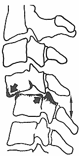
Figure 1 Schematic diagram of unilateral facet joint dislocation on X-ray
Anteroposterior views show deviation of the spinous process of the dislocated cervical vertebra from the midline, shifting toward the side of the dislocated facet joint. Oblique views can clearly reveal signs of facet joint dislocation or "locked" facets.
bubble_chart Treatment Measures
Skull traction and occipitomandibular traction are commonly used reduction methods. During traction, the head and neck should be slightly flexed (approximately 30°), with a traction weight of 5–6 kg, gradually increasing but not exceeding 10 kg to avoid or aggravate spinal cord {|###|}injury. To facilitate reduction, a sand pad can be placed under the shoulder on the dislocation side to induce grade I lateral flexion of the injured segment, separating the dislocated articular processes. The traction direction can then be adjusted to achieve reduction. As with bilateral articular process dislocation, close attention must be paid to changes in the patient's overall condition throughout the reduction process, and bedside imaging should be performed every 10 minutes to monitor the reduction. Similarly, as with bilateral articular process dislocation, the patient's systemic condition must be closely monitored throughout the reduction process, and bedside imaging should be performed every 30 minutes to track the progress and prevent additional {|###|}injury.
After reduction, maintain traction with a weight of 1–2 kg for 3–4 weeks, followed by head-neck-thorax {|###|}gypsum fixation for 2–3 months. If cervical spinal cord {|###|}injury is present, {|###|}gypsum fixation should not be used; instead, continuous traction should be maintained for 2–3 months until healing occurs.Surgical reduction and fixation: Open reduction may be considered if traction reduction fails. A posterior midline incision is made to expose the locked articular processes. The embedded joint capsule and ligamentous tissues are excised, and a periosteal elevator is used to lever and reduce the dislocation. If difficulties arise, the obstructing portion of the superior articular process of the lower vertebra can be resected, and the traction direction adjusted, which usually achieves reduction.
If spinal cord {|###|}injury is present, laminectomy decompression should be performed based on the extent of compression. To stabilize the injured segment, spinous process wiring with autologous iliac bone grafting can be performed intraoperatively. A common grafting method is interspinous "H"-shaped bone grafting with wire fixation. Alternatively, interarticular process bone grafting and fixation may also be used.
Unilateral facet joint dislocation should be differentiated from bilateral facet joint dislocation. Based on the injury and clinical manifestations, the following aspects can be used for differentiation:
1. Different injury mechanisms: Bilateral facet joint anterior dislocation is mainly caused by flexion force, while unilateral facet joint dislocation involves both flexion and torsional forces.
2. Different clinical manifestations: Bilateral facet joint dislocation mainly presents with fixed forward tilt and widespread tenderness, often accompanied by spinal cord injury. Unilateral facet joint dislocation primarily exhibits rotational fixation, with less widespread tenderness. Few cases are complicated by spinal cord injury, but local pain is severe.3. Different X-ray findings: In bilateral dislocation, the anterior displacement of the injured vertebral segment is often 2/5 or 1/2 of the anteroposterior diameter of the vertebral body, with the inferior articular process of the upper cervical vertebra located at the top or anterior to the superior articular process of the lower vertebra. In unilateral facet joint dislocation, the anterior displacement of the injured vertebral segment is about 1/3 of the anteroposterior diameter, and the anteroposterior view shows deviation of the spinous process at the dislocated segment.


