| disease | Congenital Hip Dislocation |
| alias | Hip First |
Congenital hip dislocation is one of the most common congenital malformations in children, with posterior dislocation being more frequent. It is present at birth and involves the acetabulum, femoral head, joint capsule, ligaments, and surrounding muscles, leading to joint laxity, subluxation, or dislocation. Sometimes it may be associated with other malformations, such as congenital torticollis, hydrocephalus, meningocele, congenital dislocations or contractures of other joints.
bubble_chart Epidemiology
The incidence of this disease varies greatly due to the influence of many factors, such as region, lifestyle, ethnicity, etc. The incidence is relatively high in northern Italy, southern France, and southern Germany. Mckeown et al. reported in 1960 that the incidence in Birmingham, UK, was 0.7%, while in Sweden it was 1%. The incidence is also high in Japan and among Native American tribes. Hodgson believed that the incidence in China is very low, mainly referring to southern China, because the habit of carrying children involves separating the hips and flexing the knees, as this infant posture can correct hip dislocation. In reality, the incidence varies across different regions of China, but comprehensive statistical data is lacking. However, the incidence is not too low. In Africa, the incidence is the lowest in the world.
The disease is more common in girls, with a male-to-female ratio of 5–7:1. The incidence on the left side far exceeds that on the right side, with a ratio of 10:1.
The exact cause of congenital hip dislocation remains incompletely understood. Of course, hip dislocation associated with multiple deformities should be classified as a congenital malformation. In general, most scholars in recent years believe that the cause is not singular. This means that multiple factors contribute to the development of this condition.
(1) Genetic factors: Undeniable evidence indicates a clear familial tendency for this condition, particularly evident in twin infants. The incidence rate in families with affected individuals can be as high as 20–30%, and it is even more common among sisters. The same condition can manifest in sisters as three types: hip dislocation, subluxation, and dysplasia. Without detailed, early examination and X-ray diagnosis, apart from the first type, the latter two may often go unnoticed until the age of 7 or 8, by which time the hip joint may have fully normalized.(2) Ligament laxity factors: Increasing reports in recent years suggest that joint ligament laxity is a significant factor. In animal experiments, Smith found a high percentage of hip dislocation after excising the joint capsule and round ligament in puppies. Clinically, Andren noted that the separation of the pubic symphysis in X-rays of hip dislocation cases was twice that of normal infants. He proposed that this is due to the mother requiring large amounts of endocrine hormones to relax ligaments during childbirth, and that excessive endocrine changes are a key factor in hip dislocation. Additionally, Andren and Borglin observed changes in the urinary excretion of estrone (Estrone) and estradiol-17β (Estradiol) in newborns with hip dislocation within three days of birth compared to normal infants. However, Thieme compared 16 affected infants with 19 normal infants and found no statistically significant difference in monthly measurements. Therefore, the theory that endocrine changes cause ligament laxity remains unproven.
(3) Postural and mechanical factors: Some reports indicate that breech birth accounts for 16–30% of hip dislocation cases, whereas in normal deliveries, breech births make up only 3%. Wikinson (1963) fixed the hip joints of young children in flexion, external rotation, and knee extension while administering estrogen and progesterone, resulting in hip dislocation deformity.

bubble_chart Pathological Changes
The pathological changes of congenital hip dislocation include two parts: bone changes and changes in surrounding soft tissues:
(1) Bone changes Hip dysplasia is the fundamental change, which includes the acetabulum, pelvis, femoral head, and femoral neck. In severe cases, it can also affect the spine.
1. Acetabulum In cases of congenital hip dislocation, the acetabulum is normal at birth, but there is a notch on the outer upper edge of the acetabulum. As growth progresses, the acetabulum gradually becomes narrow and shallow, taking on a triangular shape. The acetabular labrum thickens, and due to continuous pressure from the femoral head, it may invert or evert. A false acetabulum forms in the posterosuperior part of the acetabulum due to the pressure of the femoral head, and a defect is often seen on the anterosuperior edge of the acetabulum. Without the modeling effect of the femoral head, the acetabulum becomes dysplastic, gradually smaller and shallower, with the acetabular floor filled with fatty fibrous tissue. The ligamentum teres, after continuous traction, often thickens and hypertrophies, filling the acetabulum.
2. Femoral head The femoral head of a newborn is deformed, with a smooth cartilage surface. Later, due to dislocation outside the acetabulum, the shape of the femoral head gradually changes, becoming larger or smaller, conical or mushroom-shaped. The compressed area of the femoral head often shows partial flattening. The appearance of the femoral head epiphysis is delayed. Sometimes, forceful surgical reduction, due to the mismatch between the acetabulum and femoral head, can cause excessive pressure on the femoral head, leading to avascular necrosis.
4. Pelvis and spine The pelvis on the dislocated side often shows dysplasia, with a more oblique iliac wing and more separated ischial tuberosities. In cases of bilateral dislocation, in addition to the above changes, the pelvis tilts forward, increasing the lumbar lordosis, and sometimes scoliosis may occur.
(2) Soft tissue changes This refers to all the soft tissues around the hip joint, including skin, fascia, muscles, tendons, joint capsule, ligaments, and the intra-articular disc-shaped cartilage, with the intra-articular disc-shaped cartilage, joint capsule, and tendons being the most important.
1. Disc-shaped cartilage (Limbus) In a normal 14.8 mm embryo, the hip joint is a mass of mesenchymal cells. Later, a gap appears between the acetabulum and femoral head, and the mesenchymal cell mass begins to absorb, leaving only the edges. By 25 mm, the joint capsule and acetabular labrum (glenoid labrum) appear. Any mechanical stimulation during the main stage of acetabular formation can cause the normal mesenchymal cells to stop absorbing, resulting in a disc-shaped cartilage. In fact, incomplete absorption of the disc-shaped cartilage is mostly seen in the posterosuperior part of the acetabulum. Its proliferation and hypertrophy prevent the femoral head from directly pointing to the center of the acetabulum. Leveurf and Somerville believe this is the main cause of hip dislocation and the key to reduction. In surgery, for children over 3 years old, if the femoral head cannot enter the acetabulum after traction, it is often due to a thickened disc-shaped cartilage. This cartilage is completely like a disc-shaped meniscus in the knee joint, covering a large part of the joint surface and preventing contact between the femoral head and acetabulum, leading to dysplasia of both.
2. Joint Capsule The normal hip joint capsule is a layer of fibrous tissue 0.5 to 1.0 mm thick. After the femoral head dislocates from the acetabulum and shifts outward and upward, once the child bears weight, the joint capsule is stretched, elongating and thickening, sometimes reaching up to 2–3 mm in thickness. Prolonged stretching causes the joint capsule to adhere to the iliac wing above the acetabulum, and along with adhesions between the round ligament, disc-shaped cartilage, and the joint capsule, it forms a solid mass of connective tissue that obstructs the femoral head from entering the acetabulum. In the late stage [third stage], the joint capsule takes on a "bottle gourd peel" shape with a narrow neck, making it impossible for the femoral head itself to pass through. The iliopsoas tendon runs along the front of the joint capsule and sometimes develops a notch in the very early stages, hindering the reduction of the femoral head. The joint capsule attaches below the femoral head rather than between the greater and lesser trochanters.
3. Ligamentum Teres Normally, the ligamentum teres connects the fovea capitis of the femur to the inferior aspect of the acetabulum. In cases of hip dislocation, both the joint capsule and the ligamentum teres are stretched and become elongated and thickened. Over time, the ligamentum teres adheres to the joint capsule and eventually disappears. The central artery within the ligamentum teres also becomes prematurely occluded due to stretching and thickening.
4. Muscles Due to the upward displacement of the femoral head, most muscles originating from the pelvis and descending along the femur become shortened, particularly the adductor muscles and the iliopsoas. Additionally, many tendons undergo fibrous degeneration. The posterior muscle groups, including the gluteal muscles, also become shortened, leading to weakened muscle strength, compromised joint stability, and a waddling gait.
5. Fascia Although the lateral muscle groups are theoretically lengthened, fascial contractures of the gluteal fascia can be observed, preventing the patient from adducting the hip. These fascial structures often exhibit fibrous tissue hyperplasia, with severe cases showing collagen degeneration. During surgery, fascial release is essential to ensure successful reduction.
bubble_chart Clinical Manifestations
The mother of the affected child often notices abnormal limbs and seeks medical attention. If there is no history of inflammation or trauma, suspicion of this condition should be raised. The symptoms can generally be summarized as follows:
(1) Limited joint movement: In childhood, congenital hip dislocation is typically characterized by painlessness and unrestricted joint movement. However, in infants and newborns, the opposite is true, with temporary joint dysfunction presenting in a fixed posture. The typical symptom reported is that the affected limb remains flexed and unwilling to extend, with poorer movement compared to the healthy side, and weakness. When the lower limb is pulled, it can extend, but upon release, it returns to a flexed position. A few infants may have their lower limbs in an externally rotated position, abducted position, or even crossed, with some cases showing complete hip joint stiffness. A small number of children may cry when their lower limbs are pulled.
(2) Shortened limb: Unilateral hip dislocation often results in shortening of the affected limb.
(3) Other common symptoms include asymmetry of the labia majora, increased, deepened, or asymmetrical skin folds on the buttocks, inner thighs, or popliteal fossa, widened perineum, and sometimes a "clicking sound" or a sensation of snapping when moving the affected limb.
If these symptoms are detected promptly and a thorough examination is conducted, timely diagnosis and treatment can be achieved, significantly improving treatment outcomes.
The diagnosis primarily relies on signs, X-ray examinations, and measurements. The examination of newborns also focuses on the following points:
(1) Appearance and Skin Folds When multiple deformities are accompanied by hip dislocation, the examiner often finds that the proportions of the thigh and lower leg are disproportionate—the thigh is short and thick, while the lower leg is slender. The buttocks are often broad, and the inguinal folds may be short or indistinct. During the buttock examination, differences in skin folds on both sides may be observed. On the affected side, the folds are generally elevated or an additional fold is present. When the lower limb is placed flat, the affected limb often appears externally rotated by 15–20° and shortened.
(2) Inability to Palpate the Femoral Head With the hip and knee flexed at 90°, one hand holds the upper part of the lower leg while the other hand places the thumb on the inguinal ligament and the other four fingers on the buttock (around the sciatic region). When rotating the lower leg, the movement and protrusion of the femoral head can normally be felt anteriorly. In cases of dislocation, the anterior region feels empty, while the four fingers on the buttock can sense the movement of the femoral head posteriorly.
(3) Galeazzi Sign The child is placed supine with both lower limbs flexed at the knees between 85° and 90°, and the ankles positioned symmetrically flat. If a difference in knee height is observed, it is referred to as the Galeazzi sign. Femoral shortening or hip dislocation will exhibit this sign (Figure 1).
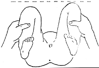
Figure 1: Galeazzi Sign
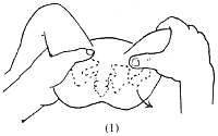
(4) Abduction Test (Ortolani Sign) The child is placed supine with the hips and knees flexed at 90°. The examiner, facing the child’s buttocks, grasps both knees and abducts them simultaneously. Normally, the knees can be laid flat and touch the examination table. However, in cases of hip dislocation, one side may not reach 90°, often stopping between 65° and 70°, with the adductor muscles visibly prominent. This is termed a positive abduction test. If a sliding or jumping sensation is felt between 75° and 80° of abduction, followed by further abduction to 90°, it is called the Ortolani click, which is a crucial diagnostic indicator. During the examination, it is essential to distinguish between the clicking sound inside or outside the acetabulum and the snapping of the knee meniscus to avoid confusion (Figures 2 and 3).
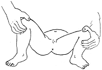
Figure 2: Abduction Test
① Stabilizing the Pelvis
② Abducting the Affected Limb
Figure 3: Ortolani Test
(5) Joint Laxity Test The prerequisite for testing joint laxity is that the soft tissues around the femoral head are loose, the muscles are not tense, and the femoral head can move up and down, entering and exiting the acetabulum. This test includes the following three methods:
1. Thomas Test In newborns, the healthy leg is flexed against the abdominal wall to eliminate lumbar lordosis. When the affected leg is extended, it should form a straight line. In normal infants, a slight flexion of about 30° remains when the leg is extended, but it can still be fully flattened into a straight line.
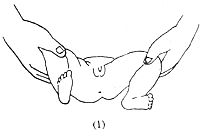
2. Barlow Test The affected limb is flexed at the knee so that the heel touches the buttock. One hand holds the ankle joint and the ipsilateral greater and lesser trochanters, while the other hand places the thumb on the pubic symphysis and the other four fingers against the sacrum. During mid-abduction, pressure from the thumb may cause the femoral head to dislocate posteriorly, and releasing the pressure allows the head to return to the joint. A positive Barlow test indicates joint laxity and susceptibility to dislocation but does not confirm hip dislocation (Figure 4).
① Thumb pressure causing femoral head dislocation
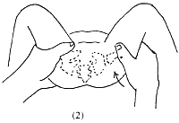
② Release thumb pressure, and the femoral head reduces spontaneously.
Figure 4: Release thumb pressure, and the femoral head reduces spontaneously.
3. Telescoping Test: The child lies supine with the hip and knee flexed at 90°. One hand holds the knee joint while the other presses on both anterior superior iliac spines of the pelvis. Pushing the knee downward, the femoral head can be felt protruding backward. When lifting upward, the femoral head reduces back into the acetabulum. This is called a positive telescoping test.
The above three sets of joint mobility tests are generally applicable to newborns and can only be accurately performed if the child cooperates without crying or fussing. Otherwise, the examination is often impossible, thus presenting certain limitations.
(6) Limping Gait: Although early diagnosis is crucial, many cases are still brought to the clinic due to limping. This gait can be identified with slight observation during walking. When the affected limb is in the stance phase, the pelvis droops, wobbles, and fails to rise; in the swing phase, this is less noticeable. Such examinations typically require the child to walk before a definitive diagnosis can be made, usually no earlier than 2 years of age, by which time treatment is relatively delayed. In bilateral hip dislocation, the child's pelvis sways markedly during walking, often described as a waddling gait. The buttocks protrude backward, and lumbar lordosis increases, making hip dislocation an obvious consideration during examination.
(7) Trendelenburg Test (Trendelenburg Sign): This is an old method rarely used today. The child stands, and when the unaffected leg is lifted, the pelvis on the same side rises. Conversely, when standing on the affected leg, due to the femoral head not being in the acetabulum and weakened gluteal muscles, the hip joint is unstable, causing the pelvis to droop.
(8) Elevated Greater Trochanter: In normal infants, a straight line connects the anterior superior iliac spine, the apex of the greater trochanter, and the ischial tuberosity, known as Nelaton's line. If the femoral head is dislocated upward and out of the acetabulum, the greater trochanter rises, disrupting this straight alignment.
X-ray Examination: Clinical examination is the first step in diagnosis, indicating only that there is a hip joint issue. Definitive diagnosis requires X-ray imaging. Within 2–3 months after birth, the ossification center of the femoral head has not yet appeared, so X-ray assessment relies on the relationship between the proximal femur and the acetabulum. Once the ossification center appears, a pelvic X-ray including both hip joints can confirm the diagnosis. For clearer and more reliable measurements, X-rays should be taken with the lower limbs together, pushing the affected limb upward and pulling it downward for comparison. Measurement methods include the following:
(1) A horizontal line connecting the bilateral Y-shaped cartilage of the acetabulum (called the Y-line or Hilgenreiner line) and a perpendicular line from the lateral ossified edge of the acetabular rim (called the Perkin line or Ombredarne line). These lines divide the acetabulum into four quadrants. Normally, the ossification center of the femoral head should lie in the lower inner quadrant. If located elsewhere, it indicates dislocation. The ossification center on the dislocated side is often smaller (Figure 5).
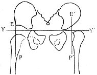
Figure 5: X-ray measurements of congenital hip dislocation.
YY′ = Y-line (Hilgenreiner line); EP, E′P′ = Perkin line; dashed line = Shenton line (continuous on the unaffected side).
(2) Acetabular Index A line is drawn from the center of the Y-shaped cartilage to the edge of the acetabulum. The angle between this line and the Hilgenreiner line is called the acetabular index, which reflects the inclination of the acetabulum and the degree of acetabular development (Figure 6). At birth, the acetabular index is 25.8°–29.4°; for infants at 6 months, it ranges from 19.4° to 23.4° (Caffey 1956). For children over 2 years old, it should be less than 20°. Most scholars consider an index exceeding 25° as abnormal, while some suggest that an index over 30° indicates a clear tendency toward dislocation. In recent years, it has been observed that the acetabular index in normal newborns can be as high as 35°–40°, with the vast majority later developing into normal hip joints. Therefore, diagnosis should not rely solely on the acetabular index. However, values exceeding the normal range indicate an increased inclination of the acetabular roof, signifying acetabular dysplasia.
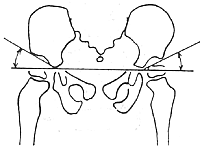
Figure 6 Measurement of Acetabular Index
(3) Epiphyseal Lateral Displacement Measurement The distance from the center of the femoral head epiphysis to the central vertical line of the pubic symphysis is called Perkins' center distance. Comparing both sides, an increased distance indicates outward displacement of the femoral head. This method is commonly used for hip joint subluxation and is particularly valuable for measuring Grade I subluxation. Before the epiphysis appears, the medial edge of the femoral neck can also be used as a reference point for measurement.
(4) Von Rosen Line With both thighs abducted 45–50° and internally rotated, an anteroposterior radiograph of the pelvis including the upper ends of both femurs is taken. The central axis lines of both femurs are drawn and extended proximally, forming the Von Rosen line. Normally, this line passes through the superolateral edge of the acetabulum; in dislocation, it passes through the anterior superior iliac spine. Before the ossification center of the femoral head appears, this method has certain diagnostic value (Figure 7).
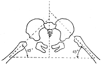
Figure 7 Von Rosen Line
Left side (normal): The femoral shaft axis passes through the superolateral edge of the acetabulum.
Right side (dislocation): The femoral shaft axis passes through the anterior superior iliac spine.
(5) Shenton's Line In a normal pelvic radiograph, the curved line of the inferior border of the pubis and the medial curved line of the femoral neck can form a continuous arc, known as Shenton's line. In cases of hip dislocation or subluxation, the continuity of this line is disrupted.
This line disappears in any type of dislocation and thus cannot differentiate between inflammatory, traumatic, or congenital conditions. Nevertheless, it remains one of the simplest diagnostic methods.
(6) Anterior Angle Radiography of the Femoral Neck Occasionally, further clarification of the anteversion angle requires radiographic examination. The simplest method involves taking an anteroposterior pelvic radiograph with the child supine and the hips elevated. Another radiograph is taken with the thigh fully internally rotated. Comparing the two images, the full length of the femoral neck becomes visible with complete internal rotation, and the femoral head appears clear. When the hip is elevated, the femoral head overlaps with the greater and lesser trochanters, allowing estimation of the anteversion angle.
(7) Arthrography Generally, arthrography is rarely necessary for diagnosis. However, in certain cases, such as identifying a discoid cartilage, joint capsule constriction, or reasons for failed reduction, arthrography may occasionally be required. Under general anesthesia, the hip joint is sterilized, and 1–3 ml of 35% iodized oil contrast agent (diodone diodast) is injected via anterior joint puncture. Fluoroscopy can reveal obstacles at the lateral edge of the acetabulum, the condition of the cartilage at the acetabular rim, and whether the joint capsule is constricted. If necessary, after manual reduction, a repeat arthrogram can confirm whether the femoral head is fully seated in the acetabulum and assess the reduction and deformation of the discoid cartilage. Due to its complexity, insufficient contrast filling, and difficulty in interpretation, arthrography has been less commonly used in recent years.
(8) Center-Edge Angle (CE Angle) Follow-up cases often require assessment of femoral head coverage by the acetabulum. Wiberg's method involves marking the center of the femoral head and the lateral edge of the acetabulum, connecting these two points with a line. A vertical line is drawn downward from the acetabular edge, and the angle formed at the acetabular edge is the center-edge angle. The normal range is 20–46°, averaging 35°; 15–19° is borderline, and less than 15° or a negative angle indicates femoral head lateral displacement, suggesting dislocation or subluxation (Figure 8).
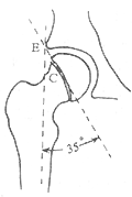
Figure 8 Measurement of the Center-Edge Angle
E = Superolateral edge of the acetabulum C = Center of the femoral head (i.e., the center of the epiphyseal line)
When available, CT or MRI examinations can provide a definitive diagnosis.
bubble_chart Treatment Measures
The treatment of congenital hip dislocation should emphasize early diagnosis. The best therapeutic results are achieved during infancy, with efficacy decreasing as age increases. It is generally believed that even if treatment after 2-3 years of age is highly successful, hip arthralgia will inevitably occur after the age of 35. Therefore, most scholars stress the importance of screening newborns to facilitate early diagnosis and treatment, which are crucial measures for achieving a complete cure. For teratologic dislocation, there is currently no effective treatment, and open reduction is usually required, but the outcomes are poor. In cases of typical congenital hip dislocation, if treated correctly and early, there is a high likelihood of developing a normal hip joint under the stimulation of normal function. For children treated before the age of 3, the cure rate is very high. As age increases, the osseous components of the femoral head and acetabulum increase, plasticity decreases, and pathological changes worsen. Even with correct treatment, normal function is difficult to achieve.
Treatment methods include closed reduction + brace, closed reduction + frog-leg plaster cast; closed reduction + rotational osteotomy to correct anteversion; open reduction, with additional acetabular reconstruction and various osteotomies depending on the specific condition. The specific treatment principles are as follows:
(1) Birth to 2 months: No traction or anesthesia is needed. Flexion of both hips to 90° followed by gradual abduction, with the thumb placed on the greater trochanter and pushed forward and inward, can achieve reduction. Avoid forceful reduction. After successful reduction, a brace can be used to fix the hip joint at 90° flexion and 70° abduction for approximately 2-3 months, depending on the age at reduction. The brace should be removed only after radiographic confirmation. There are many types of braces, including the abduction diaper pillow (Figure 9) and the Begg plastic brace (Figure 10). Both of these braces must be opened during diaper changes, which is cumbersome and less commonly used nowadays. The Barlow brace (Figure 11) and Rosen brace (Figure 12) are effective but may cause skin pressure, leading to pain and pressure sores, as well as the risk of ischemic necrosis of the femoral head. The Pavlik brace (Figure 13) avoids complications of ischemic necrosis caused by forceful reduction. It utilizes the natural position of the lower limbs flexed at 90° and the weight of the legs to achieve abduction, allowing natural reduction and maintaining the reduced position. This is beneficial for hip joint development and remodeling and allows some range of motion. The drawback is that it is made of canvas and can be stiff. If the shoulder and chest straps are too tight, breathing may be affected; if too loose, it may slip off, compromising treatment.
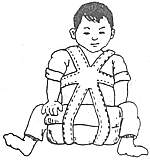
Figure 9 Abduction diaper pillow
|
|
|
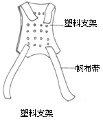
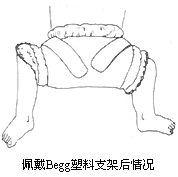
Figure 10 Begg plastic brace
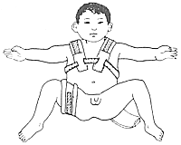
Figure 11 Barlow brace
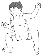
Figure 12 Rosen brace
|
|
|
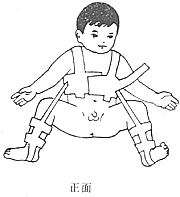
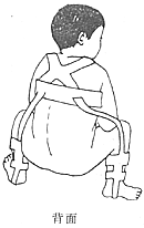
Figure 13 Pavlik Harness
(2) Over 3 months to under 2–3 years old In this group of cases, due to the prolonged dislocation, the soft tissues around the hip have varying degrees of contracture. Therefore, traction is applied before reduction, generally not exceeding 2 weeks. If muscle contracture is more pronounced, release procedures such as adductor tenotomy or iliopsoas lengthening must be performed prior to reduction. After confirmation by bedside X-ray that the femoral head has reached the level of the acetabulum, closed reduction under general anesthesia is performed. If the reduction is satisfactory, frog-leg plaster fixation is applied. To accommodate the child's growth and development, the plaster is replaced every 2–3 months, with each replacement requiring X-ray confirmation of the femoral head's position within the acetabulum. If dislocation recurs after plaster replacement, reduction must be performed again. With each plaster change, the thigh is gradually adducted until the acetabulum develops normally, at which point the plaster fixation can be removed. If reduction fails, it may indicate the presence of fibrofatty tissue hyperplasia in the acetabulum, thickened ligamentum teres, or an hourglass-shaped joint capsule obstructing the femoral head's entry into the acetabulum, necessitating open reduction.
(3) Over 3 years to 8 years old In this group, the dislocation has persisted for a long time, with more pronounced soft tissue contractures and poorer acetabular development, often being small and shallow with abundant fibrofatty tissue in the acetabular floor. Closed reduction is extremely difficult, so the vast majority require open reduction. However, 2–3 weeks of traction must be applied before open reduction until the femoral head is pulled to the level of the acetabulum. If the femoral head cannot be pulled to the acetabular level, it indicates significant soft tissue contracture. Performing open reduction at this stage carries a high risk of ischemic necrosis of the femoral head, so soft tissue release must be performed first, followed by traction. After open reduction, additional procedures may be performed based on the specific conditions:
1. Femoral head shelf procedures Generally suitable for children with subluxation and poor acetabular development where the femoral head cannot be fully covered. There are three main types of such procedures:
(1) Pelvic osteotomy (Salter procedure): Adequate reduction must be achieved before surgery. If closed reduction is difficult, open reduction must be performed during the procedure, followed by pelvic osteotomy. The distal osteotomy fragment must be pulled anteroinferiorly during surgery to increase femoral head coverage and hip joint stability (Figure 14).
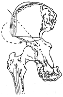
Figure 14 Salter Procedure
(2) Pelvic osteotomy with shelf augmentation (Chiari procedure): This procedure must be performed on a traction table with X-ray monitoring to ensure accurate positioning. The attachment points of the joint capsule must be clearly identified. There is a risk of sciatic nerve injury during surgery, and the chance of contamination is high, so this method is less commonly used now (Figure 15).
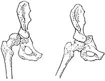
Figure 15 Chiari Procedure
(3) Pericapsular osteotomy (Pemberton procedure): This procedure involves rotating the superior part of the acetabulum anterolaterally to increase coverage. A bone graft from the ilium is inserted into the osteotomy site to stabilize the reconstructed acetabulum. Postoperative plaster fixation is applied (Figure 16).
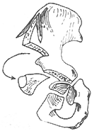
Figure 16 Pemberton Procedure
2. Zahradnick's Surgery: First, perform an open reduction and deepen the acetabulum. After reduction, due to the large femoral neck anteversion angle, the lower limb can only achieve reduction in extreme internal rotation. Therefore, a subtrochanteric rotational osteotomy must be performed, followed by fixation with a plate and screws. Postoperatively, immobilize with a cast. After 4–6 weeks, remove the anterior half of the cast to exercise hip flexion and extension functions, while continuing immobilization at night. Once X-ray confirms healing at the osteotomy site, the patient can begin functional exercises out of bed.
For children over 8 years old, open reduction is generally difficult and carries many complications, so it is usually not performed. Instead, conservative surgeries aimed at stabilizing the hip joint are preferred, such as acetabular bone grafting and shelf operation (Figure 17) or femoral osteotomy (Figure 18). In recent years, shortening the femur before performing open reduction has shown acceptable short-term results.
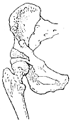
Figure 17 Acetabular Bone Grafting and Shelf Operation
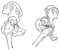
Figure 18 Subtrochanteric Bifurcation Osteotomy
For adult congenital hip dislocation, it is more commonly seen in postpartum women and is often a subluxation. Due to long-term abnormal weight-bearing on the hip joint, traumatic arthritis can easily develop, leading to hip pain. For such cases, obturator neurectomy is generally used to temporarily relieve pain. If hip joint function is already affected, total hip replacement surgery may be considered.
Complications arising from the treatment of congenital hip dislocation are mostly caused by rough manipulation, insufficient traction, failure to grasp surgical indications, unclear understanding of factors hindering reduction, and improper fixation. Most of these complications can be avoided. Common complications include:
(1) **Redislocation** — Often due to unresolved factors hindering reduction. False appearances on X-rays, carelessness during cast changes, excessive anteversion angle, or acetabular dysplasia can make redislocation more likely even after reduction.
(2) **Ischemic necrosis of the femoral head** — This complication mainly results from rough manipulation or excessive surgical trauma, damaging the blood supply to the femoral head; forceful extreme abduction during fixation; insufficient traction before reduction or failure to release the adductor and iliopsoas muscles, leading to excessive pressure on the femoral head after reduction; and some unknown causes.
(3) **Hip joint osteoarthritis** — A late-stage complication, generally occurring in older children post-surgery, and often difficult to avoid by adulthood.
(4) **Femoral head epiphyseal separation, femoral shaft fracture, sciatic nerve injury, etc.** — These are usually caused by insufficient traction, forceful reduction, or inadequate anesthesia and can generally be avoided.


