| disease | Phlyctenular Keratoconjunctivitis |
| alias | Phlyctenular Keratoconjunctivitis |
Phlyctenular keratoconjunctivitis is a delayed-type hypersensitivity disease caused by microbial proteins, primarily occurring in spring and summer. It is characterized by recurrent nodular cellular infiltrates beneath the conjunctival and corneal epithelium, with the central necrotic tissue sloughing off to form an ulcer, surrounded by localized congestion. The condition can resolve spontaneously but is highly prone to recurrence. Particularly in bilateral cases, lesions may alternate and recur over months or even years. The prognosis is generally good, but when lesions are located in the central cornea, varying degrees of visual impairment may occur.
bubble_chart Etiology
The occurrence of phlyctenular keratoconjunctivitis is now widely regarded as an infectious immune mechanism, involving a delayed-type hypersensitivity reaction triggered by various microbial proteins. These include bacterial proteins such as those from subcutaneous node bacilli, Staphylococcus aureus, common bletilla tuber fungi, Chlamydia, or proteins from Chinese Taxillus Herb parasites. When microbial proteins or other antigens enter the body, they stimulate antibody production, sensitizing T cells and promoting their proliferation, thereby placing the body in a hypersensitive state. Upon re-exposure to the same antigen, the sensitized lymphocytes directly attack cells carrying the antigen while simultaneously releasing various lymphokines, leading to a localized inflammatory response. This results in the formation of vesicles composed of monocytes, macrophages, and lymphocytes.
This condition predominantly affects children and adolescents, particularly those with malnutrition or allergic predispositions. Poor hygiene habits and damp, poorly lit living environments are also contributing factors. Patients often present with concurrent eczema on the eyelids, cheeks, ears, nose, or other body parts, as well as subcutaneous lymph nodes or subcutaneous bone nodes.
bubble_chart Clinical ManifestationsPhlyctenular keratoconjunctivitis presents only with a foreign body sensation or burning sensation. If it invades the cornea, severe photophobia, tearing, stabbing pain, and blepharospasm may occur.
Based on the site of the lesion, the condition can be clinically classified as follows: when the lesion occurs only in the conjunctiva, it is called phlyctenular conjunctivitis; when it occurs in the cornea, it is called phlyctenular keratitis; and when the lesion involves both the cornea and conjunctiva, it is called phlyctenular keratoconjunctivitis.
1. **Phlyctenular conjunctivitis**: The nodules on the bulbar conjunctiva appear grayish-red, with a diameter of about 1–4 mm, surrounded by localized conjunctival hyperemia (see figure). The nodules are prone to rupture, forming an ulcer at the apex. Subsequently, epithelial cells grow inward from the edges, and the ulcer usually heals within about a week without leaving a scar. In more severe cases, larger ulcers may form, potentially extending to the superficial sclera, leaving a scar after healing. In rare cases, phlyctenular ulcers may appear on the palpebral conjunctiva or eyelid margin, often seen in individuals with vitamin A deficiency.
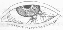
**Figure**: Phlyctenular conjunctivitis
2. **Phlyctenular keratoconjunctivitis**: The nodules are located at the limbus of the cornea, presenting as grayish-white round infiltrates with clear boundaries, which are prone to ulceration. After healing, opaque scars remain on the cornea, causing an irregular corneal limbus. Sometimes, numerous millet-seed-like tiny nodules appear along the corneal limbus and adjacent bulbar conjunctiva, arranged in a row, termed **miliary phlyctenular keratoconjunctivitis**. These nodules may disappear without ulceration or may coalesce to form ulcers.
3. **Phlyctenular keratitis and fascicular keratitis**: Refer to the chapter on corneal diseases.bubble_chart Treatment Measures
Apply 0.5% cortisone eye drops, 0.1% rifampicin eye drops, as well as yellow and white mercuric oxide ointments locally. To prevent secondary infections, broad-spectrum antibiotic eye drops can be used simultaneously. For cases involving the cornea, treat as keratitis. Administer cod liver oil, calcium supplements, and multivitamins orally. Additionally, enhance nutrition, adjust diet, increase exposure to sunlight and fresh air, and focus on physical exercise to strengthen the constitution. For stubborn and recurrent cases, subcutaneous nodular bacillus desensitization therapy may be attempted.


