| disease | Infraorbital Space Infection |
Infraorbital space infection refers to an acute suppurative infection in the infraorbital space, with main clinical manifestations including redness and increased tension in the infraorbital skin, eyelid edema, narrowing of the palpebral fissure, and disappearance of the nasolabial fold.
bubble_chart Etiology
The infraorbital space is located between the anterior wall of the maxilla and the facial expression muscles below the orbit. Its upper boundary is the infraorbital margin, the lower boundary is the maxillary alveolar process, the medial boundary is the nasal margin, and the lateral boundary is the zygomatic margin. Within the space, the infraorbital nerves, blood vessels, and infraorbital lymph nodes emerge from the infraorbital foramen. Additionally, there are the medial canthus artery, facial veins, and their communicating branches with the ophthalmic veins, infraorbital veins, and deep facial veins (Figure 2).
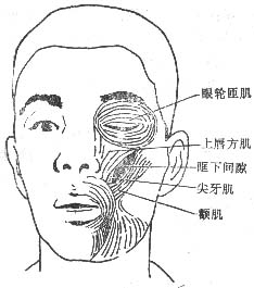
Figure 2 Anatomical location of the infraorbital space
Infections in the infraorbital space often originate from the apical suppurative inflammation or alveolar abscess of the maxillary canine and first premolar or maxillary incisors. Additionally, pus from maxillary osteomyelitis may penetrate the bone membrane, or suppurative inflammation from the base of the upper lip and nasal side may spread to the infraorbital space.
bubble_chart Clinical Manifestations
The swelling in the infraorbital region often extends to the inner canthus, eyelid, and the skin of the zygomatic area. The skin in the swollen area becomes red with increased tension, accompanied by eyelid edema, narrowing of the palpebral fissure, and disappearance of the nasolabial fold. After abscess formation, fluctuation can be palpated in the infraorbital region, with obvious swelling and tenderness in the gingival sulcus of the oral vestibule, where fluctuation is easily detected. In a few cases, the abscess may rupture spontaneously, leading to pus discharge. During the infection phase, swelling and inflammation may irritate the infraorbital nerve, causing varying degrees of pain.
Infection in the infraorbital space can spread directly upward into the orbit, leading to orbital cellulitis, or it may disseminate along the facial vein, inner canthus vein, and ophthalmic vein to the intracranial region, complicating into cavernous sinus thrombophlebitis.bubble_chart Treatment Measures
In the stage of infraorbital space cellulitis, treatment can begin with topical application of Chinese medicinals and management of the infected tooth. Once an abscess forms, timely incision and drainage should be performed. Following the principle of dependent drainage, an incision is typically made in the oral cavity at the vestibular mucosal reflection of the maxillary anterior teeth and premolar region (Figure 1). The mucoperiosteum is incised horizontally down to the bone surface, and the abscess is separated toward the canine fossa using a hemostat to ensure adequate drainage of pus. The abscess cavity is then irrigated with saline, and a rubber drain is left in place.
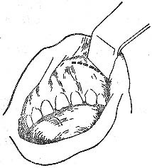
Figure 1 Incision for drainage of infraorbital space abscess.


