| disease | Femoral Hernia |
| alias | Femoral Hernia |
When organs or tissues protrude into the femoral canal through the femoral ring, and then protrude from the femoral canal into the oval fossa, it is called a femoral hernia.
bubble_chart Clinical Manifestations
Although the female pelvis is wider and the space beneath the inguinal ligament is also broader, making women more prone to femoral hernias than men, femoral hernias are still far less common than inguinal hernias.
Typically, a femoral hernia causes no specific discomfort, presenting only as a round lump near the groin below the inguinal region. Due to the narrow femoral canal and the tortuous path of the hernia, rest or lying flat does not easily reduce or completely resolve the hernial mass. The cough impulse is also not prominent.
Approximately half or more of femoral hernias may be complicated by incarceration and strangulation, often leading patients to seek medical attention due to acute abdominal pain or strangulated intestinal obstruction. Therefore, for patients with acute surgical abdominal pain, examination of the groin and femoral region should not be overlooked. The hernia contents are often the greater omentum, and parietal hernias (Richter's hernia) are also not uncommon. Femoral hernias are characterized by being irreducible and prone to incarceration and strangulation.
bubble_chart Treatment Measures
Femoral hernias should all be treated surgically, with two surgical approaches: the suprainguinal and the infrainguinal.
1. Suprainguinal approach (Figure 1) An oblique hernia incision is made, and the layers are dissected to expose the posterior wall of the inguinal canal. The round ligament/spermatic cord is retracted upward, and the transversalis fascia is incised medially and superiorly to the inguinal ligament to locate the femoral ring and the neck of the hernia sac. The neck of the hernia sac is incised, the hernia contents are reduced, and high ligation of the hernia sac is performed above the femoral ring. The distal hernia sac does not require treatment. In cases of incarcerated femoral hernia, the iliopubic tract reflection and the lacunar ligament at the medial boundary of the femoral ring must be incised to release the constriction before reducing the hernia mass. Avoid pulling the incarcerated hernia contents from the femoral canal opening.
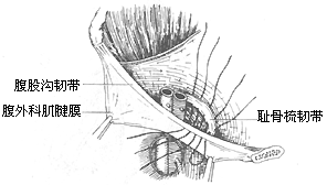
Figure 1: Suprainguinal approach for femoral hernia repair, suturing the pectineal ligament to the inguinal ligament
The repair of a femoral hernia involves suturing the inguinal ligament, iliopubic tract, lacunar ligament, and pectineal ligament to close the femoral ring, taking care to avoid injuring the femoral vein. Alternatively, the McVay method can be used, suturing the internal oblique muscle, the arched tendon of the transversus abdominis, the upper cut edge of the transversalis fascia, and the conjoint tendon to the pectineal ligament, with additional sutures placed laterally to the femoral sheath and medial to the spermatic cord.
2. Infrainguinal approach (Figure 2) A vertical incision is made over the oval fossa below the inguinal ligament. The cribriform fascia is incised to expose the hernia sac, which is then opened to reduce the hernia contents. After high ligation of the hernia sac, the inguinal ligament, iliopubic tract, lacunar ligament, pectineal ligament, and pectineal fascia are sutured to close the femoral ring.
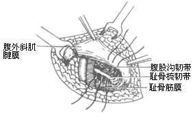
Figure 2: Infrainguinal approach for femoral hernia repair, suturing the inguinal ligament to the pectineal ligament and pectineal fascia
Although there are two surgical approaches for femoral hernia, the suprainguinal approach is more commonly used. Its advantage lies in providing clear exposure of the femoral ring, enabling true high ligation of the hernia sac and secure closure of the femoral ring. For strangulated femoral hernias, the suprainguinal approach is particularly preferred, as it allows better management of the strangulated hernia contents—something the infrainguinal approach cannot achieve. The sole advantage of the infrainguinal approach is its simplicity and lesser trauma.

The diagnosis of femoral hernia is sometimes not straightforward and requires differentiation from the following conditions.
Inguinal hernia Femoral hernia can sometimes be confused with inguinal hernia. Using the inguinal ligament as a boundary, the mass of a femoral hernia should be located below and medial to the inguinal ligament and lateral and inferior to the pubic tubercle, whereas the mass of an inguinal hernia is located above the inguinal ligament. Femoral hernias are generally smaller, difficult to reduce, and often lack a history of recurrent protrusion, whereas inguinal hernias are easier to reduce, and their reduction path differs from that of femoral hernias.
Chronic lymphadenitis Chronic lymphadenitis in the femoral triangle may present with several enlarged, mobile lymph nodes and may have a history of acute infection. Femoral hernia, in contrast, is a single irreducible mass.
Varicose veins of the great saphenous vein Varicose veins at the site where the great saphenous vein enters the fossa ovalis can form a venous mass that must be differentiated from femoral hernia. If the affected limb is elevated while lying flat, the venous mass will disappear quickly and reappear upon standing, accompanied by varicose veins in the lower limb.
Round ligament cyst Located within the inguinal canal above the inguinal ligament, this can be distinguished from femoral hernia based on its position. Additionally, the mass is round or oval, highly mobile, and has a cystic sensation.
Psoas cold abscess Cold abscesses formed by subcutaneous nodes in the lumbar spine often extend downward along the iliopsoas muscle and appear on the medial side of the thigh root. They are not actually located where femoral hernias occur, and careful identification of anatomical landmarks can facilitate differentiation. Furthermore, cold abscesses exhibit obvious fluctuation, and lumbar spine X-rays will reveal subcutaneous node lesions.


